

Abstract
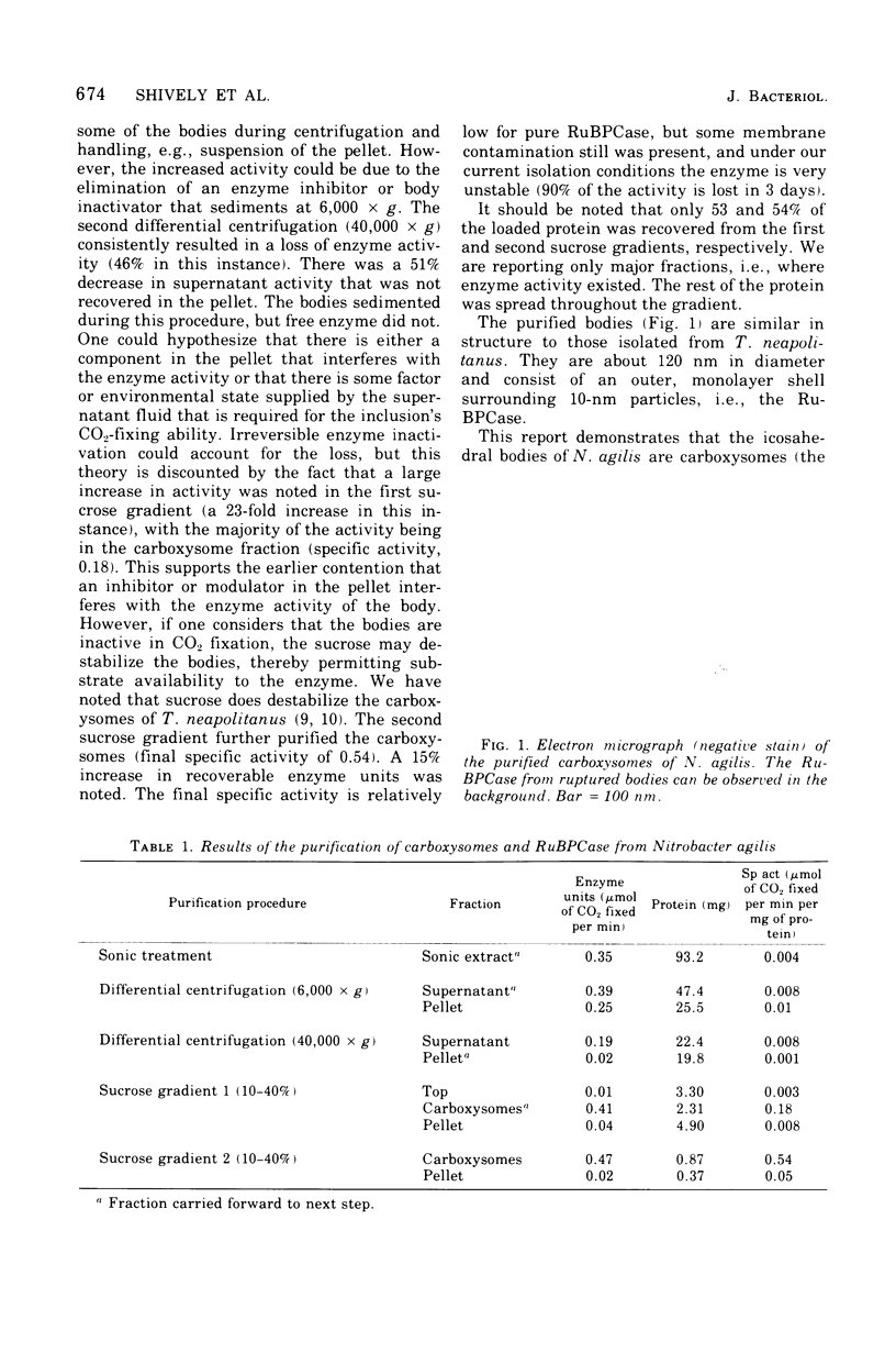
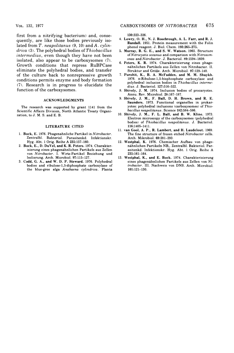
The icosahedral bodies of Nitrobacter agilis are about 120 nm in diameter and, as viewed by electron microscopy, consist of an outer shell enclosing 10-nm particles. The inner 10-nm particle is the enzyme D-ribulose 1,5-bisphosphate carboxylase. The bodies isolated from cells incubated 1 month without nitrite had a specific activity for the enzyme of 0.54 mu mol of CO2 fixed per min per mg of protein.
Full text
PDF
Images in this article
Selected References
These references are in PubMed. This may not be the complete list of references from this article.
- Bock E., Düvel D., Peters K. R. Charakterisierung eines phagenähnlichen Partikels aus Zellen von Nitrobacter. I. Wirts-Partikelbeziehung und Isolierung. Arch Microbiol. 1974 Apr 19;97(2):115–127. doi: 10.1007/BF00403051. [DOI] [PubMed] [Google Scholar]
- Bock E. Phagenähnliche Partikel in Nitrobacter. Zentralbl Bakteriol Orig A. 1976 Aug;235(1-3):157–160. [PubMed] [Google Scholar]
- LOWRY O. H., ROSEBROUGH N. J., FARR A. L., RANDALL R. J. Protein measurement with the Folin phenol reagent. J Biol Chem. 1951 Nov;193(1):265–275. [PubMed] [Google Scholar]
- MURRAY R. G., WATSON S. W. STRUCTURE OF NITROSOCYSTIS OCEANUS AND COMPARISON WITH NITROSOMONAS AND NITROBACTER. J Bacteriol. 1965 Jun;89:1594–1609. doi: 10.1128/jb.89.6.1594-1609.1965. [DOI] [PMC free article] [PubMed] [Google Scholar]
- Peters K. R. Charakterisierung eines phagenähnlichen Partikels aus Zellen von Nitrobacter. II. Struktur und Grösse. Arch Microbiol. 1974 Apr 19;97(2):129–140. doi: 10.1007/BF00403052. [DOI] [PubMed] [Google Scholar]
- Purohit K., McFadden B. A., Shaykh M. M. D-Ribulose-1,5-bisphosphate carboxylase and polyhedral inclusion bodies in Thiobacillus intermedius. J Bacteriol. 1976 Jul;127(1):516–522. doi: 10.1128/jb.127.1.516-522.1976. [DOI] [PMC free article] [PubMed] [Google Scholar]
- Shively J. M., Ball F. L., Kline B. W. Electron microscopy of the carboxysomes (polyhedral bodies) of Thiobacillus neapolitanus. J Bacteriol. 1973 Dec;116(3):1405–1411. doi: 10.1128/jb.116.3.1405-1411.1973. [DOI] [PMC free article] [PubMed] [Google Scholar]
- Shively J. M., Ball F., Brown D. H., Saunders R. E. Functional organelles in prokaryotes: polyhedral inclusions (carboxysomes) of Thiobacillus neapolitanus. Science. 1973 Nov 9;182(4112):584–586. doi: 10.1126/science.182.4112.584. [DOI] [PubMed] [Google Scholar]
- Shively J. M. Inclusion bodies of prokaryotes. Annu Rev Microbiol. 1974;28(0):167–187. doi: 10.1146/annurev.mi.28.100174.001123. [DOI] [PubMed] [Google Scholar]
- Westphal K., Bock E. Charakterisierung eines phagenähnlichen Partikels aus Zellen von Nitrobacter. Arch Microbiol. 1974;101(2):121–130. doi: 10.1007/BF00455932. [DOI] [PubMed] [Google Scholar]
- Westphal K. Chemischer Aufbau von phagenähnlichen Partikeln Nb1. Zentralbl Bakteriol Orig A. 1976 Aug;235(1-3):161–164. [PubMed] [Google Scholar]
- van Gool A. P., Lambert R., Laudelout H. The fine structure of frozen etched nitrobacter cells. Arch Mikrobiol. 1969;69(4):281–293. doi: 10.1007/BF00408570. [DOI] [PubMed] [Google Scholar]