Abstract
Only a small proportion (20%) of patients with LBP can be diagnosed based on a patho-anatomical entity. Therefore, the identification of relevant subgroups, preferably on a patoanatomical basis, is strongly needed. Modic changes have been described by several authors as being closely linked with LBP. The aims of this study were to describe the prevalence of Modic changes, their development as well as their association to LBP, previous disc contour, and surgery in patients with previous severe sciatica. This is a longitudinal cohort study where the patients were recruited from an RCT comparing two active conservative treatments, the 181 patients, who at baseline had radicular pain in or below the knee; all underwent a physical examination and MRI. MRI’s, pain history and physical examination of 166 patients were obtained at follow-up 14 months later. The prevalence of Modic changes type 1 increased from 9% at baseline to 29% at follow-up. At that time, a strong association between Modic changes and non-specific LBP was noted. Apparently, Modic changes type 1 was more strongly associated with non-specific lumbar pain than Modic changes type 2. The development of new Modic changes was closely related to the level of a previous disc herniation. A lumbar disc herniation is a strong risk factor for developing Modic changes (especially type 1) during the following year. Furthermore, Modic changes are strongly associated with LBP.
Keywords: Low back pain, Modic changes, MRI, Disc herniation, Vertebral endplates changes
Introduction
Diagnosing patients with low back pain (LBP) is a challenge for clinicians [13]. It is frequently stated that only a small proportion (approximately 20%) of patients with LBP can with certainty be diagnosed based on a patho-anatomical entity [13]. The most commonly used classification, “non-specific LBP” (80%), is not satisfactory for the LBP patient or the clinician. Therefore, the identification and diagnosis of relevant subgroups of patients with persistent LBP, preferably with a sound patoanatomical basis, is strongly needed.
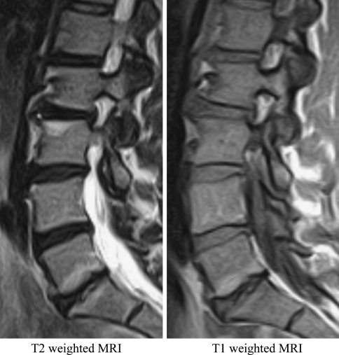
In the recent literature, Modic changes [9, 11, 14] have been described as being strongly associated with LBP. According to Modic et al. [10, 11], these changes are visible on magnetic resonance imaging (MRI) as three different types. Type 1 is seen on T2-weighted MRI as areas of increased signal intensity and on T1-weighted MRI as low signal intensity extending from the vertebral endplates. From histological studies of material harvested during surgery, Modic changes type 1 have been described as disruption and fissuring of the endplate with regions of degeneration, regeneration, and vascular granulation tissue [10]. Type 2 is observed as increased signal intensity on both T1- and T2-weighted images, portraying disruption of the end plates with increased reactive bone and granulation tissue. The hematopoetic elements in the vertebrae are replaced by abundant fat (yellow marrow) [10]. Modic changes type 3 are presumably bone sclerosis and are visualized on MRI as decreased signal intensity on both T1 and T2-weighted images [10].
Mitra et al. [9] observed Modic changes type 1 in 18% of 670 patients referred to MRI due to LBP or sciatica. These patients were rescanned after 12–72 months and in the patients whose symptoms had improved, the type 1 changes had developed into type 2. In the patients reporting a worsening of their symptoms, the type 1 changes had progressed and become worse. Similarly, Toyone et al. [12] found that 19% of 500 patients with chronic back pain had Modic changes. They also noticed that it was more common for the patients with type 1 changes to report LBP than patients with type 2 changes. Kjaer et al. [7] observed Modic changes type 1 in 15% and type 2 in 7% in a sample of 412 persons from the general population aged 40. A strong association with LBP within the past year was observed and this association was stronger for the Modic changes type 1. Apparently, Modic changes type 1 are more strongly associated with pain compared to type 2. The reason for this may be that Modic changes type 1 reflects earlier and acute stages of inflammation, whereas Modic changes type 2 are thought to be a result of previous inflammation and more progressive degeneration. Endplate changes have also been described following discectomy [3], with varying prevalence from 6 [3] to 18% [6] and as a sign of septic and aseptic discitis [4].
The aims of this paper are to describe Modic changes regarding the following: their cause and prevalence at baseline and follow-up and their relation to disc contour at baseline and localized lumbar pain and surgery.
Materials and methods
This study sample consisted of 181 patients who participated in a randomized controlled trial involving active conservative treatment of patients with severe sciatica [1]. The treatment consisted of thorough information, advise to stay active, optional medication and symptom-guided exercises or sham exercises. The setting was an outpatient back specialist centre. The study was approved by The Regional Scientific Ethical Committee No. VF 20030212, and written informed consent was obtained from all participants prior to participation.
Inclusion criteria
At baseline, all of the patients experienced well-defined radiating pain in accordance with the dermatome in one or both legs down to and at least including the knee; leg pain >3 on a 0–10 scale, duration of radiating pain was between 2 weeks and 1 year. Patients were aged 18–65.
Exclusion criteria
Cauda equine syndrome, not demonstrating proficiency in Danish, pending workers litigation claims, previous back surgery, spinal tumours, pregnancy, or inability to follow the rehabilitation protocol due to concomitant disease.
Procedures
The treatment consisted of one of two types of active conservative treatment and lasted for 8 weeks. A thorough medical history and examination of the spine and lower extremities was undertaken by the same observer at baseline and at follow-up sessions. The observer was blinded to treatment allocation and MRI results [1]. Lumbar MRI was performed immediately after the baseline examination. A standard lumbar MRI-protocol on a 0.2 T MRI-system (Siemens Open Viva) was used. All individuals were placed supine with straight legs in a psoas-tight position in order to imitate the upright lumbar lordosis. The imaging protocol consisted of one localizer and four imaging sequences:
Localizer sequence, 40/10/40 (TR/TE/flip angle), two coronal and three sagittal images in orthogonal planes, one acquisition in 32 s.
Sagittal T1-weighted spin echo, 621/26 (TR/TE), 144 × 256 matrix, 300 mm FOV, and 11 4 mm slices, distance factor 0.20, two acquisitions in 6:01 min.
Sagittal T2-weighted turbo spin echo, 4,609/134 (TR/effective TE), 210 × 256 matrix, 300 mm FOV, and 11 4 mm slices, distance factor 0.20, two acquisitions in 8:42 min.
Axial T1-weighted spin echo, 720/26 (TR/TE), 192 × 256 matrix, 240 mm FOV, and 15 5 mm slices, distance factor 0.25, two acquisitions in 8:49 min.
Axial T2-weighted turbo spin echo, 6,415/134 (TR/effective TE), 180 × 256 matrix, 250 mm FOV, and 15 5 mm slices, distance factor 0.25, one acquisition in 7:49 min.
Axial images were performed on the three lower lumbar levels. If protrusions were present at higher lumbar levels, relevant supplementing axial series were performed.
MRI evaluation was carried out by the same experienced radiologist at baseline and follow-up. Pre-defined protocols were used in evaluating MRI-changes of the five lumbar levels, counting from the last free disc. The radiologist was blinded to the patient’s treatment group, clinical signs and symptoms. The MRI findings were graded according to the latest recommendations for disc herniation [5], and defined according to the criteria of Modic et al. [10, 11] (Figs. 1, 2) and reported on a standardized sheet.
Fig. 1.
Modic changes type 1 in lower endplate of the L4 vertebrae and the upper endplate of the L5 vertebrae
Fig. 2.
Modic changes type 2 in the lower endplate of the L5 vertebrae and the upper endplate of the S1
At follow-up non-specific LBP was defined if the patient reported to have present LBP somewhere in the area from L1 to S1 and extending 15 cm to each side from the midline. Each patient marked the area of pain on a body chart. Intensity was evaluated on low back pain rating scale [8].
Statistical analyses
For the prevalence of Modic changes, 95% confidence intervals (CI) were constructed. Relevant variables were cross tabulated and differences in proportions were tested using the Chi square or Fisher’s exact test. Associations were calculated from the cross-tabulations as odds ratios (OR) with 95% confidence intervals. The statistical package STATA 8 was used.
Results
A total of 181 patients were included. One patient refused to have an MRI at baseline. Therefore, the tables include only 180 patients. During the treatment period five patients dropped out due to surgery, one had an accident and two did not complete treatment. At 14-month follow-up, six patients did not have an MRI due to pregnancy, claustrophobia or no show. In all, 166 MRI follow-up scans were obtained.
Prevalence
The prevalence rate of Modic changes type 1 increased from 17 of 180 (9%) at baseline to 48 of 166 (29%) at follow-up, the type 2 and the type 3 changes remained unchanged (Fig. 3).
Fig. 3.
The prevalence of Modic changes type 1, 2 and 3 and patients with both type 1 and 2 at baseline (n = 180) and at 14 months follow-up (n = 166) in patients with originally severe sciatica
The course of Modic changes
Development of new Modic changes type 1, were found in 29 patients at 14-month follow-up Furthermore, 19 patients who exhibited type 1 or 2 changes at baseline developed new type 1 changes. New Modic changes means; the patient had any kind of Modic changes at inclusion, where the herniation was present. 14 months later, a new Modic change type 1 was observed in another area in the same vertebrae. For example, at baseline there was a small Modic change type 2 in the very anterior part of the vertebrae, 14 months later a new Modic type 1 change was present in all of the left side of the vertebrae. In six patients with any type of Modic changes at baseline they had disappeared at follow-up (Table 1).
Table 1.
The course of different types of Modic changes and no Modic changes from baseline to the 14 months follow-up in the 166 patients
| Development of Modic changes | Number of patients | |
|---|---|---|
| From Modic changes at baseline | To Modic changes at follow-up | |
| 0 | 0 | 79 |
| 0 | 1 | 29 |
| 0 | 2 | 10 |
| 0 | 3 | 1 |
| 1 | 0 | 2 |
| 1 | New 1 | 12 |
| 1 | 2 | 2 |
| 1 | 3 | 0 |
| 2 | 0 | 4 |
| 2 | New1 | 7 |
| 2 | 2 | 9 |
| 2 | 3 | 0 |
| 3 | 0 | 0 |
| 3 | 1 | 0 |
| 3 | 2 | 1 |
| 3 | 3 | 1 |
| 0, 1, 2, to mixed types | 9 | |
| In all Modic changes at follow-up | 81 | |
| In all NO Modic changes at follow-up | 85 | |
| Total | 166 | |
Bold italic values are the ones that do not have modic changes
The new Modic changes all developed at the same vertebral level as the previously herniated disc (Fig. 4).
Fig. 4.
The total number of Modic changes in the upper and lower endplate for each lumbar vertebra, described both at baseline and at follow-up
At baseline 37 discs had Modic changes in both the upper and lower vertebrae, while 11 only at one side. At follow-up 66 discs had Modic changes in both upper and lower vertebrae, while 24 only at one side (Fig. 4).
The relationship between disc contour at baseline and Modic changes at follow-up
As seen in Table 2, none of those with normal disc contour developed Modic changes. Modic changes appear to be more closely related to sequestrated discs compared to other types of disc herniation (Table 2).
Table 2.
The development of Modic changes at 14 months follow-up in relation to the disc contour at baseline, in the 166 patients with MRI at follow-up
| Disc contour at baseline (n = 166) | Distribution (%) | Modic changes at follow-up (%) |
|---|---|---|
| Normal disc | 14 (8.4) | 0 (0) |
| Bulge | 30 (18.1) | 15 (50) |
| Focal protrusions | 62 (37.3) | 35 (56) |
| Broad based protrusions | 13 (7.8) | 4 (31) |
| Extrusions | 39 (23.5) | 22 (56) |
| Sequestrations | 8 (4.8) | 5 (63) |
Lumbar pain and Modic changes
Among the 81 patients with Modic changes 49 (60%) had self-reported non-specific LBP at follow-up and of the 85 without Modic changes only 17 (20%) suffered from non-specific LBP (P < 0.0001). Lumbar pain and Modic changes were strongly associated at follow-up with an OR of 6.1 (2.9–13.1).
The distributions of the type of Modic changes related to lumbar pain at follow-up are shown in Fig. 5. Non-specific lumbar pain was more frequent in people with Modic changes type 1 compared to those with type 2, but the difference between types was not statistically significant.
Fig. 5.
The distribution of the different types of Modic changes in relation to lumbar pain (n = 166)
Surgery and the developing Modic changes at follow up
Twelve patients underwent surgery for a lumbar disc herniation during the 1-year follow-up period, in most cases due to a new herniation or severe aggravation of previous symptoms. Of these 9 (75%) had Modic changes at follow-up, compared to 71 of the 154 (46%) (P = 0.055) who did not undergo surgery, OR 3.5 (0.8–20.8). Patients undergoing surgery for a herniated disc in the follow-up period might possibly be more likely to develop Modic changes, however, these results were not statistically significant.
Discussion
Among the patients with severe sciatica at baseline, the prevalence of Modic changes type 1 increased three-fold from baseline to 14 months follow-up, whereas the other types remained stable. Modic changes type 1 appear to be closely related to a previous disc herniation. The prevalence of Modic changes was higher in patients who had undergone surgery for lumbar disc herniation. Furthermore, Modic changes were strongly associated with LBP. Modic changes type 1 may be more strongly associated with pain compared to type 2.
Two other studies have described the development of Modic changes: Mitra et al. [9] studied a sub-sample of 44 patients with Modic type 1 from a population of 670 patients conservatively treated for LBP and/or sciatica. It is assumed that a considerable percentage of these patients with symptoms of sciatica had disc herniations. Over a period of 12–72 months 37% converted fully to type 2, 15% converted partially to type 2, 40% into more extensive type 1 changes, and 8% showed no change. This is in contrast to Modic et al.’s [10] study where Modic changes were followed in 16 patients without sciatica. Five out of six type 1 converted into type 2 while ten patients with type 2 remained unchanged after a period of 12–36 months [10].
Conclusions
A disc herniation is a strong risk factor for developing Modic changes (especially type 1) during the following year. Furthermore, both our study and others have demonstrated that Modic changes were strongly associated with LBP.
Acknowledgments
We thank Alan Jordan PhD and Charlotte Leboeuf-Yde PhD for valuable editorial assistance.
References
- 1.Albert HB (2004) Conservative treatment of patients with sciatica—a randomized controlled trial. (Dissertation) Odense, Faculty of Health Sciences, University of Southern Denmark
- 2.Babar S, Saifuddin A. MRI of the Post-discectomy lumbar spine. Clin Radiol. 2002;57:969–981. doi: 10.1053/crad.2002.1071. [DOI] [PubMed] [Google Scholar]
- 3.Boden SD, Davis DO, Dina TS, et al. Post-operative diskitis: distinguishing early MR imaging findings from normal post-operative disc space changes. Radiology. 1992;184:765–771. doi: 10.1148/radiology.184.3.1509065. [DOI] [PubMed] [Google Scholar]
- 4.Crane R. The post-operative lumbar spine. Acta Radiol Suppl. 1998;414:1–23. [PubMed] [Google Scholar]
- 5.Fardon DF, Milette PC. Nomenclature and classification of lumbar disc pathology. Spine. 2001;26:E93–E113. doi: 10.1097/00007632-200103010-00006. [DOI] [PubMed] [Google Scholar]
- 6.Grand CM, Bank WO, Baleriaux D, et al. Gadolinium enhancement of vertebral endplate following lumbar disc surgery. Neurordiology. 1993;35:503–505. doi: 10.1007/BF00588706. [DOI] [PubMed] [Google Scholar]
- 7.Kjaer P, Leboeuf-Yde C, Korsholm L, et al. Magnetic resonance imaging and low back pain in adults. A diagnostic imaging study of 40-year-old men and women. Spine. 2005;30:1173–1180. doi: 10.1097/01.brs.0000162396.97739.76. [DOI] [PubMed] [Google Scholar]
- 8.Manniche C, Asmussen K, Lauritsen B, et al. Low back pain rating scale: validation of a tool for assessment of low back pain. Pain. 1994;57:317–326. doi: 10.1016/0304-3959(94)90007-8. [DOI] [PubMed] [Google Scholar]
- 9.Mitra D, Cassar-Pullicino VN, Mitra D, et al. Longitudinal study of vertebral type 1 end-plate changes on MR of the lumbar spine. Eur Radiol. 2004;14:1574–1581. doi: 10.1007/s00330-004-2314-4. [DOI] [PubMed] [Google Scholar]
- 10.Modic MT, Steinberg PM, Ross JS, et al. Degenerative disk disease: assessment of changes in vertebral body marrow with MR imaging. Radiology. 1988;166:193–199. doi: 10.1148/radiology.166.1.3336678. [DOI] [PubMed] [Google Scholar]
- 11.Modic MT, Masaryk TJ, Ross JS, et al. Imaging of degenerative disk disease. Radiology. 1988;168:177–186. doi: 10.1148/radiology.168.1.3289089. [DOI] [PubMed] [Google Scholar]
- 12.Toyone T, Takahashi K, Kitahara H, et al. Vertebral bone-marrow changes in degenerative lumbar disc disease. An MRI study of 74 patients with low back pain. J Bone Joint Surg Br. 1994;76:757–764. [PubMed] [Google Scholar]
- 13.Waddell G. 1987 Volvo award in clinical sciences. A new clinical model for the treatment of low-back pain. Spine. 1987;12:632–644. doi: 10.1097/00007632-198709000-00002. [DOI] [PubMed] [Google Scholar]
- 14.Weishaupt D, Zanetti M, Hodler J, et al. Painful lumbar disc derangement: relevance of endplate abnormalities at MR imaging. Radiology. 2001;218:420–427. doi: 10.1148/radiology.218.2.r01fe15420. [DOI] [PubMed] [Google Scholar]


