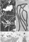
Abstract
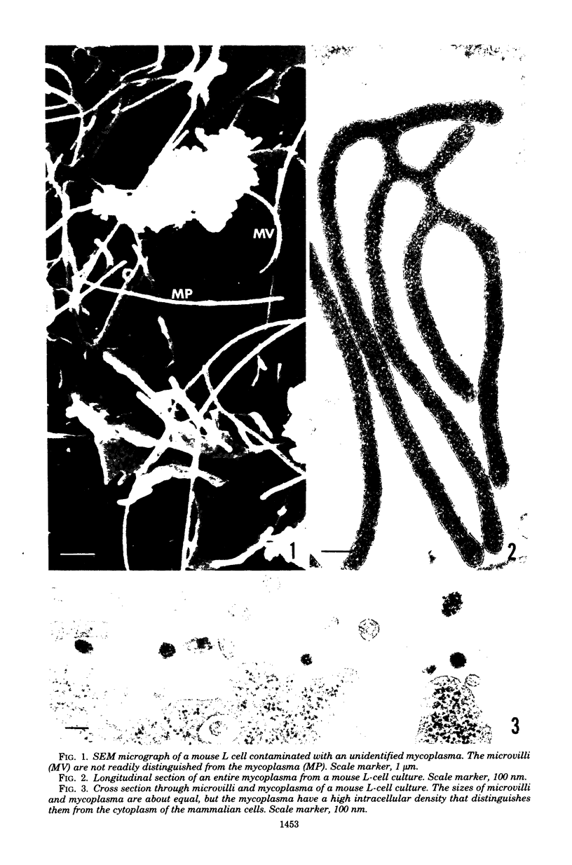
Low-level mycoplasma contamination of cell cultures is difficult to recognize with presently available techniques. This report describes the adaptation of the whole-mount technique, usually used for scanning microscopy, for transmission electron microscopy. The differentiation between microvilli and the equal-sized filamentous mycoplasma is based on the differential density obtained by the use of the method described. This method allows positive identification of mycoplasma and reduces the preparation time and the time necessary for scanning the preparation.
Full text
PDF
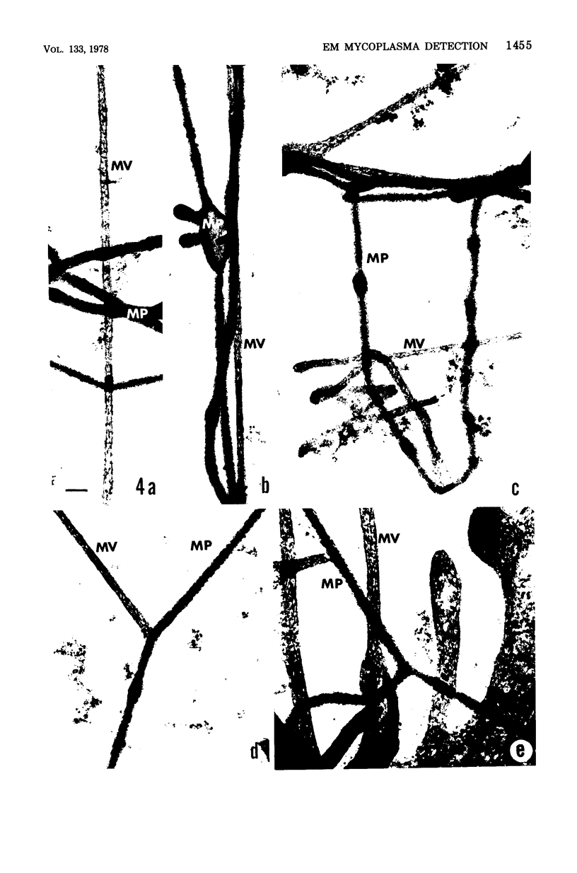
Images in this article
Selected References
These references are in PubMed. This may not be the complete list of references from this article.
- Biberfeld G., Biberfeld P. Ultrastructural features of Mycoplasma pneumoniae. J Bacteriol. 1970 Jun;102(3):855–861. doi: 10.1128/jb.102.3.855-861.1970. [DOI] [PMC free article] [PubMed] [Google Scholar]
- Brinkley B. R., Murphy P., Richardson L. C. Procedure for embedding in situ selected cells cultured in vitro. J Cell Biol. 1967 Oct;35(1):279–283. doi: 10.1083/jcb.35.1.279. [DOI] [PMC free article] [PubMed] [Google Scholar]
- Buckley I. K. Three dimensional fine structure of cultured cells: possible implications for subcellular motility. Tissue Cell. 1975;7(1):51–72. doi: 10.1016/s0040-8166(75)80007-3. [DOI] [PubMed] [Google Scholar]
- MANILOFF J., MOROWITZ H. J., BARRNETT R. J. STUDIES OF THE ULTRASTRUCTURE AND RIBOSOMAL ARRANGEMENTS OF THE PLEUROPNEUMONIA-LIKE ORGANISM A5969. J Cell Biol. 1965 Apr;25:139–150. doi: 10.1083/jcb.25.1.139. [DOI] [PMC free article] [PubMed] [Google Scholar]
- Meller S. M., Coppe M. R., Ito S., Waterman R. E. Transmission electron microscopy of critical point dried tissue after observation in the scanning electron microscope. Anat Rec. 1973 Jun;176(2):245–252. doi: 10.1002/ar.1091760210. [DOI] [PubMed] [Google Scholar]


