Abstract
Newcastle disease virus (NDV), a member of the family Paramyxoviridae, has a nonsegmented negative-sense RNA genome consisting of six genes (3′-NP-P-M-F-HN-L-5′). The first three 3′-end intergenic sequences (IGSs) are single nucleotides (nt), whereas the F-HN and HN-L IGSs are 31 and 47 nt, respectively. To investigate the role of IGS length in NDV transcription and pathogenesis, we recovered viable viruses containing deletions or additions in the IGSs between the F and HN and the HN and L genes. The IGS of F-HN was modified to contain an additional 96 nt or more or a deletion of 30 nt. Similarly, the IGS of HN-L was modified to contain an additional 96 nt or more or a deletion of 42 nt. The level of transcription of each mRNA species (NP, F, HN, and L) was examined by Northern blot analysis. Our results showed that NDV can tolerate an IGS length of at least 365 nt. The extended lengths of IGSs down-regulated the transcription of the downstream gene and suggested that 31 nt in the F-HN IGS and 47 nt in the HN-L IGS are required for efficient transcription of the downstream gene. The effect of IGS length on pathogenicity of mutant viruses was evaluated in embryonated chicken eggs, 1-day-old chicks, and 6-week-old chickens. Our results showed that all IGS mutants were attenuated in chickens. The level of attenuation increased as the length of the IGS increased. Interestingly, decreased IGS length also attenuated the viruses. These findings can have significant applications in NDV vaccine development.
Newcastle disease virus (NDV) causes a highly contagious respiratory, neurologic, or enteric disease in chickens, which leads to severe economic losses in the poultry industry worldwide. NDV isolates display a spectrum of virulence in chickens, ranging from inapparent infection to 100% mortality. Strains of NDV are classified into three major pathotypes based on the severity of the disease produced in chickens. Avirulent, intermediately virulent, and highly virulent strains are termed lentogenic, mesogenic, and velogenic strains, respectively.
NDV is a member of the genus Avulavirus in the family Paramyxoviridae. The genome of NDV is a nonsegmented, single-stranded, negative-sense RNA of 15,186 nucleotides (nt) (7, 16, 19). The genome contains at least six genes, which encode the nucleocaspid protein (NP), phosphoprotein (P), matrix protein (M), fusion protein (F), hemagglutinin-neuraminidase protein (HN), and large RNA-dependent RNA polymerase protein (L). Two additional proteins, V and W, may be produced by RNA editing during P gene transcription (24). The RNA genome is tightly encapsidated by the NP protein. This ribonucleoprotein complex serves as the template for transcription and replication by viral RNA polymerase proteins, which are L and P proteins. The NDV genes are arranged on the genomic RNA in the order 3′-NP-P-M-F-HN-L-5′. Flanking the genes are 3′ and 5′ extracistronic sequences, known as the leader and trailer, respectively. These leader and trailer regions are cis-acting regulatory elements involved in replication, transcription, and packaging of the genomic and antigenomic RNAs. The beginning and end of each gene are conserved transcriptional control sequences, known as the gene start and gene end, respectively (8, 18). Between the gene boundaries are noncoding intergenic sequences (IGSs), which vary in length from 1 to 47 nt. Each of the first three IGSs, the NP-P, P-M, and M-F gene junctions, has only 1 nt, while the other two IGSs, the F-HN and HN-L gene junctions, are 31 nt and 47 nt, respectively (4, 5, 16). The lengths of IGSs are generally conserved in most NDV strains, with the exception that the IGS in the NP-P gene junction of strain D26 is 2 nt long. However, the sequences of IGSs vary among NDV strains (12).
The transcription process for NDV is similar to that for other nonsegmented negative-sense RNA viruses (18). The viral RNA polymerase first transcribes the leader RNA at the 3′ end of the genome. Synthesis of the leader sequence terminates at the boundary of the first gene (NP) and proceeds with NP mRNA synthesis at the gene start of the NP gene. Transcription terminates at the gene end of the NP gene. The transcription complex probably passes over the NP-P IGS and begins transcription of the P gene. Therefore, IGSs are not copied into mRNAs. Transcription continues in this start-stop manner until the mRNA of the last gene, L, is synthesized. The mode of transcription leads to a gradient of mRNA abundance that is reduced according to the distance of the location of a particular gene from the 3′ end of the genome. Replication occurs when the polymerase complex ignores the transcription stop signal at the 3′ end of each gene (18). NDV RNA replication follows “the rule of six,” that is, efficient replication occurs only if the genome size is a multiple of 6 nt (7).
There are two different IGS groups among members of the order Mononegavirales. One group has short conserved or semiconserved IGSs. Members of the genera Respirovirus, Morbillivirus, and Henipavirus have IGSs that are a conserved trinucleotide. For example, Sendai virus, human parainfluenza virus type 3 (hPIV3), and Hendra virus have a conserved trinucleotide (GAA), with the exception of the HN-L IGS of Sendai virus, which is GGG (23, 28). The rhabdovirus vesicular stomatitis virus (VSV) has a semiconserved dinucleotide, GA or CA, as the IGS (11). The IGSs in the other virus group vary significantly in sequence and length. The IGSs of hPIV2 vary in length from 4 to 45 nt (14). For simian virus 5 (SV5) and respiratory syncytial virus (RSV), the IGSs vary in length from 1 to 22 and 1 to 56 nt, respectively (3, 6, 9, 20). NDV belongs to the latter group.
The roles of IGSs in transcription, replication, and viral pathogenesis of the members of the Mononegavirales are unclear. Studies using infectious recombinant RSV with various IGS lengths, from 16 to 160 nt, showed that there was no significant difference in transcription or replication in vitro or in vivo (3). Studies using a VSV minigenome system showed that some nucleotide changes in the IGS resulted in higher levels of readthrough transcription. This indicated that IGSs play a role in transcription termination in VSV (11). Studies with SV5 IGSs showed that the length of the IGS alone is not a determining factor in transcription termination/polyadenylation (10). The roles of NDV IGSs in viral transcription, replication, and pathogenicity have not been studied. It is not known why the first three 3′-end IGSs are only 1 nt long, whereas the next two IGSs are always 31 and 47 nt long.
In this paper, we investigate the role of the length of IGSs in NDV transcription and virulence. We made several constructs with either an addition or deletion of nucleotides in the F-HN and/or HN-L IGS. The lengths of IGS changes were made based on the “rule of six.” Recombinant NDVs (rNDVs) were recovered from their respective cDNA clones by a reverse genetics technique (15). The effect of IGS length on upstream and downstream gene transcription was quantified. The data indicated that addition of nucleotides to the IGS length down-regulated the transcription of the downstream gene but did not affect transcription of the upstream gene. The virulence of these recombinant viruses was examined by the mean death time (MDT) for 9-day-old embryonated chicken eggs, by the intracerebral pathogenicity index (ICPI) for 1-day-old chicks, and by the intravenous pathogenicity index (IVPI) for 6-week-old chickens. Our in vivo studies indicated that addition or deletion of nucleotides in IGSs decreased the virulence of mutant viruses.
MATERIALS AND METHODS
Cells and viruses.
DF-1 cells (chicken embryo fibroblast cell line) were maintained in Dulbecco's minimal essential medium (DMEM) with 5% fetal bovine serum (FBS). HEp2 cells (human epidermoid carcinoma cell line) were maintained in Eagle's minimal essential medium with 5% FBS. The moderately pathogenic (mesogenic) NDV strain Beaudette C (BC) and recombinant viruses generated from BC were grown in 9-day-old specific-pathogen-free (SPF) embryonated chicken eggs. A modified vaccinia virus Ankara recombinant that expresses the T7 RNA polymerase (a generous gift from Bernard Moss, National Institutes of Health) was grown in primary chicken embryo fibroblast cells.
Construction of NDV cDNAs with modified F-HN IGS and HN-L IGS.
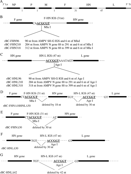
Full-length antigenomic cDNA of NDV strain BC was cloned into plasmid pBR 322 and designated pBC (15). The F-HN IGS and HN-L IGS were modified in pBC. All constructs were confirmed by dideoxynucleotide sequencing. The mutant IGSs included a deletion of 30 nt in the F-HN IGS; deletion of 30 nt or 42 nt in the HN-L IGS; additions of 96 nt, 210 nt, and 318 nt in the F-HN and HN-L IGSs; and a double deletion of 18 nt in the F-HN IGS and 30 nt in the HN-L IGS (Fig. 1).
FIG. 1.
Structures of modified IGSs between the F and HN genes and the HN and L genes of mutant NDVs. (A) Structure of NDV genome order. Between each gene, the IGS is indicated as a thin line, and the length of each IGS is shown under the line. (B) Structure of IGSs between the F and HN genes of rBC-FHN96, rBC-FHN210, and rBC-FHN318. Thirty-one nucleotides is the length of the wild-type F-HN IGS. (C) Structure of IGSs between the HN and L genes of rBC-HNL96, rBC-HNL210, and rBC-HNL318. Forty-seven nucleotides is the length of the wild-type HN-L IGS. (D) Structure of IGSs between the F and HN genes and the HN and L genes of rBC-FHNΔ18HNLΔ30. Eighteen- and 30-nt sequences were deleted from the F-HN and HN-L IGSs, respectively. (E) Structure of IGS between the F and HN genes of rBC-FHNΔ30. Thirty nucleotides were deleted from the F-HN IGS. (F) Structure of IGS between the HN and L genes of rBC-HNLΔ30. Thirty nucleotides were deleted from the HN-L IGS. (G) Structure of rBC-HNLΔ42. Forty-two nucleotides were deleted from the HN-L IGS.
For F-HN IGS insertion, different lengths (90 nt, 204 nt, and 312 nt) of avian metapneumovirus (AMPV) (GenBank accession no. AY590688) sequence, with MluI sites at both ends, were amplified by PCR. The amplified sequences were digested with MluI and inserted into the full-length cDNA clone pBC, which contains a unique MluI site in the IGS between the F and HN genes. AMPV sequences were chosen because AMPV is a closely related avian paramyxovirus. The amplified AMPV sequences were as follows: the 90-nt sequence was from the AMPV SH-G IGS, the 204-nt sequence was from AMPV N gene nt 88 to 291, and the 312-nt sequence was from AMPV N gene nt 88 to 399.
For deletion of nucleotides in the F-HN IGS, three rounds of PCR were performed. In the first-round PCR, the template pBC was amplified with primers NotI (+) (5′-CAA ATA ACA GCG GCC GCA GCT C-3′) and HN6340 (−) (5′-CTC TTA CCG TTC TAC CCG TGT TTT TTC TAA ACT CTC CGA-3′). The PCR product contained a 30-nt deletion in the F-HN IGS and a NotI site, which is present in the upstream gene F. In the second-round PCR, the template pBC was amplified with primers HN6272 (+) (5′-TCG GAG AGT TTA GAA AAA ACA CGG GTA GAA CGG TAA GAG-3′) and HN/L (−) (5′-AAG GCC TTG TCT GCT GAG AAT GAG GTG-3′). The PCR product contained an AgeI site, which is present in the HN-L IGS, and a 30-nt deletion in the F-HN IGS. In the third-round PCR, the first and second PCR products were used as templates, and primers NotI (+) and HN/L (−) were used to amplify the templates. The PCR product was digested with NotI and AgeI and then cloned into pBC, and the resulting clone was designated pBC-FHNΔ30.
For HN-L IGS insertion, sequences of different lengths (90 nt, 204 nt, and 312 nt), with AgeI sites at both ends, were amplified by PCR. These HN-L IGS insertion sequences were the same as the F-HN IGS inserted sequences. The amplified sequences were digested with AgeI and then inserted into full-length cDNA clone pBC, which contained a unique AgeI site in the IGS between the HN and L genes.
For the HN-L IGS deletion pBC-HNLΔ30, primers F6202 (+) (5′-GTG AAC ACA GAT GAG GAA CG-3′) and HNL8366 (−) (5′-TTT ACC GGT TAC ATT TTT TCT TAA TCG AGG GAC TAT TGA C-3′) were used to amplify the template pBC. The PCR product was digested with MluI and XbaI and then cloned into pBC. For pBC-HNLΔ42, three rounds of PCR were performed. In the first-round PCR, primers HN7513 (+) (5′-CGC ATA CAG CAGGCTATCTTA-3′) and HNL 8390 (−) (5′-GAG CTC GCC ATG TCC TAC CCG TAC ACA TTT TTT CTT AAT CGA GGG ACT ATT GAC-3′) were used to amplify the template pBC. The PCR product contained a SpeI site, which is present in the HN gene, and a 42-nt deletion in the HN-L IGS. In the second-round PCR, primers HNL8300(+) (5′-GTC AAT AGT CCC TCG ATT AAG AAA AAT GTG TAC GGG TAG GAC ATG GCG AGC TC-3′) and XbaI (−) (5′-AGT ACT CCG GTT ATT CTA GAA TTG TGG TTG-3′) were used to amplify the template pBC. The PCR product contained an XbaI site and a 42-nt deletion in the HN-L IGS. In the third-round PCR, primers HN7513 (+) and XbaI (−) were used to amplify the products from the first- and second-round PCRs. The PCR product was digested with SpeI and XbaI and then cloned into pBC.
For the double deletion construct, which contained deletions of 18 nt in the F-HN IGS and 30 nt in the HN-L IGS (pBC-FHNΔ18HNLΔ30), primers HN6290(+) (5′-ACT ACG CGT GAT ATA CGG GTA GAA CGG TAA GAG AGG CCG-3′) and HN8366(−) (5′-TTT ACC GGT TAC ATT TTT TCT TAA TCG AGG GAC TAT TGA C-3′) were used to amplify pBC. The PCR product was digested with MluI and AgeI and then cloned into pBC.
All manipulated regions in the NDV cDNAs were sequenced to confirm the presence of the desired mutations.
Recovery of rNDV.
rNDVs were recovered by cotransfection of each NDV cDNA mutant plasmid with support plasmids encoding NP, P, and L proteins into HEp-2 cells. Simultaneously, HEp-2 cells were infected with a recombinant vaccinia virus (MVA/T7) which is capable of synthesizing T7 RNA polymerase. Three or four days after transfection, the cell culture supernatant was used to recover the rNDV by either passage in DF-1 cells until a virus-specific cytopathic effect appeared or injection into the allantoic cavities of 9-day-old embryonated chicken eggs until the allantoic fluid became hemagglutinin positive (15).
RT-PCR and sequence analysis of modified IGSs.
Total RNAs were isolated from mutant NDV-infected allantoic fluid of 9-day-old SPF chicken embryos, using an RNeasy mini kit (Qiagen) according to the manufacturer's recommendations. Reverse transcription (RT) was performed using SuperScript II reverse transcriptase (Invitrogen). The positive-sense primers used for RT were F6202 (+) for F-HN IGS mutants and HN/L (+) (5′-TCC GCG ACA CCA AGA ATC AAA C-3′) for HN-L IGS mutants. For F-HN IGS mutants, the generated cDNA products were PCR amplified using primers F 6202 (+) and HN 6414(−) (5′-CAT GAC TGA GGA CTG CTG-3′). For HN-L IGS mutants, the primers HN/L (+) and HN/L (−) were used for PCR amplification. The RT-PCR products spanning the F-HN and HN-L IGS regions were separated in a 1% agarose gel, and the sequences were confirmed by sequencing.
Characterization of rNDVs.
The growth kinetics of recombinant mutant viruses were evaluated by a multiple-step growth assay. DF-1 cells in duplicate wells of six-well plates were infected with viruses at a multiplicity of infection (MOI) of 0.01 PFU. After 1 hour of adsorption, the cells were washed with DMEM and then covered with DMEM containing 5% FBS at 37°C in 5% CO2. Supernatant was collected and replaced with an equal volume of fresh medium at 8-h intervals until 64 h postinfection. The titer of virus in the sample was quantified by plaque assay with DF-1 cells. All plaque assays were performed in six-well plates. Briefly, monolayers of DF-1 cells were infected with 0.2 ml of 10-fold-diluted fresh virus. After 1 h of adsorption, cells were covered with DMEM containing 2% FBS and 1% methylcellulose and then incubated at 37°C in 5% CO2. Four days later, the cells were fixed with methanol and stained with crystal violet. The average plaque diameter was calculated by measurement of 10 plaques for each virus.
Northern blot hybridization.
The total intracellular RNAs were isolated from virus-infected DF-1 cells by using an RNeasy mini kit (Qiagen). The RNAs were electrophoresed in denaturing 1.5% agarose gels containing 0.5 M formaldehyde, transferred to a nitrocellulose membrane, and then hybridized with a 32P-labeled double-stranded cDNA probe specific to the NDV NP, F, HN, or L gene. The bands corresponding to the individual NDV mRNAs were quantified by a Fuji phosphorimager. In each agarose gel, 28S and 18S RNAs were used as loading controls (data not shown).
Pathogenicity studies.
The pathogenicity of the mutant viruses was determined by three different internationally accepted pathogenicity tests (1). These included the MDT test for 9-day-old embryonated chicken eggs, the ICPI test for 1-day-old chicks, and the IVPI test for 6-week-old chickens.
Briefly, for MDT, a series of 10-fold (10−6 to 10−9) dilutions of fresh infective allantoic fluid was made with sterile phosphate-buffered saline, and 0.1 ml of each diluent was inoculated into the allantoic cavities of five 9-day-old SPF embryonated chicken eggs (Bee Eggs Company, PA) and incubated at 37°C. Each egg was examined three times daily for 7 days, and times of embryo death were recorded. The minimum lethal dose is the highest virus dilution that causes all embryos inoculated with the dilution to die. The MDT is the mean time in hours for the minimum lethal dose to kill all inoculated embryos. The MDT has been used to classify NDV strains into the following groups: velogenic strains (taking less than 60 h to kill), mesogenic strains (taking 60 to 90 h to kill), and lentogenic strains (taking more than 90 h to kill).
For ICPI, 0.05 ml (1:10 dilution) of fresh infective allantoic fluid of each virus was inoculated into groups of 10 1-day-old SPF chicks via the intracerebral route. The inoculation was done using a 27-gauge needle attached to a 1-ml stepper syringe dispenser that was set to dispense 0.05 ml of solution per inoculation. The birds were inoculated by inserting the needle up to the hub into the right or left rear quadrant of the cranium. The birds were observed for clinical symptoms and mortality once every 8 h for a period of 8 days. At each observation, the birds were scored as follows: 0, healthy; 1, sick; and 2, dead. The ICPI is the mean score per bird per observation over the 8-day period. Highly virulent (velogenic) viruses give values approaching 2, and avirulent (lentogenic) viruses give values close to 0.
For IVPI, 0.1 ml (1:10 dilution) of fresh infective allantoic fluid of each virus was inoculated intravenously into groups of 10 6-week-old SPF chickens. The birds were observed for clinical symptoms and mortality once every 8 h for a period of 10 days. At each observation, the birds were scored as follows: 0, healthy; 1, sick; 2, paralyzed; and 3, dead. The IVPI is the mean score per bird per observation over a 10-day period. Highly virulent (velogenic) viruses give values approaching 3, and avirulent (lentogenic) viruses give values close to 0.
Each experiment had mock-inoculated controls that received a similar volume of sterile phosphate-buffered saline by the respective routes. The MDT, ICPI, and IVPI values were calculated as described by Alexander (1).
RESULTS
Construction of rNDVs with F-HN and HN-L IGSs of different lengths.
We manipulated the lengths of the IGSs between the F and HN genes and the HN and L genes in a full-length NDV strain BC antigenomic cDNA clone, pBC. The reason for selecting the F-HN and HN-L IGSs is that these two IGSs include 31 nt and 47 nt, respectively, but the other three IGSs (NP-P, P-M, and M-F) have only 1 nt. We wanted to know why the IGSs of F-HN and HN-L are longer than other IGSs of NDV and what effect the IGS length has on the level of mRNA transcription, virus replication, and pathogenicity of the virus. We made a 30-nt deletion in the F-HN IGS, 30-nt and 42-nt deletions in the HN-L IGS, and a double deletion that included an 18-nt deletion in the F-HN IGS and a 30-nt deletion in the HN-L IGS. We also made six additional constructs in which 96 nt, 210 nt, and 318 nt were added to the F-HN and HN-L IGSs. The numbers of deleted or added nucleotides were adjusted so that the entire genome length of NDV obeyed the “rule of six” (19). Schematics of the constructs are shown in Fig. 1.
The recombinant viruses were recovered by a reverse genetics technique as described previously (15). The sequences of modified IGSs were confirmed by RT-PCR and sequencing. There was no noticeable difference in the recovery of recombinant viruses from different constructs.
Modified IGSs are stable in rNDVs.
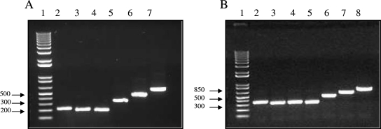
Mutant viruses were passaged five times in 9-day-old embryonated chicken eggs to examine the stability of the modified IGSs. Allantoic fluid from each passage was collected and analyzed by RT-PCR, using primers that flanked the F-HN and HN-L IGSs. The lengths of PCR products from rNDVs with modified F-HN and HN-L IGSs are shown on an agarose gel (Fig. 2). The integrity of the RT-PCR products was confirmed by sequence analysis. Our results did not show any change in length or sequence of rNDV IGSs, even after five passages, indicating that the modified F-HN and HN-L IGSs were stable.
FIG. 2.
RT-PCR analysis of F-HN and HN-L IGSs from mutant NDVs. Total RNAs isolated from allantoic fluid after the fifth passage of mutant NDVs in 9-day-old SPF chicken embryos were analyzed by RT-PCR, using primers spanning the F-HN and HN-L IGSs. The RT-PCR products were separated in 1% agarose gels. (A) RT-PCR of recombinant viruses with mutant F-HN IGS. Lanes: 1, 1-kb DNA ladder; 2, rBC; 3, rBC-FHNΔ18HNLΔ30; 4, rBC-FHNΔ30; 5, rBC-FHN96; 6, rBC-FHN210; and 7, rBC-FHN318. (B) RT-PCR of recombinant viruses with mutant HN-L IGS. Lanes: 1, 1-kb DNA ladder; 2, rBC; 3, rBC-FHNΔ18HNLΔ30; 4, rBC-HNLΔ30; 5, rBC-HNLΔ42; 6, rBC-HNL96; 7, rBC-HNL210; and 8, rBC-HNL318.
Increased IGS length affects viral replication in vitro and results in reduced plaque size.
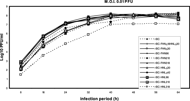
Assays of multistep growth kinetics were performed in order to analyze the replication of rNDVs in vitro. DF-1 cell monolayers were infected at an MOI of 0.01 PFU per cell, and supernatant samples were taken at 8-h intervals until 64 h postinfection. Samples were then quantified by plaque assay. There was no significant difference in growth kinetics between the various mutant viruses and the parental virus rBC, with the exception that rBC-HNL318 showed slightly delayed growth kinetics (Fig. 3).
FIG. 3.
Kinetics of replication of mutant NDVs in DF-1 cells. DF-1 cell monolayers were infected in duplicate with the indicated viruses at an MOI of 0.01 PFU per cell for 1 h. The cells were washed with DMEM and then covered with DMEM containing 5% FBS at 37°C in 5% CO2. Aliquots of the supernatant medium were taken at 8-h intervals until 64 h postinfection, replaced with an equal volume of fresh medium, flash frozen, and analyzed in a single plaque assay.
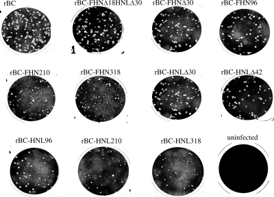
The plaques formed by the various rNDVs in the DF-1 cell monolayer were visualized by staining with crystal violet (Fig. 4). The plaque size was measured in several independent experiments. Viruses that had deletions in either the F-HN or HN-L IGS were indistinguishable from rBC on the basis of plaque size. Interestingly, the double deletion virus, rBC-FHNΔ18HNLΔ30, produced slightly larger plaques than those of parental rBC. The viruses with additions of 96 nt or more in either the F-HN or HN-L IGS produced progressively smaller plaques than those of rBC. The average sizes of 10 plaques of various rNDVs in a typical experiment were as follows: rBC, 1.48 mm (standard deviation [SD], 0.19); rBC-FHNΔ18HNLΔ30, 1.54 mm (SD, 0.16); rBC-FHNΔ30, 1.44 mm (SD, 0.24); rBC-FHN96, 1.25 mm (SD, 0.24); rBC-FHN210, 0.96 mm (SD, 0.19); rBC-FHN318, 0.68 mm (SD, 0.21); rBC-HNLΔ30, 1.41 mm (SD, 0.23); rBC-HNLΔ42, 1.40 mm (SD, 0.24); rBC-HNL96, 1.19 mm (SD, 0.26); rBC-HNL210, 0.93 mm (SD, 0.08); and rBC-HNL318, 0.66 mm (SD, 0.12).
FIG. 4.
Plaque morphology of mutant NDVs containing F-HN and HN-L IGSs with increased or decreased lengths. Recovered viruses were titrated in duplicate in six-well plates. Supernatants collected from virus-inoculated samples were serially diluted, and 0.2 ml of each diluent per well was added to confluent DF-1 cells. After 1 h of adsorption, cells were overlaid with DMEM containing 2% FBS and 1% methylcellulose and incubated at 37°C for 4 days. The cells were then fixed with methanol and stained with crystal violet.
Transcription of the downstream gene is down-regulated by increased IGS length.
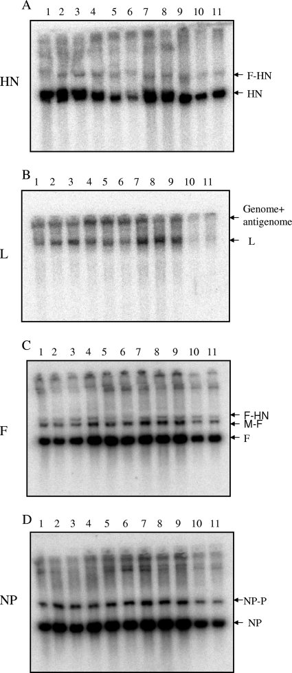
Transcription of the upstream or downstream gene from the IGS in the recombinant viruses was analyzed by Northern blot hybridization. DF-1 cells were infected with viruses at an MOI of 5 PFU per cell and harvested at 24 h postinfection. The total intracellular RNA was isolated and analyzed by Northern blot hybridization with a double-stranded radiolabeled cDNA probe specific to the NP, F, HN, or L gene. The results are shown in Fig. 5, and accumulation of transcripts of the downstream and upstream genes was quantified with a Fuji phosphorimager (data not shown). F-HN IGS mutants rBC-FHN210 and rBC-FHN318 showed decreased transcription of the downstream gene, HN (Fig. 5A, lanes 5 and 6), but no significant change in the transcription level of the upstream gene, F (Fig. 5C, lanes 5 and 6). Similarly, HN-L IGS mutants rBC-HNL210 and rBC-HNL318 showed decreased transcription of the downstream gene, L (Fig. 5B, lanes 10 and 11), but no significant change in the transcription level of the upstream gene, HN (Fig. 5A, lanes 10 and 11). These results indicated that extending the IGS length probably affected the initiation of downstream gene transcription but had no significant effect on the transcription of the immediate upstream gene. In addition, there was no consistent difference in the level of NP gene expression between the viruses (Fig. 5D), indicating that increasing IGS length has no effect on upstream gene transcription.
FIG. 5.
Northern blot analysis of RNAs synthesized by mutant NDVs bearing modified F-HN and HN-L IGSs. (A to D) DF-1 cells were infected with the indicated viruses (MOI of 5 PFU), incubated for 24 h, and harvested, and then total intracellular RNA was extracted. RNAs were separated by electrophoresis in formaldehyde agarose gels, transferred to nitrocellulose membranes, and hybridized with a 32P-labeled double-stranded cDNA probe specific to the HN, L, F, or NP gene, as indicated. Positions of the specific gene, readthrough, genomic, and antigenomic RNAs are indicated. Lanes: 1, rBC; 2, rBC-FHNΔ18HNLΔ30; 3, rBC-FHNΔ30; 4, rBC-FHN96; 5, rBC-FHN210; 6, rBC-FHN318; 7, rBC-HNLΔ30; 8, rBC-HNLΔ42; 9, rBC-HNL96; 10, rBC-HNL210; and 11, rBC-HNL318.
Pathogenicity studies of rNDVs containing decreased or increased F-HN and HN-L IGS lengths.
We wanted to determine the effect of increased or decreased IGS length on the pathogenicity of recombinant viruses. At present, a definitive assessment of NDV virulence is based on the following in vivo tests: MDT test with embryonated SPF chicken eggs, ICPI test with 1-day-old chicks, and IVPI test with 6-week-old chickens. We examined the virulence of parental rNDV and rNDVs containing modified IGS lengths by all these tests (Table 1).
TABLE 1.
Pathogenicities of mutant viruses in chicken embryos, chicks, and chickens
| Virus | MDT (h)a | ICPIb | IVPIc |
|---|---|---|---|
| rBC | 60 | 1.49 | 2.06 |
| rBC-FHNΔ18HNLΔ30 | 60 | 1.28 | 0.72 |
| rBC-FHNΔ30 | 58 | 1.36 | 1.04 |
| rBC-FHN96 | 59 | 1.44 | 0.34 |
| rBC-FHN210 | 60 | 1.31 | 0.20 |
| rBC-FHN318 | 60 | 1.06 | 0.14 |
| rBC-HNLΔ30 | 64 | 1.45 | 0.99 |
| rBC-HNLΔ42 | 59 | 1.44 | 0.44 |
| rBC-HNL96 | 58 | 1.11 | 0.54 |
| rBC-HNL210 | 60 | 0.61 | 0.00 |
| rBC-HNL318 | 68 | 0.47 | 0.00 |
For 9-day-old embryonated chicken eggs. Viruses were classified as follows: values of >90, lentogen; values of 60 to 90, mesogen; and values of <60, velogen (1).
For 1-day-old chicks. Viruses were classified as follows: values of 0.0 to 0.5, lentogen; values of 1.0 to 1.5, mesogen; and values of 1.5 to 2.0, velogen (1).
For 6-week-old chickens. Viruses were classified as follows: value of 0.0, lentogen; values of 0.0 to 0.5, mesogen; and values of 2.0 to 3.0, velogen (1).
The MDT test results showed that the parental strain rBC took 60 h to cause embryo mortality. All recombinant viruses showed MDTs similar to that of rBC, except for rBC-HNLΔ30 and rBC-HNL318, which took 4 h and 8 h longer, respectively, in causing the death of embryos (Table 1).
The ICPI value for parental rBC was 1.49 (of a maximum of 2.00) (Table 1). The ICPI values for rNDVs containing decreased IGS lengths (rBC-FHNΔ30, rBC-HNLΔ30, and rBC-HNLΔ42) were similar to that of the parental strain rBC. However, the ICPI value for rBC-FHNΔ18HNLΔ30 was 1.28, which was lower than that of parental rBC. Interestingly, the ICPI values for recombinant viruses decreased as the length of the IGS increased. In the case of increased F-HN IGS length, the ICPI values for rBC-FHN96, rBC-FHN210, and rBC-FHN318 were 1.44, 1.31, and 1.06, respectively. In the case of increased HN-L IGS length, the ICPI values for rBC-HNL96, rBC-HNL210, and rBC-HNL318 were 1.11, 0.61, and 0.47, respectively. These results indicated that increased HN-L IGS length reduces the pathogenicity of rNDV more than increased F-HN IGS length does. To determine the amount of each recombinant virus required to cause the death of 1-day-old chicks by the intracerebral route, the brain tissues from the dead chicks were plaque assayed in DF-1 cells. Our results showed that all rNDVs grew to similar titers (104 to 105 PFU/g brain tissue) in the brain at the time of chick death, but the time to cause death was longer for viruses with increased IGS lengths. The viruses recovered from the brain tissues of dead chicks were sequenced to determine the stability of the modified IGSs. Our results showed that the modified IGSs in rNDVs were stable after their growth in the chick brain.
The results obtained for the mutant viruses by IVPI tests with 6-week-old chickens were quite surprising (Table 1). The parental rBC virus had an IVPI value of 2.06 (of a maximum of 3.00), but the IVPI values for all mutant viruses were much lower (ranging from 0.00 to 1.04), indicating that both decreased and increased IGS lengths reduced the virulence of rNDVs. All rNDVs containing decreased IGS lengths showed reduced virulence. The IVPI values for rBC-FHNΔ18HNLΔ30, rBC-FHNΔ30, rBC-HNLΔ30, and rBC-HNLΔ42 were 0.72, 1.04, 0.99, and 0.44, respectively. Again, interestingly, the IVPI values for rNDVs decreased as the length of the IGS increased. In the case of increased F-HN IGS length mutants, the IVPI values for rBC-FHN96, rBC-FHN210, and rBC-FHN318 were 0.34, 0.20, and 0.14, respectively. In the case of increased HN-L IGS length mutants, the IVPI values for rBC-HNL96, rBC-HNL210, and rBC-HNL318 were 0.54, 0.00, and 0.00, respectively. These results confirmed that increased HN-L IGS length reduced the pathogenicity of rNDVs in adult chickens more than increased F-HN IGS length did. Furthermore, our results showed that the IVPI test is more sensitive than the ICPI test in determining the pathogenicity of NDV strains. The MDT test is the least sensitive test for assessing the pathogenicity of NDV.
DISCUSSION
The lengths and sequence compositions of IGSs vary among the members of the Mononegavirales. In some viruses, such as VSV, Sendai virus, and measles virus, the IGSs are a conserved dinucleotide or trinucleotide, whereas in some other viruses, such as NDV, RSV, and SV5, the IGSs vary in length and in sequence composition. Previous studies have indicated that in viruses with conserved IGSs, the IGS plays an important role in termination of upstream mRNA transcription and initiation of downstream mRNA synthesis (2, 25, 26). In contrast, in some other viruses, such as RSV, the IGS has little effect on either upstream or downstream mRNA transcription (3, 17). In NDV, the first three 3′-proximal IGSs are single nucleotides, whereas the last two 5′-proximal IGSs, the F-HN and HN-L IGSs, are nonconserved 31- and 47-nt sequences, respectively. In this study, we analyzed the effect of decreasing or increasing length of the F-HN or HN-L IGS on mRNA transcription and on the eventual pathogenicity of the virus in its natural host, the chicken.
Increasing either the F-HN or HN-L IGS length had no significant effect during multistep growth kinetics of the mutant NDVs in cell culture, except that the rBC-HNL318 virus showed decreased growth kinetics. However, the sizes of plaques varied among the mutant viruses. Based on plaque size, the level of attenuation of in vitro growth increased as the length of the IGS increased. Our research confirmed the previous observation of the RSV IGS study that plaque size was remarkably sensitive to small changes in IGS length (3). It is unknown whether the effect of increased in vitro attenuation was due to the effect of increased genome length of the rNDV or due to alteration of the mRNA transcription level resulting from the increased IGS length.
Decreasing either the F-HN or HN-L IGS length had no discernible effect during multistep growth kinetics of mutant NDVs in cell culture. However, when the plaque sizes of IGS deletion viruses were compared, we found that the plaque sizes of single IGS deletion viruses were similar to or slightly smaller than that of the parental virus. This result indicated that the decreased plaque size observed with viruses of increased or decreased IGS length was probably not due to the effect of a simple increase in genome length but could be due to the alteration of the mRNA transcription level. Surprisingly, the plaque size of the double IGS deletion virus rBC-FHNΔ18HNLΔ30 was increased. Although it is unclear why the plaque size was increased, it is possible that the augmentation effect was due to the ratio of mRNAs produced as a result of double 5′-proximal IGS deletions.
In the present study, we demonstrated that NDV can tolerate an IGS length of at least 365 nt. The IGS length was stable after five passages in chickens and chicken embryos. Among the members of the Mononegavirales, the IGSs of Ebola virus vary from 3 to 143 nt (22), rabies virus has a 423-nt G-L intergenic region which is thought to be a pseudogene (27), and human metapneumovirus IGSs vary from 2 to 126 nt (13). Recombinant RSV was shown to tolerate an IGS length of 160 nt (3). Therefore, to our knowledge, this is one of the largest IGSs in the Mononegavirales.
During virus recovery studies, we came across an interesting observation. When the F-HN IGS was replaced with random nonviral sequences, we were unable to recover viable NDV, even after five attempts. In all those attempts, the parental virus and the viruses in which the F-HN or HN-L IGS was replaced with avian metapneumovirus sequences were recovered. Although these experiments need to be repeated with additional viral and nonviral random sequences, our research indicated that certain nonviral sequences cannot be used to replace NDV IGSs. It is possible that the NDV polymerase recognizes sequences from a distantly related virus but fails to recognize randomly generated nonviral sequences.
Our observations of diminution of plaque size with either increased or decreased IGS length indicated that it was probably not due to the result of the change in genome length but was associated with a change in the efficiency of sequential transcription. As expected, our Northern blot analysis showed that increased F-HN or HN-L IGS length resulted in decreased transcription of the downstream gene. We were unable to detect any consistent effect on the transcription of the upstream gene. Our results were consistent with a study of hPIV3 (23). hPIV3 has a conserved trinucleotide, GAA or GCG, as the IGS. Studies showed that insertion of an additional nucleotide into the IGS can decrease the efficiency of transcription of the downstream gene (23). In contrast, for RSV, in which IGSs vary in length and in sequence composition, studies with a minigenome and infectious recombinant virus showed that RSV IGSs did not modulate transcription (3, 17).
The pathogenicity studies showed that all IGS mutants were attenuated in ICPI and IVPI tests. The results showed that the level of attenuation increased as the length of the IGS increased. For example, rBC-HNL210 and rBC-HNL318 were completely attenuated in the IVPI test. These results are in agreement with studies of RSV, where increasing IGS length affected viral pathogenicity in mice (17). Interestingly, our results showed that decreasing the lengths of IGSs also affected viral pathogenicity. These findings suggested that either increasing or decreasing the length of an IGS alters the level of downstream gene transcription, which changes the ratios of viral transcripts, and these ratios are probably very important for virus growth and pathogenicity. It is possible that the most effective ratios of transcripts in NDV are obtained by having 1 nt in the first three 3′-end IGSs and 31 and 47 nt in the last two IGSs. Therefore, NDV has maintained these IGS lengths over the years without any changes. We also observed that the HN-L IGS was more amenable to attenuation than the F-HN IGS. This result indicated that the level of L protein is probably more important for virus growth and that any minor change in its level can have a dramatic effect on virus growth and pathogenicity.
Previously, it was suggested that the ability of NDV strains to produce large plaques is related to their virulence for chickens (21). Seemingly, our data differ from this suggestion in that the double IGS deletion virus rBC-FHNΔ18HNLΔ30 produced slightly larger plaques than those of the parental rBC virus, despite its attenuation in chickens. Therefore, plaque size may not always be a correct indicator of NDV virulence.
Our pathogenicity studies showed that the IVPI test was more sensitive than the ICPI test, which in turn was more sensitive than the MDT test to assess the pathogenicity of NDV IGS mutants. These results indicate that the immune system of the host also plays an important role in the pathogenicity of NDV. The virus has to overcome a more mature immune system in a 6-week-old chicken in the IVPI test than the less developed immune system in a 1-day-old chick in the ICPI test or in a 9-day-old embryo in the MDT test. Therefore, any minor difference in the pathogenicity of an NDV mutant could be amplified in the IVPI test compared to the ICPI or the MDT test.
The results presented in this paper can have significant applications in the development of live attenuated NDV vaccines. One of the major problems encountered in current live attenuated NDV vaccines is the undesirable pathogenicity of the vaccine virus. Since field strains of low virulence are currently used as NDV vaccines, the pathogenicity of these strains cannot be changed. There is a great need to develop a live NDV vaccine that is immunogenic but less pathogenic. Our research shows a new method of attenuating NDV strains. We hope that by changing the lengths of the IGSs, the pathogenicity of an NDV strain can be adjusted to the desired level without affecting the immunogenicity of the vaccine virus, which will greatly benefit the poultry industry throughout the world.
Acknowledgments
We thank Daniel Rockemann, Peter Savage, Subrat Rout, Madhuri Subbiah, and Sachin Kumar for their excellent technical assistance and Ireen Dryburgh-Barry for her critical reading of the manuscript.
Footnotes
Published ahead of print on 21 November 2007.
REFERENCES
- 1.Alexander, D. J. 1989. Newcastle disease, p. 114-120. In H. G. Purchase, L. H. Arp, C. H. Domermuth, and J. E. Pearson (ed.), A laboratory manual for the isolation and identification of avian pathogens, 3rd ed. The American Association of Avian Pathologists, Kendall/Hunt Publishing Company, Dubuque, IA.
- 2.Barr, J. N., S. P. J. Whelan, and G. W. Wertz. 1997. Role of the intergenic dinucleotide in vesicular stomatitis virus RNA transcription. J. Virol. 711794-1801. [DOI] [PMC free article] [PubMed] [Google Scholar]
- 3.Bukreyev, A., B. R. Murphy, and P. L. Collins. 2000. Respiratory syncytial virus can tolerate an intergenic sequence of at least 160 nucleotides with little effect on transcription or replication in vitro and in vivo. J. Virol. 7411017-11026. [DOI] [PMC free article] [PubMed] [Google Scholar]
- 4.Chambers, P., N. S. Millar, and P. T. Emmerson. 1986. Nucleotide sequence of the gene encoding the fusion glycoprotein of Newcastle disease virus. J. Gen. Virol. 672685-2694. [DOI] [PubMed] [Google Scholar]
- 5.Chambers, P., N. S. Millar, R. W. Bingham, and P. T. Emmerson. 1986. Molecular cloning of complementary DNA to Newcastle disease virus, and nucleotide sequence analysis of the junction between the genes encoding the haemagglutinin-neuraminidase and the large protein. J. Gen. Virol. 67475-486. [DOI] [PubMed] [Google Scholar]
- 6.Collins, P. L., L. E. Dickens, A. Buckler-White, R. A. Olmsted, M. K. Spriggs, E. Camargo, and K. V. W. Coelingh. 1986. Nucleotide sequences for the gene junctions of human respiratory syncytial virus reveal distinctive features of intergenic structure and gene order. Proc. Natl. Acad. Sci. USA 834594-4598. [DOI] [PMC free article] [PubMed] [Google Scholar]
- 7.de Leeuw, O., and B. Peeters. 1999. Complete nucleotide sequence of Newcastle disease virus: evidence for the existence of a new genus within the subfamily Paramyxovirinae. J. Gen. Virol. 80131-136. [DOI] [PubMed] [Google Scholar]
- 8.Galinski, M. S., and S. L. Wechsler. 1991. The molecular biology of the Paramyxovirus genus, p. 68-70. In D. W. Kingsbury (ed.), The paramyxoviruses. Plenum Press, New York, NY.
- 9.Hardy, R. W., S. B. Harmon, and G. W. Wertz. 1999. Diverse gene junctions of respiratory syncytial virus modulate the efficiency of transcription termination and respond differently to M2-mediated antitermination. J. Virol. 73170-176. [DOI] [PMC free article] [PubMed] [Google Scholar]
- 10.He, B., and R. A. Lamb. 1999. Effect of inserting paramyxovirus simian virus 5 gene junctions at the HN/L gene junction: analysis of accumulation of mRNAs transcribed from rescued viable viruses. J. Virol. 736228-6234. [DOI] [PMC free article] [PubMed] [Google Scholar]
- 11.Hinzman, E. E., J. N. Barr, and G. W. Wertz. 2002. Identification of an upstream sequence element required for vesicular stomatitis virus mRNA transcription. J. Virol. 767632-7641. [DOI] [PMC free article] [PubMed] [Google Scholar]
- 12.Ishida, N., H. Taira, T. Omata, K. Mizumoto, S. Hattori, K. Iwasaki, and M. Kawakita. 1986. Sequence of 2,617 nucleotides from the 3′ end of Newcastle disease virus genome RNA and the predicted amino acid sequence of viral NP protein. Nucleic Acids Res. 146551-6564. [DOI] [PMC free article] [PubMed] [Google Scholar]
- 13.Ishiguro, N., T. Ebihara, R. Endo, X. Ma, H. Kikuta, H. Ishiko, and K. Kobayashi. 2004. High genetic diversity of the attachment (G) protein of human metapneumovirus. J. Clin. Microbiol. 423406-3414. [DOI] [PMC free article] [PubMed] [Google Scholar]
- 14.Kawano, M., K. Okamoto, H. Bando, K. Kondo, M. Tsurudome, H. Komada, M. Nishio, and Y. Ito. 1991. Characterizations of the human parainfluenza type 2 virus gene encoding the L protein and the intergenic sequences. Nucleic Acids Res. 192739-2745. [DOI] [PMC free article] [PubMed] [Google Scholar]
- 15.Krishnamurthy, S., Z. Huang, and S. K. Samal. 2000. Recovery of a virulent strain of Newcastle disease virus from cloned cDNA: expression of a foreign gene results in growth retardation and attenuation. Virology 278168-182. [DOI] [PubMed] [Google Scholar]
- 16.Krishnamurthy, S., and S. K. Samal. 1998. Nucleotide sequences of the trailer, nucleocapsid protein gene and intergenic regions of Newcastle disease virus strain Beaudette C and completion of the entire genome sequence. J. Gen. Virol. 792419-2424. [DOI] [PubMed] [Google Scholar]
- 17.Kuo, L., R. Fearns, and P. L. Collins. 1996. The structurally diverse intergenic regions of respiratory syncytial virus do not modulate sequential transcription by a dicistronic minigenome. J. Virol. 706143-6150. [DOI] [PMC free article] [PubMed] [Google Scholar]
- 18.Lamb, R. A., and D. Kolakofsky. 1996. Paramyxoviridae: the viruses and their replication, p. 1171-1204. In B. N. Fields, D. M. Knipe, and P. M. Howley (ed.), Fields virology, 3rd ed. Lippincott-Raven, Philadelphia, PA.
- 19.Phillips, R. J., A. C. R. Samson, and P. T. Emmerson. 1998. Nucleotide sequence of the 5′ terminus of Newcastle disease virus and assembly of the complete genomic sequence: agreement with the “rule of six.” Arch. Virol. 1431993-2002. [DOI] [PubMed] [Google Scholar]
- 20.Rassa, J. C., and G. D. Parks. 1999. Highly diverse intergenic regions of the paramyxovirus simian virus 5 cooperate with the gene end U tract in viral transcription termination and can influence reinitiation at a downstream gene. J. Virol. 733904-3912. [DOI] [PMC free article] [PubMed] [Google Scholar]
- 21.Reeve, P., and G. Poste. 1971. Studies on the cytopathogenicity of Newcastle disease virus: relation between virulence, polykaryocytosis and plaque size. J. Gen. Virol. 1117-24. [DOI] [PubMed] [Google Scholar]
- 22.Sanchez, A., M. Kiley, B. Holloway, and D. Auperin. 1993. Sequence analysis of the Ebola virus genome: organization, genetic elements, and comparison with the genome of Marburg virus. Virus Res. 29215-240. [DOI] [PubMed] [Google Scholar]
- 23.Skiadopoulos, M. H., S. R. Surman, A. P. Durbin, P. L. Collins, and B. R. Murphy. 2000. Long nucleotide insertions between the HN and L protein coding regions of human parainfluenza virus type 3 yield viruses with temperature-sensitive and attenuation phenotypes. Virology 272225-234. [DOI] [PubMed] [Google Scholar]
- 24.Steward, M., I. B. Vipond, N. S. Millar, and P. T. Emmerson. 1993. RNA editing in Newcastle disease virus. J. Gen. Virol. 742539-2547. [DOI] [PubMed] [Google Scholar]
- 25.Stillman, E. A., and M. A. Whitt. 1998. The length and sequence composition of vesicular stomatitis virus intergenic regions affect mRNA levels and the site of transcript initiation. J. Virol. 725565-5572. [DOI] [PMC free article] [PubMed] [Google Scholar]
- 26.Stillman, E. A., and M. A. Whitt. 1997. Mutational analyses of the intergenic dinucleotide and the transcriptional start sequence of vesicular stomatitis virus (VSV) define sequences required for efficient termination and initiation of VSV transcripts. J. Virol. 712127-2137. [DOI] [PMC free article] [PubMed] [Google Scholar]
- 27.Tordo, N., O. Poch, A. Ermine, G. Keith, and F. Rougeon. 1986. Walking along the rabies genome: is the large G-L intergenic region a remnant gene? Proc. Natl. Acad. Sci. USA 833914-3918. [DOI] [PMC free article] [PubMed] [Google Scholar]
- 28.Wang, L., M. Yu, E. Hansson, L. I. Pritchard, B. Shiell, W. P. Michalski, and B. T. Eaton. 2000. The exceptionally large genome of Hendra virus: support for creation of a new genus within the family Paramyxoviridae. J. Virol. 749972-9979. [DOI] [PMC free article] [PubMed] [Google Scholar]


