Abstract
In this study, we explored the molecular basis determining the virulence of H5N1 avian influenza viruses in mammalian hosts by comparing two viruses, A/Duck/Guangxi/12/03 (DK/12) and A/Duck/Guangxi/27/03 (DK/27), which are genetically similar but differ in their pathogenicities in mice. To assess the genetic basis for this difference in virulence, we used reverse genetics to generate a series of reassortants and mutants of these two viruses. We found that a single-amino-acid substitution of serine for proline at position 42 (P42S) in the NS1 protein dramatically increased the virulence of the DK/12 virus in mice, whereas the substitution of proline for serine at the same position (S42P) completely attenuated the DK/27 virus. We further demonstrated that the amino acid S42 of NS1 is critical for the H5N1 influenza virus to antagonize host cell interferon induction and for the NS1 protein to prevent the double-stranded RNA-mediated activation of the NF-κB pathway and the IRF-3 pathway. Our results indicate that the NS1 protein is critical for the pathogenicity of H5N1 influenza viruses in mammalian hosts and that the amino acid S42 of NS1 plays a key role in undermining the antiviral immune response of the host cell.
H5N1 highly pathogenic avian influenza virus (HPAIV) is not only a catastrophic pathogen for poultry, but it poses a severe threat to the public health and may cause a future influenza pandemic. In 1997, highly pathogenic H5N1 avian influenza virus caused outbreaks in chickens in Hong Kong and was transmitted to humans, causing the deaths of 6 of 18 people infected (4, 31). The H5N1 outbreaks in poultry, which became widespread in late 2003, affected at least 10 Asian countries initially, but since then, H5N1 viruses have been isolated from wild birds (3) and poultry in multiple countries in Asia, Europe, and Africa (http://www.oie.int). H5N1 influenza virus infections have occurred in several mammalian species, such as pigs, domestic cats, tigers, and leopards (http://www.oie.int). More importantly, human cases of H5N1 infections have been reported in many countries (http://www.who.int), with greater than 50% mortality caused by H5N1 viruses among infected humans. Such findings have sparked great interest in pandemic preparedness as well as in understanding the genetic determinants of influenza virus pathogenicity and the ability of the virus to cross species barriers to mammalian hosts.
The pathogenicity of influenza viruses is determined by many factors, including virus-specific determinants encoded within the virus genome. In the H5 and H7 subtypes of influenza viruses, the multiple basic amino acids adjacent to the cleavage site of the hemagglutinin (HA) glycoprotein are a prerequisite for lethality in chickens and mice (12, 13, 30). For H5N1 influenza viruses, a reverse genetics study demonstrated that a single-amino-acid substitution at position 627 of the PB2 protein from glutamic acid to lysine is responsible for virulence in mammalian species (12). Moreover, the amino acid at position 701 in PB2 plays a crucial role in the ability of H5N1 viruses of duck origin to replicate and be lethal in mice (16). This same PB2 amino acid residue contributes to the increased lethality of an H7N1 avian influenza virus in a mouse model (9).
Several studies have reported that the NS1 protein is also associated with the virulence and host range of influenza viruses in different animal models (17, 23, 27, 28). Influenza viruses in which the NS1 gene was deleted exhibited an attenuated phenotype in mice and pigs (23, 28). The glutamic acid at position 92 of the NS1 protein of the H5N1 influenza virus that transmitted to humans in 1997 was shown to be critical in conferring virulence and resistance to antiviral cytokines in pigs (27). However, H5N1 virus with this amino acid residue is no longer circulating in nature and glutamic acid is not found in the NS1 proteins of other influenza viruses. Another amino acid substitution at position 149 of the NS1 protein from valine to alanine was shown to be responsible for the replication of a goose H5N1 influenza virus in chickens (17); however, this mutation did not affect virus virulence in mammals (H. Chen, unpublished data). Thus, the specific amino acid residues in avian NS1 that are responsible for conferring high virulence in mammals remain unclear.
Host factors, such as the immune responses, also play a role in determining influenza virus pathogenicity (14). The interferon (IFN) response represents an early host defense mechanism against viral infections and is an important component of innate immunity (33). The presence of double-stranded RNA (dsRNA) is a signal to the host cell that virus infection and replication are occurring and triggers a plethora of antiviral host defense mechanisms (5, 29). The presence of dsRNA induces the synthesis of alpha/beta IFN (IFN-α/β) proteins through the activation of several transcription factors, including IRF-3, IRF-7, NF-κB, and c-Jun/ATF2. Influenza viruses have dsRNA species of replication intermediates that elicit the host IFN response. The secreted IFN-α/β induces an antiviral state in influenza virus-infected and uninfected neighboring cells by stimulating the transcription of IFN-stimulated response element promoter-containing genes via the JAK/STAT pathway (29). However, influenza and other viruses have developed strategies to counteract host IFN-α/β production, through inhibiting the activation of transcription factors involved in IFN activation (10, 18) and by attenuating host gene expression (20). Antagonism of the innate response by influenza virus is a property of the NS1 protein (7, 11, 20). Although data in this area of research are growing, there remain facets of host range and virulence determination that need further examination.
In this study, we characterized two H5N1 avian influenza viruses that were isolated from ducks, A/Duck/Guangxi/12/2003 (DK/12) and A/Duck/Guangxi/27/2003 (DK/27), in the Guangxi province of China in 2003. These two viruses are highly pathogenic for chickens but differ in their virulences in mice. We used reverse genetics to determine the molecular basis for the difference in virulence in mice and to explore the possible underlying mechanisms. We found a specific amino acid in NS1 that confers lethality in mice to an avian H5N1 virus.
MATERIALS AND METHODS
Cells and viruses.
Human embryonic kidney cells (293T) and Vero cells were grown in Dulbecco's modified Eagle's medium supplemented with 10% fetal bovine serum plus antibiotics. Human lung epithelial cells (A549) were grown in nutrient mixture F-12 Ham Kaighn's modified medium with 10% fetal bovine serum. The cells were incubated at 37°C in 5% CO2. Recombinant vesicular stomatitis virus (VSV) expressing green fluorescent protein (VSV-GFP) was generated by inserting the G protein gene of VSV into the VSVΔG*GFP vector by use of reverse genetics as described previously (15, 17).
Construction of plasmids.
The construction of plasmids for virus rescue was performed as described previously (16). Mutations were introduced into the NS1 gene by site-directed mutagenesis (Invitrogen) with the set of primers shown in Table 1. We also generated plasmids expressing two wild-type and two mutant NS1 protein sequences by use of the pCAGGS plasmid vector for the reporter gene assay. These plasmids were designated as p12NS1 and p27NS1 for the wild-type NS1 proteins and p12NS1P42S and p27NS1S42P for the mutant NS1 proteins. To investigate the IRF-3-dependent promoter activation, we synthesized the mouse ISG-54 promoter fragment based on the available sequence information (GenBank accession number X77259) and inserted it between the NheI and BglII sites of the pLuc 3-enhancer plasmid (Promega) in front of the firefly luciferase open reading frame and designate the construct as pISG54-Luc. All of the constructs were completely sequenced to ensure the absence of unwanted mutations.
TABLE 1.
Primers used for pBD cDNA construction to amplify the full-length cDNAs of the viruses and for introducing the mutations in the NS1 gene
| Purpose | Primer (5′-3′)a
|
|
|---|---|---|
| Forward | Reverse | |
| For PB2 amplification | CCAGCAAAAGCAGGTCAAATATATTCA | 5′TTAGTAGAAACAAGGTCGTT |
| For PB1 amplification | CCAGCAAAAGCAGGCAAACCA | 5′TTAGTAGAAACAAGGCATTTTTTC |
| For PA amplification | CCAGCAAAAGCAGGTACTGATC | 5′TTAGTAGAAACAAGGTACTTTTTTGGAC |
| For HA amplification | CCAGCAAAAGCAGGGGTCCAATC | 5′TTAGTAGAAACAAGGGTGTTTTTAACTAC |
| For NP amplification | CCAGCAAAAGCAGGGTAGATAATC | 5′TTAGTAGAAACAAGGGTATTTTTC |
| For NA amplification | CCAGCAAAAGCAGGAGTTCAAAATGAAT | 5′TTAGTAGAAACAAGGAGTTTTTTGAACAA |
| For M amplification | CCAGCAAAAGCAGGTAGATGTTGAAAGATG | 5′TTAGTAGAAACAAGGTAGTTTTTTACTC |
| For NS amplification | CCAGCAAAAGCAGGGTGACAA | 5′TTAGTAGAAACAAGGGTGTTTTTTATCAT |
| For DK/12NS1P42S NS mutation | TCAGAAGTCCCTAAGAGGAAGAGGC | 5′GCCTCTTACTCTTAGGGACTTCTGA |
| For DK/12NS1N48S NS mutation | TAAGAGGAAGAGGCAGCACCCTTGG | 5′CCAAGGGTCCTGCCTCTTCCTCTTA |
| For DK/27NS1S42P NS mutation | TCAGAAGCCCCTAAGAGGAAGAGGC | 5′GCCTCTTCCTCTTAGGGGCTTCTGA |
| For DK/27NS1S48N NS mutation | TAAGAGGAAGAGGCAACACCCTTGG | 5′CCAAGGGTTCTGCCTCTTCCTCTTA |
| For GX27NS1R38A NS mutation | 5′ACCGGCTTCGCGCAGATCAGAAGTC | 5′TAGGGACTTCTGATCTGCGCGAAGC |
| For GX27NS1K41A NS mutation | 5′CGCCGAGATCAGGCGTCCCTAAGAG | 5′CTCTTAGGGACGCCTGATCTCGGCG |
| For GX27NS1R38AK41A NS mutation | 5′TTCGCGCAGATCAGGCGTCCCTAAG | 5′CTTAGGGACGCCTGATCTGCGCGAA |
The nucleotides that have been changed are underlined and in boldface.
Generation of reverse genetic reassortant viruses.
Reassortant viruses were generated by reverse genetics as described previously (12, 16). The rescued viruses were detected by hemagglutination assay, and RNA was extracted and analyzed by reverse transcription-PCR (RT-PCR). Each viral segment was sequenced to confirm the identity of the reassortant viruses.
Animal experiments.
Determination of the intravenous pathogenicity index value (IVPI) with chickens was performed according to the recommendations of the Office International Des Epizooties (21). For the mouse study, groups of 11 6-week-old female BALB/c mice (Beijing Experimental Animal Center) were lightly anesthetized with CO2 and inoculated intranasally with 106.0 50% egg infectious doses (EID50) of H5N1 influenza virus in a volume of 50 μl. Three mice in each group were euthanized on days 4 and 6 postinoculation (p.i.). Organs were collected and titrated for virus infectivity in eggs as described previously (2). The remaining mice were monitored for 14 days for weight loss and mortality. The 50% mouse lethal dose (MLD50) was determined by inoculating groups of five mice with 10-fold serial dilutions containing 101 to 106 EID50 of the virus in a 50-μl volume and calculated by using the method of Reed and Muench (24). The chicken and mouse studies have been approved by the Review Board of Harbin Veterinary Research Institute, Chinese Academy of Agricultural Sciences.
Sequence analysis.
Reassortant viruses and the plasmids used for virus rescue were fully sequenced to confirm the absence of unwanted mutations. Viral RNA was extracted from allantoic fluid and was reverse transcribed. A set of fragment-specific primers (primer sequences available on request) were used for the PCR amplification and sequence analysis. The sequence data for the two viruses used in these studies are available in GenBank (accession no. pending).
Detection of IFN secretion.
Monolayers of 80% confluent A549 cells were infected at a multiplicity of infection (MOI) of 2. Following infection, cells were incubated with Opti-MEM (Gibco/BRL) and the supernatants were harvested 24 h p.i. Viruses present in the supernatants were UV inactivated by placing samples on ice 70 cm below a 30-W UV lamp for 20 min, and inactivation of the virus was confirmed by egg propagation. The UV-inactivated supernatants were then added to A549 cells and incubated for 24 h. The cells were then infected with 0.001 MOI of VSV-GFP. At 14 h p.i., cells expressing GFP were visualized by fluorescence microscopy.
Analysis of IFN-α/β mRNA by RT-PCR.
A549 cells were infected with influenza viruses at an MOI of 2, and at 20 h p.i., total RNA was extracted and digested with DNase I (Roche). RT-PCR was performed by using primer pairs specific for human IFN-α and human IFN-β mRNA (GenBank accession numbers M54886, BC112302, and NM002176). A 550-bp fragment of human β-actin was amplified as a control. The products were sequenced and confirmed to be derived from the expected mRNAs.
Quantification of IFN-α/β production by ELISA.
For quantification of secreted IFN-α/β, A549 cells were either mock infected or infected with different H5N1 viruses at an MOI of 2. The supernatants were harvested 24 h p.i. Viruses present in the supernatants were UV inactivated by placing samples on ice 70 cm below a 30-W UV lamp for 20 min. Production of IFN-α/β in culture supernatants was measured using a human IFN-α or IFN-β enzyme-linked immunosorbent assay (ELISA) kit (human IFN-α and human IFN-β ELISA kit; Adlitteram Diagnostic laboratories Inc.) according to the manufacturer's instructions. For each treatment, three sets of sample were collected, and each sample was tested in duplicate by ELISA.
Western blot analysis of the viral protein levels in the virus-infected A549 cells.
Six-well plates of 90% confluent A549 cells were mock infected or infected with viruses at an MOI of 2. Cells were lysed 12 h p.i. and the lysates subjected to Western blot analysis using mouse anti-truncated A/Goose/Guangdong/1/96 NS1 and chicken anti-A/Goose/Guangdong/1/96 NP antibodies and monoclonal anti-β actin antibody (Sigma) as the control.
Reporter gene assay.
293T cells were used for investigating NF-κB promoter activation, and Vero cells were used for the IRF-3-dependent promoter, ISG54, activation assay. Cells were transfected with an NF-κB-responsive promoter-driven firefly luciferase reporter plasmid, pNF-κB-Luc (Stratagene, La Jolla, CA) or pISG54-Luc. In addition, an internal control plasmid to normalize transfection efficiency, pTK-RL (Promega), encoding the Renilla luciferase protein, was transfected into the cells. The reporter gene plasmids pNF-κB-Luc or pISG-54K-Luc and the pTK-RL plasmids were cotransfected at 0.5 μg along with 4.0 μg of the NS1 (wild-type or mutant) plasmid into 80% confluent cells by using Lipofectamine 2000 (Invitrogen). At 24 h posttransfection, the cells were mock treated or transfected with 40 μg poly(I:C) (Amersham Pharmacia) by using Lipofectamine 2000. At 24 h posttreatment, the cells were lysed and luciferase activities were determined with a dual-luciferase reporter assay system (Promega) and normalized on the basis of the Renilla luciferase activities.
Nucleotide sequence accession numbers.
The sequence data for the two viruses used in these studies have been deposited in GenBank under accession numbers EV263342 to EV263357.
RESULTS
Biological properties of the two H5N1 avian influenza viruses isolated from ducks.
We isolated two H5N1 viruses, DK/12 and DK/27, from apparently healthy ducks in the Guangxi province of China in 2003 during routine surveillance. The pathogenicity analysis of these two viruses in chickens (following the recommendation by the Office International Des Epizooties [21]) revealed that the DK/27 virus killed all 10 chickens within 24 h and yielded an IVPI of 3 (with 3.0 being the most pathogenic and 0 being the least pathogenic). Although the DK/12 virus was milder than the DK/27 virus, it killed 7 of 10 chickens within 10 days, and its IVPI value was 1.4. Therefore, both of the viruses were highly pathogenic for chickens.
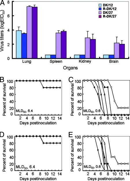
We then tested the virulence of these two viruses in a mammalian mouse model described previously (2). Three mice from each group were killed on day 4 and day 6 after intranasal inoculation of 106 EID50 of virus, and their organs were collected for virus titration in eggs. The DK/12 virus replicated in the mouse lungs without adaptation, and the mean titer reached 3.9 log EID50 on day 4 p.i.; however, the virus was not detected in any other organ tested (Fig. 1A). DK/12 virus caused 5% body weight loss by day 9 p.i. at which point the mice started to regain the weight over the course of the remaining observation period (data not shown). In contrast, DK/27 caused a systemic infection, replicated to the high titer of 7.3 log EID50 in the lungs and to the titers of 3.6, 2.9, and 2.2 log EID50 in the spleen, kidney, and brain, respectively (Fig. 1A; Table 2). Infection with DK/27 virus caused a more than 30% reduction in body weight, and all of the mice died before day 8 p.i. The two H5N1 viruses markedly differed in the dose required to kill 50% of infected mice (MLD50): 6.4 log EID50 for DK/12 and 0.6 log EID50 for DK/27 (Fig. 1B and C).
FIG. 1.
Replication and lethality of the DK/12 and DK/27 viruses in mice. (A) Six-week-old SPF BALB/c mice (three/group) were inoculated intranasally with 106 EID50 of each virus in a 50-μl volume and killed on day 3 p.i., and organs were collected for virus titration in eggs. Data shown are the mean virus titers ± standard deviation. (B to E) Death patterns of the mice infected with different H5N1 viruses, DK/12 (B), DK/27(C), R-DK/12 (D), and R-DK/27 (E), with the doses of 101 to 106 EID50 (101 EID50, •; 102 EID50, ○; 103 EID50, ▵; 104 EID50, ▴; 105 EID50, ⋄; 106 EID50, ⧫).
TABLE 2.
Replication of transfectant viruses in micea
| Virus | Mean virus titer (log10 EID50/ml ± SD) at indicated day p.i. in:
|
MLD50 (log EID50) | |||||||
|---|---|---|---|---|---|---|---|---|---|
| Lung
|
Spleen
|
Kidney
|
Brain
|
||||||
| Day 4 p.i. | Day 6 p.i. | Day 4 p.i. | Day 6 p.i. | Day 4 p.i. | Day 6 p.i. | Day 4 p.i. | Day 6 p.i. | ||
| DK/12 | 3.9 ± 0.5 | 3.6 ± 0.7 | < | < | < | < | < | < | 6.4 |
| DK/27 | 7.3 ± 0.1 | 6.9 ± 0.4 | 3.6 ± 0.3 | 2.0 ± 0.9 | 2.9 ± 0.9 | 3.0 ± 0.1 | 2.2 ± 0.8 | 3.1 ± 0.1 | 0.6 |
| R-DK/12 | 3.5 ± 0.1 | 3.5 ± 0.6 | < | < | < | < | < | < | 6.4 |
| R-DK/27 | 7.2 ± 0.3 | 6.9 ± 0.4 | 3.8 ± 0.3 | 1.8 ± 0.6 | 2.7 ± 0.7 | 2.9 ± 0.1 | 2.0 ± 0.5 | 2.8 ± 0.3 | 0.6 |
| DK/12-27PB2 | 4.5 ± 0.5 | 4.5 ± 0.7 | < | < | < | < | < | < | 6.4 |
| DK/12-27PA | 5.9 ± 0.4c | 4.1 ± 1.0 | < | < | < | < | < | < | 6.4 |
| DK/12-27HA | 3.8 ± 0.3 | 4.5 ± 0.4 | < | < | < | < | < | < | 6.4 |
| DK/12-27NP | 5.1 ± 0.3c | 5.5 ± 0.5b | < | < | < | < | < | < | 6.4 |
| DK/12-27NS | 5.8 ± 0.6c | 6.9 ± 0.4c | 2.3 ± 0.1 | 2.6 ± 0.3 | 1.6 ± 0.3 | 1.6 ± 0.5 | < | 3.1 ± 0.3 | 2.0 |
| DK/27-12PB2 | 5.6 ± 0.5e | 5.9 ± 0.5 | 2.2 ± 0.8e | 2.8 ± 0.4 | 2.0 ± 0.5 | 2.9 ± 0.5 | 1.5 ± 0.1 | 2.9 ± 0.5 | 0.6 |
| DK/27-12PA | 5.7 ± 0.4e | 6.4 ± 0.1 | 2.1 ± 0.6d | 2.3 ± 0.8 | 1.3 ± 0.1d | 1.9 ± 0.4d | 1.9 ± 0.7 | 2.5 ± 0.4 | 1.5 |
| DK/27-12HA | 6.8 ± 0.3 | 5.8 ± 0.5d | 3.5 ± 0.7 | 3.0 ± 0.5d | 2.4 ± 0.1 | 2.5 ± 0.4 | 1.4 ± 0.1 | 2.4 ± 0.9 | 1.5 |
| DK/27-12NP | 6.5 ± 0.4 | 6.3 ± 0.6 | 3.1 ± 0.6 | 2.5 ± 0.4 | 2.4 ± 0.1 | 2.2 ± 0.1 | 1.4 ± 0.1 | 2.9 ± 0.4 | 0.6 |
| DK/27-12NS | 3.9 ± 0.5e | 4.9 ± 0.5e | < | < | < | < | < | < | 6.4 |
| DK/12NS1P42S | 6.5 ± 0.4c | 6.8 ± 0.3c | 2.0 ± 0.7 | 2.0 ± 0.7 | 1.3 ± 0.1 | 1.3 ± 0.1 | < | 3.3 ± 0.8 | 2.2 |
| DK/12NS1N48S | 3.4 ± 0.1 | 4.5 ± 0.4 | < | < | < | < | < | < | 6.4 |
| DK/27NS1S42P | 4.5 ± 0.4e | 5.8 ± 0.3d | < | < | < | < | < | < | 6.4 |
| DK/27NS1S48N | 7.3 ± 0.1 | 7.5 ± 0.1 | 2.5 ± 1.1 | 2.8 ± 0.8 | 1.7 ± 0.7 | 3.1 ± 0.8 | 2.2 ± 0.9 | 3.1 ± 0.3 | 0.8 |
| DK/27NS1R38A | 7.6 ± 0.3 | 7.3 ± 0.8 | 2.1 ± 0.8 | 1.4 ± 0.1 | 2.1 ± 0.6 | 2.1 ± 0.6 | 1.4 ± 0.1 | 2.4 ± 0.1 | 0.8 |
| DK/27NS1K41A | 6.6 ± 0.3 | 6.5 ± 0.4 | 1.7 ± 0.4 | 1.4 ± 0.1 | 1.6 ± 0.3 | 2.2 ± 0.3 | 1.6 ± 0.5 | 2.9 ± 0.4 | 0.6 |
| DK/27NS1R38AK41A | 5.0 ± 0.5d | 4.6 ± 0.7e | < | < | < | < | < | < | 6.4 |
Six-week-old SPF BALB/c mice were inoculated intranasally with 106 EID50 of each virus in a 50-μl volume. Three mice from each group were killed on days 4 and 6 p.i., and virus titers were determined in samples of lung, spleen, kidney, and brain in eggs. <, no virus was isolated from the sample.
P value was <0.05 compared with the titers in the corresponding organs of the DK/12- or R-DK/12-inoculated mice.
P value was <0.01 compared with the titers in the corresponding organs of the DK/12- or R-DK/12-inoculated mice.
P value was <0.05 compared with the titers in the corresponding organs of the DK/27- or R-DK/27-inoculated mice.
P value was <0.01 compared with the titers in the corresponding organs of the DK/27- or R-DK/27-inoculated mice.
To determine the genetic relationship between the two viruses, we sequenced their genomes and compared them with the available sequences of H5N1 viruses. We found that the two viruses are closely related, with all of their eight segments sharing over 99% homology with the previously reported A/Duck/Fujian/01/02 virus (2). DK/12 and DK/27 share the same PB1, NP, M1, M2, and NS2 genes at the amino acid levels. At the amino acid level, we mapped a total of eight differences between the two viruses in their PB2, PA, HA, NP, and NS1 genes (Table 3). These data suggest that single- or multiple-amino-acid combinations among these eight different amino acids contribute to the difference in virulence in mice of the two viruses.
TABLE 3.
Amino acid differences between the DK/12 and DK/27 viruses
| Gene segment | Position of amino acid | Amino acid in:
|
|
|---|---|---|---|
| DK/12 virus | DK/27 virus | ||
| PB2 | 497 | Ser (S) | Asn (N) |
| 607 | Leu (L) | Val (V) | |
| 612 | Ala (A) | Thr (T) | |
| PA | 44 | Glu (G) | Val (V) |
| HA | 216 | Val (V) | Ile (I) |
| NP | 105 | Met (M) | Val (V) |
| NS1 | 42 | Pro (P) | Ser (S) |
| 48 | Asn (N) | Ser (S) | |
Rescued DK/12 and DK/27 viruses maintained the biological properties of the wild-type viruses.
To investigate the genetic basis of the virulence of the DK/12 and DK/27 viruses, we established a reverse genetics system for the two viruses. We inserted cDNAs of each full-length RNA segment of DK/12 and DK/27 into the viral RNA-mRNA bidirectional expression plasmid pBD, as described in Materials and Methods. Using these plasmids, we generated the DK/12 and DK/27 viruses from cloned cDNA, designated R-DK/12 and R-DK/27, respectively. After confirmation by sequence analysis, we prepared virus stocks by use of 10-day-old specific-pathogen-free (SPF) eggs and tested the replications and lethalities of these viruses in mice. R-DK/12 and R-DK/27 exhibited properties similar to those of their respective original viruses in terms of virus titers in organs and with respect to MLD50s (Fig. 1D and E; Table 2).
The NS gene plays a major role in the difference in pathogenicities in mice between the DK/12 and DK/27 viruses.
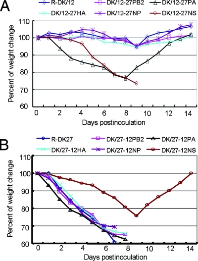
To identify the genes responsible for the difference in pathogenicities between the DK/12 and DK/27 viruses, we generated five single-gene recombinant viruses, each bearing the PB2, PA, HA, NP, or NS gene from DK/27 and the other seven genes from DK/12. The recombinant viruses that contained the PB2, PA, HA, or NP gene of DK/27 (designated DK/12-27PB2, DK/12-27PA, DK/12-27HA, or DK/12-27NP, respectively) displayed the same low pathogenicity in mice as the wild-type DK/12 virus (MLD50, 6.4 log EID50) and replicated only in the lungs (Table 2), although the virus titers in the lungs of mice infected with DK/12-27PA or DK/12-27NP virus were significantly higher than that of the DK/12 virus-infected mice. DK/12-27PA also caused about 20% weight loss in inoculated mice, whereas DK/12-27PB2, DK/12-27HA, and DK/12-27NP viruses caused only a transient reduction in body weight (Fig. 2A). In contrast to the other single-gene reassortants, the single-gene recombinant containing the NS gene of DK/27 (DK/12-27NS) caused systemic infection (Table 2). The MLD50 of the DK/12-27NS virus was over 104-fold higher than that of the DK/12 virus (MLD50, 2.0 versus 6.4 log EID50).
FIG. 2.
Comparison of weight changes in mice infected with different H5N1 avian influenza viruses. Mice (five mice/group) were intranasally infected with 106 EID50 of virus. (A) Mice infected with R-DK/12 and the reassortants in the DK/12 background. (B) Mice infected with R-DK/27 and the reassortants in the DK/27 background.
The effect of individual genes derived from the DK/12 virus on the virulence of DK/27 virus was also examined by generating five single-gene recombinant viruses, each containing the PB2, PA, HA, NP, or NS gene from DK/12 virus and the remaining segments from DK/27 virus. The viruses that carried the PB2, PA, HA, and NP gene of DK/12 (DK/27-12PB2, DK/27-12PA, DK/27-12HA, and DK/27-12NP, respectively) replicated in all four organs tested and caused rapid weight loss. The mice died within 9 days of inoculation (Fig. 2B; Table 2), although the virus titers in the lungs of DK/27-12PB2- and DK/27-12PA-inoculated mice were significantly lower than that of the DK/27 virus-inoculated animals. The MLD50s of these four recombinant viruses were similar to that of the DK/27 virus (ranging from 0.6 to 1.5 log EID50). The recombinant virus containing the NS gene of DK/12 in a background of genes from the DK/27 virus (DK/27-12NS), however, replicated only in the lungs, and the virus was dramatically attenuated in mice (MLD50, 0.6 versus 6.4 log EID50). In addition, DK/27-12NS caused a reduction in the body weight of the mice over the first 10 days after infection, but the mice regained weight over the remaining observation period (Fig. 2B). These results indicated that the NS gene plays a major role in the difference in pathogenicities in mice between the DK/12 and DK/27 viruses.
Amino acid substitution at position 42 in the NS1 protein changes the pathogenicity of the DK/12 and DK/27 viruses in mice.
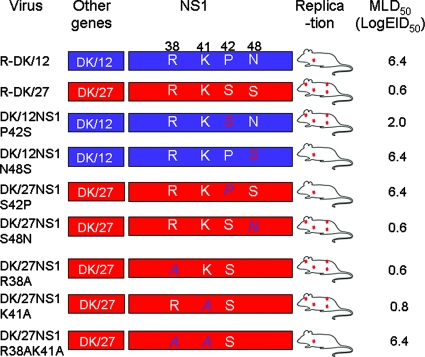
There are only two amino acid differences in the NS gene between DK/12 and DK/27, and both of the changes are located in the NS1 protein; that is, at positions 42 and 48 (Table 3). To elucidate the molecular basis of the virulence and replication discrepancy between the DK/12 and DK/27 viruses, we generated four mutant viruses each containing a substitution of the NS1 amino acid residues at position 42 or 48 and tested their pathogenicities in mice. A mutant DK/12 virus, designated DK/12NS1P42S and containing a substitution at amino acid position 42 that reflected the sequence found in the DK/27 NS1 protein (a Pro-to-Ser substitution), was highly pathogenic in mice, in contrast to the DK/12 virus (MLD50, 2.2 versus 6.4 log EID50). This mutant caused systemic infection in animals (Fig. 3; Table 2). A mutant DK/12 virus encoding an Asn-to-Ser mutation at position 48 of NS1 (DK/12NS1N48S) was not lethal and replicated only in the lung of mice (MLD50, 6.4 log EID50) (Fig. 3; Table 2). Similarly, a Ser-to-Pro substitution at position 42 in the NS1 protein of DK/27 that reflected the sequence found in the DK/12 virus resulted in marked attenuation of the DK/27 virus (MLD50, 0.6 versus 6.4 log EID50). This mutant, DK/27NS1S42P, replicated only in the lung and caused a transient reduction in the body weight of the mice (Table 2; Fig. 3). However, the mutant virus DK/27NS1S48N, containing a Ser-to-Asn mutation introduced at position 48 of the NS1 protein, was not attenuated with respect to the DK/27 virus. These results suggest that the amino acid at position 42 of NS1 protein is critical for the difference in virulence between the DK/12 and DK/27 viruses in mice.
FIG. 3.
NS1 mutant viruses and their virulences in mice. The color of the bar indicates the origin of the gene as follows: blue, DK/12; red, DK/27. The corresponding amino acids are shown as single-letter abbreviations with the positions numbered at the top. The red dots in the mouse figures indicate tissue tropism (upper left, brain; lower left, lung; upper right, kidney; lower right, spleen). The mutated amino acids are shown in red or blue and italics. Amino acid abbreviations: R, Arg; K, Lys; P, Pro; S, Ser; A, Ala.
The genomes of the viruses recovered on days 4 and 6 p.i. from the lungs of mice infected with the NS reassortants or mutant viruses were sequenced. No amino acid residue change was found in any of the gene segments except for PB2, for which we found that a mutation corresponding to a change from Glu to Lys at position 627 occurred in about 30% of the DK/12-27NS and DK/12NS1P42S viruses isolated from inoculated mice on day 6 p.i. but not from any samples recovered from other virus-inoculated mice (Table 4).
TABLE 4.
Amino acids at the key positions of the PB2 and NS1 genes of the viruses recovered from mice
| Virus | Amino acid at indicated key position of the viruses recovered from the mice on:
|
|||||||
|---|---|---|---|---|---|---|---|---|
| Day 4 p.i
|
Day 6 p.i.
|
|||||||
| PB2
|
NS1
|
PB2
|
NS1
|
|||||
| 627 | 701 | 42 | 48 | 627 | 701 | 42 | 48 | |
| R-DK/12 | E | D | P | N | E | D | P | N |
| R-DK/27 | E | D | S | S | E | D | S | S |
| DK/12-27NS | E | D | S | S | E (67%) + K (33%) | D | S | S |
| DK/27-12NS | E | D | P | N | E | D | P | N |
| DK/12-NS1P42S | E | D | S | N | E (67%) + K (33%) | D | S | N |
| DK/12-NS1N48S | E | D | P | S | E | D | P | S |
| DK/27-NS1S42P | E | D | P | S | E | D | P | S |
| DK/27-NS1S48N | E | D | S | N | E | D | S | N |
The amino acid at position 42 of the NS1 protein affects DK/12 and DK/27 virus antagonization of IFN-α/β action in human lung epithelial A549 cells.
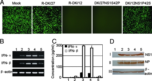
The amino-terminal 73 amino acids of the NS1 protein of influenza virus are responsible for binding to RNAs, in particular dsRNA, conferring upon the virus the ability to escape the IFN-α/β response by inhibiting the activation of transcription factors involved in IFN activation (10, 18). To determine whether the difference in the replication and virulence of DK/12 and DK/27 was directly correlated with the abilities of these viruses to inhibit the IFN-α/β system, the IFN-inducing properties of influenza viruses expressing wild-type or mutant NS1 proteins were investigated. Human epithelial lung A549 cells were infected at an MOI of 2 with wild-type DK/27, wild-type DK/12, mutant DK/27NS1S42P virus, or mutant DK/12NS1P42S virus. At 24 h postinfection, supernatants from infected A549 cells were used to determine the levels of secreted IFN-α/β in a bioassay based on the inhibition of VSV-GFP viral replication. The results are shown in Fig. 4. Supernatants from mock-infected cells caused no inhibition of GFP expression by VSV-GFP in A549 cells. However, VSV-GFP replication was completely abolished in cells pretreated with the supernatant of wild-type DK/12- and mutant DK/27NS1S42P-infected cells. By contrast, the supernatant of wild-type DK/27- or mutant DK/12NS1P42S-infected cells did not inhibit VSV-GFP replication (Fig. 4A).
FIG. 4.
Induction of IFN-α/β synthesis in A549 cells infected with influenza A viruses expressing wild-type or mutant NS1 proteins. (A) IFN-α/β bioassay. A549 cells were pretreated for 24 h with UV-inactivated supernatants from A549 cells infected with the indicated influenza viruses. The pretreated A549 cells were then infected with VSV-GFP, and, 14 h p.i., the cells expressing GFP were monitored by fluorescence microscopy. (B) RT-PCR analysis of IFN-α/β mRNA levels in virus-infected A549 cells. Cells were infected at an MOI of 2, and, 20 h p.i., total RNA was extracted and RT-PCR was done using primer pairs specific for human IFN-α/β and β-actin mRNA. The fragments of IFN-α, IFN-β, and β-actin are shown on the left. (C) Quantification of IFN-α/β production. A549 cells were either mock infected or infected at an MOI of 2. Twenty-four hours postinfection, the amounts of IFN-α and IFN-β released into the culture supernatant were measured by ELISA. (D) Levels of virus protein expression in infected cells. A549 cells were infected with virus at an MOI of 2 and lysed 12 h postinfection. Lysates of mock-infected cells or of cells infected with the indicated influenza viruses were incubated with mouse anti-truncated NS1 antiserum (I) or with chicken antiserum that was generated by inoculating SPF chickens with inactivated GS/GD/1/96 virus (II). Expression of β-actin protein was also examined as a control (III). Binding was visualized with DAB (3,3′-diaminobenzidine) reagent after incubation with peroxidase-conjugated secondary antibodies. The NS1, NP, and β-actin proteins are indicated on the right. Numbers 1 to 5 in panels B, C, and D indicate the type of infection as follows: 1, mock; 2, R-DK/27; 3, R-DK/12; 4, DK/27NS1S42P; 5, DK/12NS1P42S.
To determine whether the induction of the antiviral state in the treated cells correlated with the level of IFN-α/β induction in A549 cells infected with different viruses, the relative levels of IFN-α/β mRNA in the infected A549 cells were examined by RT-PCR. A549 cells were infected with various influenza viruses, and the cells were harvested for RNA extraction 20 h after infection. Total RNA was digested with DNase I to remove DNA from RNA samples. RT-PCR analysis confirmed that infection with wild-type DK/12 or mutant DK/27NS1S42P virus induces higher levels of IFN-α/β mRNA than infection with wild-type DK/27 or mutant DK/12NS1P42S virus (Fig. 4B). The PCR products were sequenced and confirmed to be derived from the expected mRNAs. We further investigated the expression and secretion of the IFN-α/β for A549 cells infected by different viruses. The culture supernatants were harvested 24 h after infection, and the amounts of IFN-α and IFN-β were measured by ELISA. The results confirmed that infection with wild-type DK/12 or mutant DK/27NS1S42P virus induces higher levels of IFN-α/β expression and secretion than infection with wild-type DK/27 or mutant DK/12NS1P42S virus (Fig. 4C).
Since previous reports indicated that the ability of the influenza virus to antagonize IFN-α/β induction by the host was related to NS1 protein expression levels in the infected cells (17, 28), we compared the levels of NS protein expressed in virus-infected cells by Western blotting (Fig. 4D). No significant difference in the levels of viral proteins, including NS1 and NP, was detected among the samples.
The amino acid at position 42 is critical for the NS1 protein to inhibit dsRNA-mediated NF-κB-responsive promoter activation.
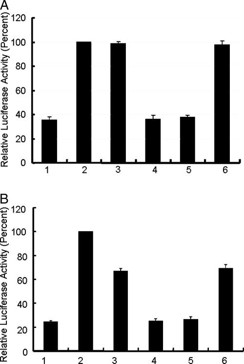
Activation of NF-κB and IRF-3 is required for the induction of the IFN-β promoter (6, 25, 26, 32, 36). NF-κB has been shown to bind to the positive regulatory domain II of the IFN-β promoter and to play an essential role in regulating IFN-β transcription (1, 8). Expression of the NS1 protein of influenza A virus is known to prevent virus- and/or dsRNA-mediated activation of the NF-κB pathway (35). To determine whether the contribution of the NS1 protein to the pathogenicities of the DK/12 and DK/27 viruses is associated with blocking the activation of the NF-κB pathway, we used an NF-κB reporter gene, pNF-κB-Luc, which contains a luciferase reporter gene under the control of an NF-κB-responsive promoter. Cotransfection of pNF-κB-Luc with expression plasmid p12NS1 or p27NS1S42P did not inhibit the expression of dsRNA [poly(I:C)]-induced reporter gene activity in 293T cells, whereas cotransfection of p27NS1 or p12NS1P42S substantially reduced it (Fig. 5A). These results demonstrate that the NS1 proteins of DK/12 and DK/27 viruses differ in their abilities to prevent the dsRNA-mediated activation of the NF-κB pathway, and the amino acid at position 42 in NS1 is critical for this function. The difference in reporter gene induction was not due to differences in NS1 protein levels, as Western blotting demonstrated that wild-type and mutant NS1 proteins were expressed in 293T cells at similar levels (data not shown).
FIG. 5.
Prevention of poly(I:C)-induced activation of an NF-κB promoter and the IRF-3-depedent promoter by NS1 protein. (A) NF-κB promoter assay. 293T cells were cotransfected with pNF-κB-Luc and pTK-RL plasmids along with the specified NS1 plasmids, with or without subsequent poly(I:C) transfection. (B) ISG-54 promoter assay. Vero cells were cotransfected with pISG-54-Luc and pTK-RL plasmids along with the specified NS1 plasmids, with or without subsequent poly(I:C) transfection. Bars: 1, pCAGGS; 2, pCAGGS with poly(I:C); 3, p12NS1 with poly(I:C); 4, p27NS1 with poly(I:C); 5, p12NS1P42S with poly(I:C); 6, p27NS1S42P with poly(I:C).
The amino acid at position 42 is critical for the NS1 protein to inhibit dsRNA-mediated IRF-3-dependent promoter activation.
NS1 protein of influenza A virus has been reported to inhibit the activation of IRF-3, which is a key regulator of IFN gene expression (32). The ISG54 promoter is one of the promoters that can be directly activated by the IRF-3, and it could also be activated by IFN produced in response to dsRNA treatment or to viral infection. To investigate if the amino acid change in 42 affects the ability of NS1 protein to inhibit the activation of the IRF-3-dependent promoter, we transfected the Vero cells, which do not produce IFN-α/β, with a ISG54-Luc reporter plasmid plus a constitutively expressed Renilla luciferase plasmid and a mammalian expression plasmid. The expression plasmid was empty vector pCAGGS or the pCAGGS plasmid expressing the wild-type or mutant NS1 gene. Cotransfection of pISG54-Luc with the plasmid p12NS1 or p27NS1S42P did not inhibit the expression of dsRNA [poly(I:C)]-induced reporter gene activity in Vero cells, whereas cotransfection of p27NS1 or p12NS1P42S substantially reduced it (Fig. 5B). These results demonstrate that the NS1 proteins of DK/12 and DK/27 viruses differ in their abilities to inhibit the dsRNA-mediated activation of the IRF-3-dependent promoter, and the amino acid at position 42 in NS1 is critical for this function. Western blotting demonstrated that wild-type and mutant NS1 proteins were expressed in Vero cells at similar levels (data not shown).
At least one of the two basic amino acids at position R38 or K41 in the NS1 protein is required for the virulence of the DK/27 virus in mice.
Amino acids R38 and K41 in the RNA-binding domain of the NS1 protein have previously been shown to be important in the inhibition of IFN production and the virulence of the influenza viruses (6, 19). Both of our H5N1 avian influenza viruses contain R38 and K41 in the NS1 protein. To investigate whether these two basic amino acids contribute to virulence, we generated three NS1 mutant viruses in the DK/27 background, DK/27R38A, DK/27K41A, and DK/27R38AK41A, and tested their replications and virulences in mice. As shown in Table 2 and Fig. 3, the mutants with a single-amino-acid substitution, DK/27R38A and DK/27K41A, replicate systemically and were highly virulent in mice, with MLD50s of 0.8 and 0.6 log EID50, respectively. However, the double mutant DK/27R38AK41A replicated only in the lungs and was highly attenuated in this animal (MLD50, 6.4 log EID50) (Fig. 3; Table 2). DK/27R38AK41A still has Ser at position 42 of NS1. These results demonstrate that the amino acid S42 and at least one of the basic amino acids at position R38 or K41 in NS1 are required for the virulence of DK/27 in mice.
DISCUSSION
H5N1 avian influenza viruses have caused the deaths of more than half of the humans they have infected since 1997 and clearly represent a threat to public health. Although different strains of H5N1 virus are known to have substantially different pathotypes, the effects of specific amino acid changes on the host ranges and virulences of H5N1 HPAIV remain largely unexplored. Here, we characterized two H5N1 HPAIV, DK/12 and DK/27, which have similar genomes but show markedly different pathogenicities in mice. Using this pair of viruses and single-gene reassortant viruses created from them, we demonstrated that the NS1 protein contributes to the different pathogenicities of these two viruses in mice and that the amino acid residue at position 42 in the NS1 protein of DK/27 is important for its ability to replicate and cause lethality in mice and to antagonize IFN-α/β production in A549 cells. We further demonstrated that the amino acid at position 42 of NS1 is critical for the ability of these viruses to inhibit dsRNA-mediated activation of the NF-κB pathway and the IRF-3 pathway. This is the first demonstration that a single amino acid at position 42 of the NS1 gene plays a major role in the determination of virulence of H5N1 avian influenza virus in a mammalian host.
Mutation or deletions within the effector domain of the NS1 gene have previously been shown to affect the ability of influenza viruses to antagonize IFN-α/β production, and the decreased ability of the virus to antagonize IFN-α/β production is linked with reduced expression levels of the NS1 protein in infected cells (17, 28). In this study, the mutation of amino acid 42 within the RNA-binding domain of NS1 affected the ability of the virus to antagonize IFN-α/β production in A549 cells, suggesting that the RNA-binding domain also contributes to the ability of the NS1 protein to antagonize IFN-α/β production in host cells. However, NS1 protein levels were not significantly different among the four viruses tested (Fig. 4D), suggesting that the mechanisms by which the NS1 effector domain and the NS1 RNA-binding domain antagonize host IFN-α/β production are different. In addition, the mutation of amino acid 42 within the RNA-binding domain of NS1 blocked the virus from preventing the dsRNA-mediated activation of the NF-κB pathway and the IRF-3-dependent promoter, demonstrating the importance of the ability of NS1 to counteract the host cell antiviral immune response.
The amino acid at position 42 of NS1 varies among avian influenza viruses; however, the serine at this position is highly conserved in the human, swine, and equine influenza viruses (2,561 of 2,564 influenza viruses isolated from mammalian hosts have S42 in their NS1 genes). Based on the available sequence information, all 135 H5N1 influenza viruses that have been isolated from humans and other mammals have serine at position 42 in the NS1 protein. The amino acid at position 42 is located within the second α-helix spanning amino acids 30 to 50 of the RNA-binding domain of NS1 protein (34). Two basic amino acids, R38 and K41, in this same region have previously been reported to be important for inhibiting host IFN induction and virulence of influenza A viruses (6). Using WSN virus, Donelan et al. demonstrated that the basic amino acid arginine at position 38 and lysine at position 41 of NS1 play a critical role in the inhibition of IFN production and in the virulence of the virus in mice, and an attenuated mutant containing NS1, R38AK41A, acquired increased virulence in mice as a result of the mutation of NS1 S42G (6). A recent study (19) reported that a mutant virus designated A/Udorn/72 and containing alanine instead of asparagine at position 38 was highly attenuated in MDCK cells. Our data indicate that DK/27 viruses containing the single substitution of alanine for either of the basic amino acids at positions 38 and 41 alone were not attenuated in mice, but substitution of alanine at both positions led to complete attenuation. Together with our finding of the importance of the amino acid at position 42 in virulence, these data emphasize the critical role of the second α-helix structure of the RNA-binding domain of NS1 for IFN antagonism and for virulence.
A recent study reported that the RNA helicase enzymes retinoic acid-inducible gene I (RIG-I) acts as a single-stranded RNA sensor and a potential target of viral immune evasion, and NS1 protein of influenza A virus blocks the RIG-I activation mediated by viral genomic single-stranded RNA bearing 5′ phosphates (22). It remains to be investigated if the contribution of the NS1 protein to the pathogenicities of the DK/12 and DK/27 viruses is associated with blocking the activation of RIG-I induced by the viral genomic 5′-phosphorylated RNA.
In summary, we demonstrate here that the amino acid at position 42 of NS1 plays an important role in the ability of H5N1 influenza viruses to antagonize the host IFN response and for the virulence of H5N1 avian influenza virus in a mammalian host. We also attest that the RNA-binding domain of the NS1 protein plays an important role in preventing the dsRNA-mediated activation of the NK-κB pathway and the IRF-3 pathway. Our results provide strong additional evidence that the NS1 protein is a virulence factor for H5N1 avian influenza viruses and that multiple domains within this protein may be suitable targets for the development of antiviral drugs and attenuated vaccines.
Acknowledgments
We thank Gloria Kelly and Susan Watson for editing the manuscript, Michael Whitt for providing the reverse genetics system for generating the recombinant VSV-GFP, and Nancy Cox and Kanta Subbarao for providing the plasmid pBD.
This work was supported by Chinese National S&T Plan Grant 2004BA519A-57, the Chinese National Key Basic Research Program (973) 2005CB523005 and 2005CB523200; by grants-in-aid and a contract research fund from the Ministry of Education, Culture, Sports, Science and Technology, Japan, Program of Founding Research Centers for Emerging and Reemerging Infectious Diseases; and by National Institute of Allergy and Infectious Diseases Public Health Service research grants.
Footnotes
Published ahead of print on 21 November 2007.
REFERENCES
- 1.Algarté, M., H. Nguyen, C. Heylbroeck, R. Lin, and J. Hiscott. 1999. IκB-mediated inhibition of virus-induced beta interferon transcription. J. Virol. 732694-2702. [DOI] [PMC free article] [PubMed] [Google Scholar]
- 2.Chen, H., G. Deng, Z. Li, G. Tian, Y. Li, P. Jiao, L. Zhang, Z. Liu, R. G. Webster, and K. Yu. 2004. The evolution of H5N1 influenza viruses in ducks in southern China. Proc. Natl. Acad. Sci. USA 10110452-10457. [DOI] [PMC free article] [PubMed] [Google Scholar]
- 3.Chen, H., Y. Li, Z. Li, J. Shi, K. Shinya, G. Deng, Q. Qi, G. Tian, S. Fan, H. Zhao, Y. Sun, and Y. Kawaoka. 2006. Properties and dissemination of H5N1viruses isolated during an influenza outbreak in migratory waterfowl in western China. J. Virol. 805976-5983. [DOI] [PMC free article] [PubMed] [Google Scholar]
- 4.Claas, E. C., A. Osterhaus, R. van Beek, J. C. De Jong, G. F. Rimmelzwaan, D. A. Senne, S. Krauss, K. F. Shortridge, and R. G. Webster. 1998. Human influenza A H5N1 virus related to a highly pathogenic avian influenza virus. Lancet 351472-477. [DOI] [PubMed] [Google Scholar]
- 5.Daly, C., and N. C. Reich. 1993. Double-stranded RNA activates novel factors that bind to the interferon-stimulated response element. Mol. Cell. Biol. 133756-3764. [DOI] [PMC free article] [PubMed] [Google Scholar]
- 6.Donelan, N., C. F. Basler, and A. García-Sastre. 2003. A recombinant influenza A virus expressing an RNA-binding defective NS1 protein induces high levels of beta interferon and is attenuated in mice. J. Virol. 7713257-13266. [DOI] [PMC free article] [PubMed] [Google Scholar]
- 7.Egorov, A., S. Brandt, S. Sereinig, J. Romanova, B. Ferko, D. Katinger, A. Grassauer, G. Alexandrova, H. Katinger, and T. Muster. 1998. Transfectant influenza A viruses with long deletions in the NS1 protein grow efficiently in Vero cells. J. Virol. 726437-6441. [DOI] [PMC free article] [PubMed] [Google Scholar]
- 8.Flory, E., M. Kunz, C. Scheller, C. Jassoy, R. Stauber, U. R. Rapp, and S. Ludwig. 2000. Influenza virus-induced NF-kB-dependent gene expression is mediated by overexpression of viral proteins and involves oxidative radicals and activation of IkB kinase. J. Biol. Chem. 2758307-8314. [DOI] [PubMed] [Google Scholar]
- 9.Gabriel, G., B. Dauber, T. Wolff, O. Planz, H.-D. Klenk, and J. Stech. 2005. The viral polymerase mediates adaptation of an avian influenza virus to a mammalian host. Proc. Natl. Acad. Sci. USA 10218590-18595. [DOI] [PMC free article] [PubMed] [Google Scholar]
- 10.Garcıa-Sastre, A. 2006. Antiviral response in pandemic influenza viruses. Emerg. Infect. Dis. 1244-47. [DOI] [PMC free article] [PubMed] [Google Scholar]
- 11.Garcıa-Sastre, A., A. Egorov, D. Matassov, S. Brandt, D. E. Levy, J. E. Durbin, P. Palese, and T. Muster. 1998. Influenza A virus lacking the NS1 gene replicates in interferon-deficient systems. Virology 252324-330. [DOI] [PubMed] [Google Scholar]
- 12.Hatta, M., P. Gao, P. Halfmann, and Y. Kawaoka. 2001. Molecular basis for high virulence of Hong Kong H5N1 influenza A viruses. Science 2931840-1842. [DOI] [PubMed] [Google Scholar]
- 13.Kawaoka, Y., and R. G. Webster. 1988. Sequence requirements for cleavage activation of influenza virus hemagglutinin expressed in mammalian cells. Proc. Natl. Acad. Sci. USA 85324-348. [DOI] [PMC free article] [PubMed] [Google Scholar]
- 14.Krug, R. M., W. Yuan, D. L. Noah, and A. G. Latham. 2003. Intracellular warfare between human influenza viruses and human cells: the roles of the viral NS1 protein. Virology 309181-189. [DOI] [PubMed] [Google Scholar]
- 15.Lawson, N., E. Stillmans, M. Whit, and J. K. Rose. 1995. Recombinant vesicular stomatitis viruses from DNA. Proc. Natl. Acad. Sci. USA 924477-4481. [DOI] [PMC free article] [PubMed] [Google Scholar]
- 16.Li, Z., H. Chen, P. Jiao, G. Deng, G. Tian, Y. Li, E. Hoffmann, R. G. Webster, Y. Matsuoka, and K. Yu. 2005. Molecular basis of replication of duck H5N1 influenza viruses in a mammalian mouse model. J. Virol. 7912058-12064. [DOI] [PMC free article] [PubMed] [Google Scholar]
- 17.Li, Z., Y. Jiang, P. Jiao, A. Wang, F. Zhao, G. Tian, X. Wang, K. Yu, Z. Bu, and H. Chen. 2006. The NS1 gene contributes to the virulence of H5N1 avian influenza viruses. J. Virol. 8011115-11123. [DOI] [PMC free article] [PubMed] [Google Scholar]
- 18.Mibayashi M., L. Martinez-Sobrido, Y. M. Loo, W. B. Cardenas, M. Gale, Jr., and A. Garcia-Sastre. 2007. Inhibition of retinoic acid-inducible gene I-mediated induction of beta interferon by the NS1 protein of influenza A virus. J. Virol. 81514-524. [DOI] [PMC free article] [PubMed] [Google Scholar]
- 19.Min, J. Y., and R. M. Krug. 2006. The primary function of RNA binding by the influenza A virus NS1 protein in infected cells: inhibiting the 2′-5′ oligo (A) synthetase/RNase L pathway. Proc. Natl. Acad. Sci. USA 1037100-7105. [DOI] [PMC free article] [PubMed] [Google Scholar]
- 20.Noah, D. L., K. Y. Twu, and R. M. Krug. 2003. Cellular antiviral responses against influenza A virus are countered at the posttranscriptional level by the viral NS1A protein via its binding to a cellular protein required for the 3′ end processing of cellular pre-mRNAS. Virology 307386-395. [DOI] [PubMed] [Google Scholar]
- 21.Office International des Epizooties. 2004. OIE manual of diagnostic tests and vaccines for terrestrial animals. Office International des Epizooties, Paris, France.
- 22.Pichlmair, A., O. Schulz, C. Tan, T. Näslund, P. Liljeström, F. Weber, and C. Sousa. 2006. RIG-I-mediated antiviral responses to single-stranded RNA bearing 5′-phosphates. Science 314997-1001. [DOI] [PubMed] [Google Scholar]
- 23.Quinlivan, M., D. Zamarin, A. Garcia-Sastre, A. Cullinane, T. Chambers, and P. Palese. 2005. Attenuation of equine influenza viruses through truncations of the NS1 protein. J. Virol. 798431-8439. [DOI] [PMC free article] [PubMed] [Google Scholar]
- 24.Reed, L. E. L., and H. Muench. 1938. A simple method of estimating fifty percent endpoints. Am. J. Hyg. 27493-497. [Google Scholar]
- 25.Sato, M., N. Tanaka, N. Hata, E. Oda, and T. Taniguchi. 1998. Involvement of the IRF family transcription factor IRF-3 in virus-induced activation of the IFN-beta gene. FEBS Lett. 425112-116. [DOI] [PubMed] [Google Scholar]
- 26.Schafer, S. L., R. Lin, P. A. Moore, J. Hiscott, and P. M. Pitha. 1998. Regulation of type I interferon gene expression by interferon regulatory factor-3. J. Biol. Chem. 2732714-2720. [DOI] [PubMed] [Google Scholar]
- 27.Seo, S. E., E. Hoffmann, and R. G. Webster. 2002. Lethal H5N1 influenza viruses escape host antiviral cytokine responses. Nat. Med. 8950-954. [DOI] [PubMed] [Google Scholar]
- 28.Solorzano, A., R. J. Webby, K. M. Lager, B. H. Janke, A. Garcia-Sastre, and J. A. Richt. 2005. Mutations in the NS1 protein of swine influenza virus impair anti-interferon activity and confer attenuation in pigs. J. Virol. 797535-7543. [DOI] [PMC free article] [PubMed] [Google Scholar]
- 29.Stark, G. R., I. M. Kerr, B. R. Williams, R. H. Silverman, and R. D. Schreiber. 1998. How cells respond to interferons. Annu. Rev. Biochem. 67227-264. [DOI] [PubMed] [Google Scholar]
- 30.Stech, J., and H. D. Klenk. 2006. A new approach to an influenza life vaccine: haemagglutinin cleavage site mutants generated by reverse genetics. Berl. Munch. Tierarztl. Wochenschr. 119186-191. [PubMed] [Google Scholar]
- 31.Subbarao, K., A. Klimov, J. Katz, H. Regnery, W. Lim, H. Hall, M. Perdue, D. Swayne, C. Bender, J. Huang, M. Hemphill, T. Rowe, M. Shaw, X. Xu, K. Fukuda, and N. Cox. 1998. Characterization of an avian influenza A (H5N1) virus isolated from a child with a fatal respiratory illness. Science 279393-396. [DOI] [PubMed] [Google Scholar]
- 32.Talon, J., C. M. Horvath, R. Polley, C. F. Basler, T. Muster, P. Palese, and A. García-Sastre. 2000. Activation of interferon regulatory factor 3 is inhibited by the influenza A virus NS1 protein. J. Virol. 747989-7996. [DOI] [PMC free article] [PubMed] [Google Scholar]
- 33.Vilcek, J., and G. C. Sen. 1996. Orthomyxoviruses, p. 375-399. In B. N. Fields, D. M. Knipe, P. M. Howley, R. M. Chanock, J. L. Melnick, T. P. Monath, B. Roizman, and S. E. Straus (ed.), Fields virology. Lippincott-Raven Publishers, Philadelphia, PA.
- 34.Wang, W., K. Riedel, P. Lynch, C. Y. Chien, G. T. Montelione, and R. M. Krug. 1999. RNA binding by the novel helical domain of the influenza virus NS1 protein requires its dimer structure and a small number of specific basic amino acids. RNA 5195-205. [DOI] [PMC free article] [PubMed] [Google Scholar]
- 35.Wang, X., M. Li, H. Zheng, T. Muster, P. Palese, A. A. Beg, and A. García-Sastre. 2000. Influenza A virus NS1 protein prevents activation of NF-κB and induction of alpha/beta interferon. J. Virol. 7411566-11573. [DOI] [PMC free article] [PubMed] [Google Scholar]
- 36.Yoneyama, M., W. Suhara, Y. Fukuhara, M. Fukuda, E. Nishida, and T. Fujita. 1998. Direct triggering of the type I interferon system by virus infection: activation of a transcription factor complex containing IRF-3 and CBP/p300. EMBO J. 171087-1095. [DOI] [PMC free article] [PubMed] [Google Scholar]


