Abstract
The spores of six strains of Bacillus anthracis (four virulent and two avirulent) were compared with those of four other types of spore-forming bacteria for their resistance to four liquid chemical sporicides (sodium hypochlorite at 5,000 ppm available chlorine, 70,000 ppm accelerated H2O2, 1,000 ppm chlorine dioxide, and 3,000 ppm peracetic acid). All test bacteria were grown in a 1:10 dilution of Columbia broth (with manganese) incubated at 37°C for 72 h. The spore suspensions, heat treated at 80°C for 10 min to rid them of any viable vegetative cells, contained 1 × 10⁸ to 3 × 10⁸ CFU/ml. The second tier of the quantitative carrier test (QCT-2), a standard of ASTM International, was used to assess for sporicidal activity, with disks (1 cm in diameter) of brushed and magnetized stainless steel as spore carriers. Each carrier, with 10 μl (≥106 CFU) of the test spore suspension in a soil load, was dried and then overlaid with 50 μl of the sporicide being evaluated. The contact time at room temperature ranged from 5 to 20 min, and the arbitrarily set criterion for acceptable sporicidal activity was a reduction of ≥106 in viable spore count. Each test was repeated at least three times. In the final analysis, the spores of Bacillus licheniformis (ATCC 14580T) and Bacillus subtilis (ATCC 6051T) proved to be generally more resistant than the spores of the strains of B. anthracis tested. The use of one or both of the safe and easy-to-handle surrogates identified here should help in developing safer and more-effective sporicides and also in evaluating the field effectiveness of existing and newer formulations in the decontamination of objects and surfaces suspected of B. anthracis contamination.
Bacillus anthracis, the etiologic agent of anthrax, is a spore-forming zoonotic pathogen (3). Its spores are hardy enough for weaponization and environmental dispersal (10); viable spores of the organism have been recovered after many decades from deliberately contaminated sites (6). The more-recent and malicious use of the spores of B. anthracis in the United States (14) clearly attests to the potential of this organism to cause mortality, morbidity, and general societal disruption and reemphasizes the importance of having available suitable remedial measures for countering any subsequent accidental or deliberate release of such spores.
The biohazardous nature of B. anthracis mandates its handling in laboratories at biosafety level 3, also called containment level 3 (CL-3), in Canada. The availability of only a very limited number of such facilities seriously hampers experimentation with this increasingly significant pathogen, a recognized biothreat agent. This is especially true for developmental studies on decontamination agents, as such work is often conducted in settings without biosafety level 3 facilities. This study evaluated several potential bacterial surrogates for the spores of B. anthracis to enable a wider search for safer and more-effective liquid chemical sporicides to deal with the threat of anthrax.
MATERIALS AND METHODS
Preparation of spore suspensions.
Details on the bacteria used are given in Table 1. All Bacillus species other than the B. anthracis strains were acquired from the American Type Culture Collection (ATCC, Manassas, VA) and housed at the National Microbiology Lab (NML), Winnipeg, Manitoba, Canada, in a lyophilized state. All work with them was done in CL-2. The B. anthracis strains were gifts from the Animal Diseases Research Institute (ADRI), Lethbridge, Alberta, Canada, except for the avirulent Sterne strain 34F2, which was received from Jody Berry of the NML; they were handled in a CL-3 facility.
TABLE 1.
Organisms selected for testing by QCT-2a
| Organism (source or description) | Reference no. | Spore titer |
|---|---|---|
| Bacillus licheniformis | ATCC 14580T | 1.80 × 10⁸ |
| Bacillus subtilis | ATCC 6051T | 6.00 × 10⁸ |
| Bacillus atrophaeus | ATCC 49337T | 8.00 × 10⁸ |
| Bacillus atrophaeus (formerly “B. globigii”) | ATCC 51189 | 3.20 × 10⁸ |
| Bacillus anthracis (caprine) | ADRI Lethbridge no. 2001-50 | 8.50 × 10⁸ |
| Bacillus anthracis (bovine) | ADRI Lethbridge no. 9610 | 2.92 × 10⁸ |
| Bacillus anthracis (bison) | ADRI Lethbridge no. 93-189C | 3.90 × 10⁸ |
| Bacillus anthracis (Pasteur strain; avirulent) | ATCC 4229 | 2.10 × 10⁸ |
| Bacillus anthracis (bovine) | NML 03-0191 | 2.45 × 10⁸ |
| Bacillus anthracis (Sterne; avirulent) | 34F2 | 2.19 × 10⁸ |
All organisms came from the National Microbiology Lab, Winnipeg, Manitoba, Canada.
Spore preparations were made as described previously (17, 18). Briefly, after their initial recovery on plates of 5% sheep blood agar, all organisms were grown aerobically for 72 h at 36°C ± 1°C (mean ± standard deviation) with ∼150 rpm agitation in a 1:10 dilution of Columbia broth (Difco [Becton-Dickinson], Sparks, MD) containing a final concentration of 10 mM MnSO4·4H2O/ml; the Sterne strain required 96 h of incubation to produce the desired level of sporulation. The broth cultures were transferred to 50-ml high-clarity polypropylene tubes (BD Biosciences, Mississauga, Ontario, Canada) and centrifuged (Beckman Coulter KR4 centrifuge; VWR, Mississauga, Ontario, Canada) at 5,000 × g for 20 min at 4°C, and each spore pellet was washed using 20 ml of cold, sterile, distilled water. The washing step was repeated twice, with centrifugation in between, and the spores were resuspended in a final volume of 10 ml sterile distilled water. The suspensions were heated at 80°C in a water bath for 10 min to inactivate any vegetative forms. The titer in all such concentrated and heat-treated suspensions was >108 CFU/ml. Only one such suspension was produced for each bacterial species tested; the suspension was stored at 4°C and used throughout this investigation.
The carrier tests.
The second tier of the quantitative carrier test (QCT-2), standard E-2197-02 of ASTM International (formerly, the American Society for Testing and Materials) (2), was used in all testing, with metal disks (1 cm in diameter) as the spore carriers. The disks, purchased from Muzeen & Blythe (Winnipeg, Manitoba, Canada), were cut from sheets (0.75-mm thickness) of American Iron and Steel Institute grade #430 brushed stainless steel. They were first soaked overnight in a detergent solution (#7; Fisher Scientific, Ottawa, Ontario, Canada) to degrease them and then washed in tap water and rinsed thoroughly in double-distilled water prior to autoclave sterilization (121°C for 15 min). The disks were used only once and discarded.
HW.
When a disinfectant under test required dilution to prepare its use concentration, water with a hardness of 400 ppm calcium carbonate (CaCO3) was the diluent. The hard water (HW) was prepared as specified by AOAC International (formerly, the Association of Official Analytical Chemists) (1).
Sporicides tested.
Four different chemicals were evaluated in this study. Locally purchased liquid domestic bleach (Clorox Co., Oakland, CA) containing 5.25% sodium hypochlorite was diluted 10-fold in HW. The pH of the diluted bleach was 9.6, and the free available chlorine (FAC) and total chlorine levels in it were measured by using a Hach colorimetric chlorine detection kit (ClearTech, Winnipeg, Manitoba, Canada). A product based on 7% accelerated hydrogen peroxide (AHP) was obtained from Virox Technologies (Oakville, Ontario, Canada) and tested undiluted. An aqueous solution of chlorine dioxide (ClO2) was prepared by dissolving one Exterm (ChlorDiSys, Inc., Lebanon, NJ) tablet in 360 ml HW in a plastic squirt bottle; the FAC in the solution was at least 1,000 ppm, as determined by using test strips (Selective Micro Technologies, Beverly, MA). Peracetic acid (PAA; FMC, Tonawanda, NJ) was diluted to 0.3% (wt/vol) by mixing 100 μl of the stock solution with 4.9 ml HW.
Soil load.
A soil load was included to simulate the presence of residual body fluids or accumulated surface dirt, increasing the demand on the disinfectants. The tripartite soil load was prepared as follows, according to previously described methods (18, 19). A 340-μl volume of each spore suspension was mixed with 35 μl 5% tryptone (Difco), 25 μl of 5% bovine serum albumin (Difco), and 100 μl of 0.4% mucin (type 1) from bovine submaxillary glands (Sigma, St. Louis, MO), all in sterile normal saline (pH 7.2).
Disinfectant neutralizers and their effect on spore viability.
The AHP-based formulation was neutralized with a solution of Letheen broth (Difco) with 0.1% (wt/vol) sodium thiosulfate (Na2S2O3) and 0.1% (vol/vol) Tween 80 in sterile normal saline (13). Chlorine-based formulations (bleach and ClO2,) and PAA were neutralized with a solution of 0.1% sodium thiosulfate and 0.1% Tween 80 in saline (9).
To ensure that the disinfectant neutralization procedure itself was not detrimental to spore viability, spore-contaminated carriers were treated with either saline alone or saline separately containing the appropriate neutralizer. After an exposure time of 10 min, the carriers were eluted and the eluates assayed for CFU.
Determining sporicidal activity.
Ten microliters of the test spore suspension was carefully placed on each disk by using a positive-displacement pipette. The inoculated carriers were held in a laminar flow hood to dry the inoculum, and they were then placed at room temperature in a desiccator under vacuum for no less than two hours. One disk each with the dried inoculum was placed in a 15-ml-capacity Teflon vial (Cole Parmer, Vernon Hills, IL) with the inoculated side up, overlaid with 50 μl of the disinfectant under test, and held at room temperature for the desired contact time; control carriers received an equivalent volume of normal saline. At the end of the contact time, 9.95 ml of the appropriate neutralizer in saline was added to stop the sporicidal action.
The vial with its contents was then vortexed for 45 seconds to recover the inoculum; 1 ml of the eluate was placed in 9 ml of saline and subjected to at least four additional 10-fold dilutions in saline. The eluate remaining in the vial and several rinses of the vial with sterile saline were passed through a membrane filter (47-mm diameter) with a pore diameter of 0.22 μm (Pall, Ann Arbor, MI). Each 10-fold dilution of eluate was also separately membrane filtered, using one filter per dilution. One filter each was placed on the surface of a plastic petri plate (100 mm in diameter) of tryptic soy agar (Difco) and incubated at 36°C ± 1°C for a maximum of 5 days; CFU were counted, and log10 reductions in spore titers calculated.
Selection of potential surrogates.
The selection of potential surrogate species and strains of spore formers used here was based on (i) safety for handling; (ii) availability of type strains from a credible source, such as the ATCC; (iii) ability to yield relatively high titers of spores in a liquid medium from one or more commercial sources; (iv) representation from genera other than Bacillus; and (v) prior history of use as a surrogate/biological indicator (4, 11, 12, 15). Review of the use of Bacillus subtilis and Bacillus atrophaeus strains as surrogates was complicated by the significant changes these taxa have undergone over the years, whereby strains now designated as B. atrophaeus have been referred to in the literature as “B. subtilis var. subtilis,” “Bacillus globigii,” “Bacillus subtilis var. niger,” the “red strain,” “Bacillus niger,” or “Bacillus atrophaeus subsp. globigii” (5, 7, 20). In this study, the type strains of B. subtilis and B. atrophaeus, as well as B. atrophaeus ATCC 51189, formerly called “B. globigii,” were selected for QCT-2 study (Table 1).
This investigation started with 13 types of nonanthrax spore formers, which included not only Brevibacillus brevis (ATCC 8246T) and Virgibacillus pantothenticus (ATCC 14576T) but also seven additional members of the genus Bacillus, including B. cereus (ATCC 14579T), B. megaterium (ATCC 14581T), B. sphaericus (ATCC 14577T), and B. thuringiensis (ATCC 13367). However, after an initial screening of the organisms for their susceptibility to a 1:10 dilution of domestic bleach (about 5,000 ppm available chlorine), the four nonanthrax species of Bacillus were selected for further study based on their higher resistance.
Microbicide performance criterion.
Each formulation tested was arbitrarily required to inactivate at least 106 spores in QCT to be considered sporicidal (17).
Statistical analyses.
Each experiment included three control carriers, and all contact time determinations were repeated three times, with two test carriers at each. The results were examined by using Prism4 (GraphPad Software, Inc., San Diego, CA). One-way analysis of variance (ANOVA) for each individual contact time between the surrogates was examined. ANOVA was also carried out at 10 and 20 min of contact between the formulations and each Bacillus surrogate (comparison of microbicide at a specific contact time). When performing ANOVA analysis with Prism4 software, the option of Tukey's posttest was selected from a list of posttest operations to further distinguish and identify statistically significant differences between data sets. The results for the four disinfectants tested against B. anthracis spores were then taken to Prism4 statistical software for analysis of the mean log10 reductions at the 5, 10, and 20 min contact times for FAC, AHP, chlorine dioxide, and PAA. Analyses using one-way ANOVA were carried out for each individual contact time in the B. anthracis spore inactivation data. Statistical ANOVA comparison was conducted at 10 and 20 min of contact for the four surrogates and the seven B. anthracis strains against each of the four sporicidal agents to determine significant differences between surrogate and B. anthracis responses to sporicidal treatments.
RESULTS
Standardization of conditions for bacterial growth and sporulation.
As shown in Table 1, all organisms used in this study achieved a spore titer of at least 108/ml in the heat-treated suspensions. However, when the titers were much higher, the stock solutions were diluted with saline to the desired level of 1 × 10⁸ to 3 × 10⁸ CFU/ml. This was to ensure that the minimum 6-log10 reduction could be demonstrated for the effective formulations without wide variations in the starting titers of the various bacterial species tested.
Validating effectiveness of sporicide neutralization.
In testing for disinfectant activity, it is necessary to validate that the microbicidal activity of the formulation is successfully neutralized immediately at the end of the contact time. In this study, saline containing one or more chemicals known to neutralize the target chemical was added to the carrier soon after the required time of exposure to the test formulation was over. The neutralization step had to be validated as well, to determine that the neutralizer itself had no deleterious effects on the recovery and viability of the test organisms. The results (Table 2) demonstrate that the addition of a mixture of neutralizer and test formulation to spore-inoculated carriers did not significantly reduce the recoverable titer compared to the results for saline-treated control carriers and neutralizer alone. Therefore, all neutralizers used could successfully arrest the action of the respective formulation, with no deleterious effects on the viability of the spores tested.
TABLE 2.
Neutralization to stop sporicidal activity
| Species tested | Sporicide (concn) | Neutralizer | Log10 CFU recovered
|
||
|---|---|---|---|---|---|
| Control carrier | Neutralizer + sporicide | Neutralizer alone | |||
| B. licheniformis (ATCC 14580) | Sodium hypochlorite (5,000 ppm FAC) | 1% sodium thiosulfate + 0.1% Tween 80 in saline | 6.75 | 6.71 | 6.80 |
| B. atrophaeus (ATCC 51189) | Sodium hypochlorite (5,000 ppm FAC) | 1% sodium thiosulfate + 0.1% Tween 80 in saline | 6.90 | 6.98 | 6.86 |
| B. subtilis (ATCC 6051) | Sodium hypochlorite (5,000 ppm FAC) | 1% sodium thiosulfate + 0.1% Tween 80 in saline | 6.60 | 6.50 | 6.53 |
| B. atrophaeus (ATCC 51189) | AHP (70,000 ppm) | Letheen broth + 1% sodium thiosulfate | 6.71 | 6.71 | 6.96 |
| B. licheniformis (ATCC 14580) | AHP (70,000 ppm) | Letheen broth + 1% sodium thiosulfate | 7.05 | 6.98 | 7.03 |
| B. subtilis (ATCC 6051) | AHP (70,000 ppm) | Letheen broth + 1% sodium thiosulfate | 6.69 | 6.59 | 6.65 |
| B. anthracis (ATCC ADRI 9610) | AHP (70,000 ppm) | Letheen broth + 1% sodium thiosulfate | 6.49 | 6.52 | 6.56 |
| B. subtilis (ATCC 6051) | PAA (3,000 ppm) | 1% sodium thiosulfate + 0.1% Tween 80 in saline | 6.43 | 6.41 | 6.36 |
| B. licheniformis (ATCC 14580) | PAA (3,000 ppm) | 1% sodium thiosulfate + 0.1% Tween 80 in saline | 6.84 | 6.80 | 6.82 |
| B. atrophaeus (ATCC 51189) | PAA (3,000 ppm) | 1% sodium thiosulfate + 0.1% Tween 80 in saline | 6.63 | 6.63 | 6.65 |
| B. atrophaeus (ATCC 51189) | Chlorine dioxide (1,000 ppm) | 1% sodium thiosulfate + 0.1% Tween 80 in saline | 6.68 | 6.66 | 6.73 |
| B. subtilis (ATCC 6051) | Chlorine dioxide (1,000 ppm) | 1% sodium thiosulfate + 0.1% Tween 80 in saline | 6.25 | 6.36 | 6.32 |
Effect of contact time on sporicidal activity.
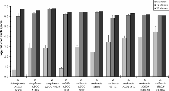
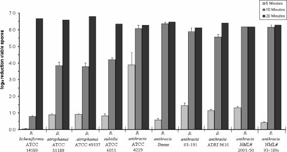
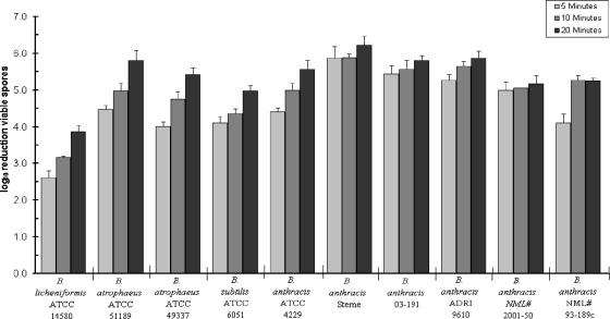
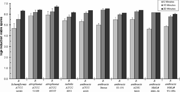
All formulations were tested at contact times of 5, 10, and 20 min, and the findings with the diluted bleach, AHP, chlorine dioxide, and PAA are presented in Fig. 1, 2, 3, and 4, respectively. The bleach (Fig. 1) required no longer than 10 min to reduce the viability of the spores of all species tested by ≥6 log10. There was considerable variation in its sporicidal action at 5 min of contact, with Bacillus licheniformis and B. subtilis (ATCC 6051) showing the highest levels of resistance. The AHP-based formulation (Fig. 2) showed generally low sporicidal activity at 5 min of contact, with virtually no reduction in the viability of the spores of B. licheniformis. At 10 min, it could reduce the spore titers of all six strains of B. anthracis by ≥6 log10 while being unable to do so for the four surrogates tested. After 20 min of dwell time, it could inactivate ≥6 log10 of the spores across the board. At the level tested here, chlorine dioxide showed the least degree of consistency and power in its sporicidal action (Fig. 3). It could inactivate the spores of only the Sterne strain of B. anthracis by ≥6 log10 at all of the contact times tested. B. licheniformis exhibited the highest level of resistance to this chemical even at 20 min of contact. In general, PAA (Fig. 4) showed a faster level of sporicidal activity, because, even at 5 min, it achieved higher levels of spore kill than the other three formulations tested. Its activities at 10 and 20 min were roughly comparable to those of the diluted bleach and AHP.
FIG. 1.
Comparative resistance of Bacillus spores to sodium hypochlorite (5,000 ppm FAC). Error bars show standard deviations.
FIG. 2.
Comparative resistance of Bacillus spores to AHP (70,000 ppm H2O2). Error bars show standard deviations.
FIG. 3.
Comparative resistance of Bacillus spores to aqueous chlorine dioxide (1,000 ppm available chlorine). Error bars show standard deviations.
FIG. 4.
Comparative resistance of Bacillus spores to 0.3% PAA. Error bars show standard deviations.
This study successfully demonstrated the higher resistance of B. licheniformis to all the formulations, notably, at shorter contact times (5 and 10 min), in comparison to the resistance of B. atrophaeus (P < 0.05, using Tukey's multiple comparison testing). With minimal exceptions, the resistance profiles of the two strains of B. atrophaeus (ATCC 51189 and 49337) to FAC, AHP, chlorine dioxide, and PAA were similar at 20 min of contact, and both were more susceptible than B. subtilis ATCC 6051 (P < 0.05, Tukey's test).
DISCUSSION
Microbicidal chemicals are crucial in interrupting the spread of a variety of infectious agents in many settings, but particularly so in dealing with infectious bioagents where the risk of failure can be unacceptably high. Therefore, the findings of any lab-based evaluation of a chemical for potential application in the remediation of sites known or suspected to be contaminated with hard-to-kill bioagents, such as the spores of B. anthracis, must generate data with a high level of confidence for success under field situations. While it is virtually impossible to recreate in a laboratory the unlimited variations that may occur in natural settings, protocols can be designed to incorporate several levels of stringency to present the test microbicide with a reasonable level of challenge. QCT-2 is one such method, which entails the use of carriers with an uneven surface, applies a relatively small volume of the test formulation for the surface area to be decontaminated, adds a soil load to the microbial test suspension, and avoids any wash-off of the test organism during all manipulations (17).
Any meaningful comparison of the microbicide susceptibility/resistance of two or more organisms must ensure that they are grown and processed using virtually identical procedures. This important condition was met in this study by culturing all test organisms in the same medium and processing the spores using standardized procedures (17). The length of incubation for sporulation was also the same, except in the case of the Sterne strain, which required 96 h of incubation as opposed to 72 h for all the others. The organisms that failed to grow or sporulate well in diluted Columbia broth in 72 to 96 h at 37°C were excluded from further consideration.
The formulations tested in this study are all known for their relatively rapid sporicidal activity (9). This was an important selection criterion because contact times of longer than a few minutes are generally not feasible or practical in dealing with the decontamination of environmental surfaces. While formaldehyde and glutaraldehyde can also be sporicidal, they would require several hours at room temperature to inactivate the levels of spores selected for the sporicide performance criterion in this investigation.
The sporicidal activity of a given formulation is only one among a set of features to be considered in dealing with any decontamination scenario. Human and environmental safety, as well as materials compatibility, are among the other factors to keep in mind. Though bleach is fast acting, inexpensive, and generally readily available, its main drawbacks are off-gassing inactivation in the presence of organic loads and high corrosivity. In contrast, AHP-based formulations are reasonably fast acting and also have much-higher materials compatibility, while being less toxic to humans and the environment (8).
While disinfectants have been tested previously against the spores of B. anthracis and their potential surrogates (16), the wide variations in the test protocols used make meaningful comparisons between the findings virtually impossible. The use of suspension test protocols for this purpose (4) also makes it difficult to extrapolate the findings to the inactivation of the spores on environmental surfaces. A recent study used a carrier test for this purpose (13). However, certain manipulations in the test method could inadvertently lead to the wash-off of viable spores and thus give erroneously higher levels of spore kills.
Evidence for microbicidal activity using QCT-2 is now recognized for the registration and marketing of disinfectants in Canada (http://www.hc-sc.gc.ca/dhp-mps/prodpharma/applic-demande/guide-ld/disinfect-desinfect/disinf_desinf_e.html). The Environmental Protection Agency, which registers environmental surface disinfectants for sale in the United States, is currently working with the Organization for Economic Cooperation and Development (OECD) to adopt a test guide based on QCT-2 (http://www.oecd.org/dataoecd/28/7/2494153.pdf).
To our knowledge, this is the first study using a fully quantitative carrier test and standardized conditions for spore production and processing to identify one or more suitable bacterial surrogates for B. anthracis. While B. licheniformis spores showed resistance that was only similar to the resistance of spores of the other potential surrogates and B. anthracis in some treatments, an overall trend of spore survival higher than or equivalent to that of B. anthracis to liquid sporicidal treatment is only one of the qualities of a good surrogate. B. licheniformis also satisfies the other requirements of a good surrogate (safe to work with, high-titer yield, and availability of type strain). Based upon this and the results of this study, it is recommended that spores of B. licheniformis ATCC 14580T, either alone or together with B. subtilis ATCC 6051T, be considered in testing liquid microbicides for their potential use against the spores of B. anthracis. Both of these organisms are safe and easy to work with, thus requiring no more than the basic skills and facilities that are more likely to be available in company or contract labs.
Acknowledgments
This study was supported by grants 02-0067RD32 and 04-0019-TD from the CBRN Research and Technology Initiative (CRTI) of Canada's Department of National Defense and by a stipend from the University of Manitoba.
We thank Thomas Hassard, Department of Community Health Sciences, University of Manitoba, for his help with statistical analyses; Michelle Alfa, St. Boniface General Hospital, Winnipeg, Manitoba, Canada, and Sola Adegbunrin, CREM, University of Ottawa, for technical expertise; L. Nakamura, U.S. Department of Agriculture, for the V. pantothenticus strain; and Greg Tiffin, ADRI, for five of the six strains of B. anthracis.
Footnotes
Published ahead of print on 14 December 2007.
REFERENCES
- 1.AOAC International. 1998. AOAC official methods of analysis, 16th ed., p. 1-16. AOAC International, Gaithersburg, MD.
- 2.ASTM International. 2007. Standard quantitative disk carrier test method for determining the bactericidal, virucidal, fungicidal, mycobactericidal and sporicidal activities of liquid chemical germicides. ASTM Book of Standards, vol. 11.05. Document E2197-02. ASTM International, West Conshohocken, PA.
- 3.Atlas, R. M. 2002. Responding to the threat of bioterrorism: a microbial ecology perspective—the case of anthrax. Int. Microbiol. 5:161-167. [DOI] [PubMed] [Google Scholar]
- 4.Brazis, A. R., J. E. Leslie, P. W. Kabler, and R. L. Woodward. 1958. The inactivation of spores of Bacillus globigii and Bacillus anthracis by free available chlorine. Appl. Microbiol. 6:338-342. [DOI] [PMC free article] [PubMed] [Google Scholar]
- 5.Fritze, D., and R. Pukall. 2001. Reclassification of bioindicator strains Bacillus subtilis DSM 675 and Bacillus subtilis DSM 2277 as Bacillus atrophaeus. Int. J. Syst. Evol. Microbiol. 51:35-37. [DOI] [PubMed] [Google Scholar]
- 6.Manchee, R. J., M. G. Broster, A. J. Stagg, and S. E. Hibbs. 1994. Formaldehyde solution effectively inactivates spores of Bacillus anthracis on the Scottish island of Gruinard. Appl. Environ. Microbiol. 60:4167-4171. [DOI] [PMC free article] [PubMed] [Google Scholar]
- 7.Nakamura, L. K. 1989. Taxonomic relationship of black-pigmented Bacillus subtilis strains and a proposal for Bacillus atrophaeus. Int. J. Syst. Bacteriol. 39:295-300. [Google Scholar]
- 8.Omidbukhsh, N., and S. A. Sattar. 2006. Broad-spectrum microbicidal activity, toxicological assessment and materials compatibility of a new generation of accelerated hydrogen peroxide (AHP)-based environmental surface disinfectant. Am. J. Infect. Control 34:251-257. [DOI] [PMC free article] [PubMed] [Google Scholar]
- 9.Perez, J., V. S. Springthorpe, and S. A. Sattar. 2005. Activity of selected oxidizing microbicides against the spores of Clostridium difficile: relevance to environmental control. Am. J. Infect. Control 33:320-325. [DOI] [PubMed] [Google Scholar]
- 10.Raber, E., A. Jin, K. Noonan, R. McGuire, and R. D. Kirvel. 2001. Decontamination issues for chemical and biological warfare agents: how clean is clean enough? Int. J. Environ. Health Res. 11:128-148. [DOI] [PubMed] [Google Scholar]
- 11.Rogers, J. V., C. L. Sabourin, Y. W. Choi, W. R. Richter, D. C. Rudnicki, K. B. Riggs, M. L. Taylor, and J. Chang. 2005. Decontamination assessment of Bacillus anthracis, Bacillus subtilis and Geobacillus stearothermophilus spores on indoor surfaces using a hydrogen peroxide gas generator. J. Appl. Microbiol. 99:739-748. [DOI] [PubMed] [Google Scholar]
- 12.Sagripanti, J. L., and A. Bonfacino. 1996. Comparative sporicidal effects of liquid chemical agents. Appl. Environ. Microbiol. 62:545-551. [DOI] [PMC free article] [PubMed] [Google Scholar]
- 13.Sagripanti, J. L., M. Carrera, J. Insalaco, M. Ziemski, J. Rogers, and R. Zandomeni. 2007. Virulent spores of Bacillus anthracis and other Bacillus species deposited on solid surfaces have similar sensitivity to chemical decontaminants. J. Appl. Microbiol. 102:11-21. [DOI] [PubMed] [Google Scholar]
- 14.Salerno, R. M., and L. T. Hickok. 2007. Strengthening bioterrorism prevention: global biological materials management. Biosecur. Bioterror. 5:107-116. [DOI] [PubMed] [Google Scholar]
- 15.Serry, F. M., A. A. Kadry, and A. A. Adelrahman. 2003. Potential biological indicators for glutaraldehyde and formaldehyde sterilization processes. J. Ind. Microbiol. Biotechnol. 135:140. [DOI] [PubMed] [Google Scholar]
- 16.Spotts Whitney, E. A., M. E. Beatty, T. H. Taylor, R. Weyant, J. Sobel, M. J. Arduino, and D. A. Ashford. 2003. Inactivation of Bacillus anthracis spores. Emerg. Infect. Dis. 9:623-627. [DOI] [PMC free article] [PubMed] [Google Scholar]
- 17.Springthorpe, V. S., and S. A. Sattar. 2003. Quantitative carrier tests to assess the germicidal activities of chemicals: rationales and procedures. Centre for Research on Environmental Microbiology (CREM), University of Ottawa, Ottawa, ON, Canada.
- 18.Springthorpe, V. S., and S. A. Sattar. 2005. Carrier tests to assess microbicidal activities of chemical disinfectants for use on medical devices and environmental surfaces. J. AOAC Int. 88:182-201. [PubMed] [Google Scholar]
- 19.Springthorpe, V. S., and S. A. Sattar. 2007. Application of a quantitative carrier test to evaluate microbicides against mycobacteria. J. AOAC Int. 90:817-823. [PubMed] [Google Scholar]
- 20.Szabo, J. G., E. W. Rice, and P. L. Bishop. 2007. Persistence and decontamination of Bacillus atrophaeus subsp. globigii spores on corroded iron in a model drinking water system. Appl. Environ. Microbiol. 73:2451-2457. [DOI] [PMC free article] [PubMed] [Google Scholar]


