Abstract
The epithelial Na+ channel (ENaC), located in the apical membrane of tight epithelia, allows vectorial Na+ absorption. The amiloride-sensitive ENaC is highly selective for Na+ and Li+ ions. There is growing evidence that the short stretch of amino acid residues (preM2) preceding the putative second transmembrane domain M2 forms the outer channel pore with the amiloride binding site and the narrow ion-selective region of the pore. We have shown previously that mutations of the αS589 residue in the preM2 segment change the ion selectivity, making the channel permeant to K+ ions. To understand the molecular basis of this important change in ionic selectivity, we have substituted αS589 with amino acids of different sizes and physicochemical properties. Here, we show that the molecular cutoff of the channel pore for inorganic and organic cations increases with the size of the amino acid residue at position α589, indicating that αS589 mutations enlarge the pore at the selectivity filter. Mutants with an increased permeability to large cations show a decrease in the ENaC unitary conductance of small cations such as Na+ and Li+. These findings demonstrate the critical role of the pore size at the αS589 residue for the selectivity properties of ENaC. Our data are consistent with the main chain carbonyl oxygens of the αS589 residues lining the channel pore at the selectivity filter with their side chain pointing away from the pore lumen. We propose that the αS589 side chain is oriented toward the subunit–subunit interface and that substitution of αS589 by larger residues increases the pore diameter by adding extra volume at the subunit–subunit interface.
Keywords: ion channel, molecular sieving, pore, Xenopus oocyte, epithelial Na+ channel
INTRODUCTION
The epithelial Na+ channel (ENaC) is expressed in the apical membrane of epithelial cells of the distal nephron, the colon, and the lung, where it mediates vectorial transepithelial Na+ absorption. The Na+ ions enter the cell through ENaC at the apical membrane and are transported out of the cell by the Na+/K+ ATPase at the basolateral membrane. In the distal nephron, aldosterone and vasopressin regulate ENaC activity, serving to maintain Na+ balance, extracellular volume, and blood pressure (Garty and Palmer 1997).
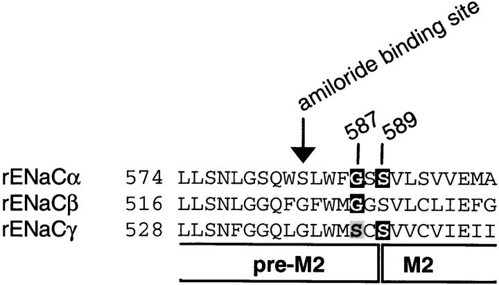
ENaC belongs to the ENaC/degenerin (DEG) family of ion channels. Members of this family are present in nematodes, flies, snails, and mammals, where they are involved in Na+ transport, neurotransmission, mechanotransduction, and nociception (Mano and Driscoll 1999). Members of the ENaC/DEG gene family are cation channels that share a common membrane topology characterized by intracellular NH2- and COOH termini, two transmembrane domains, and a large extracellular loop. ENaC differs from the other members of the ENaC/DEG family by its high selectivity for Na+ and Li+ over K+ ions and its high affinity for amiloride, a channel pore blocker. ENaC is a heterotetramer made of two α, one β, and one γ homologous subunits arranged around the central channel pore (Canessa et al. 1994; Firsov et al. 1998). All three subunits are involved in basic channel functions such as ionic selectivity, channel gating, and channel block by amiloride. The amiloride binding site has been localized in the short segment (preM2 segment) preceding the second transmembrane segment M2 of the ENaC subunits by mutations of either the α (S583), β (G525), or γ subunit (G537) that decrease channel affinity for amiloride by two to three orders of magnitude (Schild et al. 1997; Fig. 1). Mutations of αG587 and homologous residues in β and γ subunits (βG529 and γS541; Fig. 1), four residues downstream of the amiloride binding site affect channel conductance for Na+ and Li+ ions by changing the channel affinity for these permeant ions. These findings suggest that at this site the pore is narrow and the ions interact tightly with the Gly and Ser residues lining the pore (Kellenberger et al. 1999b). Mutations of the conserved Ser residue αS589 and the homologous residue in γENaC result in channels that are permeable to cations larger than Na+ and Li+ such as K+, Rb+, and Cs+ (Kellenberger et al. 1999a; Sheng et al. 1999; Snyder et al. 1999). The effects of mutations of residues 587–589 on ENaC conductance and ion selectivity identify this short stretch of amino acid residues in the preM2 segment as the narrow ion-selective region of the channel pore.
Figure 1.
Sequence alignment of the preM2/M2 segments of rat α, β, and γ ENaC. The number of the first residue shown is indicated. The putative start of M2 and the position of the amiloride binding site (αS583, βG525, and γG537) are indicated. Mutations that change ionic selectivity are mutations of αG587 and αS589 as well as residues at the homologous positions in β and γ subunits. Residues shown in white on a dark background change Na+/K+ selectivity when mutated, and mutation of αG587 and the homologous residues in β and γ subunits change Li+/Na+ selectivity. Mutation of γS541 (shown in black on gray background) changes Li+/Na+, but not Na+/K+ selectivity.
To gain further information on the role of the residue αS589 in maintaining the channel pore structure necessary for ion discrimination, we have substituted αS589 with amino acids of different volumes. We have analyzed channel selectivity properties of the αS589 mutants for inorganic and organic cations of varying diameter and shape. We found that the K+ permeability as well as the molecular cutoff of the channel permeability for large cations increases with the size of the amino acid residue at position αS589. This is consistent with an increase of the pore diameter at the selectivity filter. The consequences of this enlargement of the pore are an increase in channel permeation to larger cations, and at the same time, a decrease in the unitary conductance of these mutant channels to small cations such as Na+ and Li+. The observation that mutant channels with large side chains of residue α589 form a wide pore indicates that the side chain of the residue at position 589 points away from the ion permeation pathway. We propose that it points toward the interface between the subunits lining the pore, and that substitutions of αS589 with large amino acid residues increase the diameter of the narrowest part of the channel pore by adding extra volume to the subunit–subunit interface. The largest amino acid substitutions cause structural changes that are incompatible with channel expression at the cell surface.
MATERIALS AND METHODS
Site-directed Mutagenesis, Expression in Xenopus laevis Oocytes and Electrophysiological Analysis
Site-directed mutagenesis was performed on rat ENaC cDNA as described previously (Schild et al. 1997). Complementary RNAs of each α, β, γ subunit were synthesized in vitro. Healthy stage V and VI Xenopus oocytes were pressure-injected with 100 nl of a solution containing equal amounts of αβγ ENaC subunits at a total concentration of 100 ng/μl. For simplicity, mutants are named by the mutated subunit only, although all three subunits (α, β, and γ) always were coexpressed.
Electrophysiological measurements were taken at 16–48 h after injection. Macroscopic amiloride-sensitive currents, defined as the difference between ionic currents obtained in the presence and absence of 100 μM amiloride (Sigma-Aldrich) in the bath were recorded using the two-electrode voltage-clamp technique. All macroscopic currents shown are amiloride-sensitive currents as defined above. Currents were recorded with an amplifier (model TEV-200; Dagan Corp.) equipped with two bath electrodes. The standard bath solution contained 110 mM NaCl, 1.8 mM CaCl2, and 10 mM HEPES-NaOH, pH 7.35. For selectivity measurements, Na+ was replaced with Li+, K+, Rb+, Cs+, or the organic cations at the same concentration. Pulses for current-voltage curves were applied, and data were acquired using a PC-based data acquisition system (Pulse; HEKA Electronic). Single-channel currents were measured in the outside-out configuration of the patch-clamp technique essentially as described previously (Kellenberger et al. 1999b). The bath solution in patch-clamp experiments was the standard bath solution described above, with Li+ or Na+ as the cation. The pipette solution contained 75 mM CsF, 17 mM NMDG-HCl, 10 mM EGTA, and 10 mM HEPES, pH 7.35. Pipettes were pulled from Borosilicate glass (World Precision Instruments). In patch-clamp experiments, currents were recorded with a List EPC-9 patch-clamp amplifier (HEKA Electronic) and filtered at 100 Hz for analysis. Aqueous stock solutions of MTS ethylammonium (MTSEA; Toronto Research Chemicals) were prepared just before the experiment, maintained on ice, and diluted into the bath solution immediately before use.
Binding Experiments
The FLAG reporter octapeptide that had been introduced in β and γ subunits is recognized by the anti-FLAG M2 (M2Ab) mouse mAb (Sigma-Aldrich). M2Ab was iodinated as described by Firsov et al. 1996. Iodinated M2Ab had a specific activity of 5–20 · 1017 cpm/mol, and were used up to 2 months after synthesis. On the day after mRNA injection, oocytes were transferred to a 2-ml Eppendorf tube containing modified Barth's saline ([in mM] 10 NaCl, 90 NMDG-HCl, 0.8 MgSO4, 0.4 CaCl2, and 5 HEPES, pH 7.2) supplemented with 10% heat-inactivated calf serum, and incubated for 30 min on ice. The binding was started upon addition of 12 nM 125I-M2Ab (final concentration) in a volume of 5–6 μl/oocyte. After 1 h of incubation on ice, the oocytes were washed eight times with 1 ml modified Barth's saline supplemented with 5% heat-inactivated calf serum, and then transferred individually into tubes for γ counting containing 250 μl of the same solution. The samples were counted, and nonspecific binding was determined from parallel assays of noninjected oocytes.
Immunoprecipitation
Oocytes were injected with FLAG-tagged and untagged subunits, as specified. After overnight incubation of the injected oocytes, microsomal membranes were obtained as described previously (Geering et al. 1989) and solubilized in a Triton X-100 buffer (150 mM NaCl, 1.5 mM MgCl2,1 mM EGTA, 10% glycerol, 1% Triton X-100, 50 mM HEPES, pH 7.5, 1 mM phenylmethylsulfonylfluoride, and 10 μg/ml each of leupeptin, pepstatin A, and aprotinin).
Immunoprecipitations were performed in Triton X-100 buffer overnight at 4°C with the anti-FLAG M2 antibody (M2Ab) and protein G–Sepharose. After SDS-PAGE, polypeptides were transferred for immunostaining onto nitrocellulose membranes (Schleicher & Schuell) and immunoblotted using anti–rat αENaC antibody (May et al. 1997) as primary and goat anti–rabbit horseradish peroxidase (Amersham) as secondary antibody and visualized using Super Signal® West Dura Extended Duration Substrate (Pierce Chemical Co.).
RESULTS
Amino Acid Substitutions of Ser 589 in α ENaC
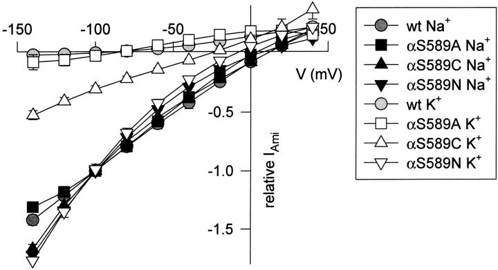
We have substituted the amino acid αS589 with residues of different sizes and physicochemical properties. The mutant α subunits were coexpressed with wild-type (wt) βγ ENaC subunits in Xenopus oocytes. Ionic currents through wt and mutant channels were completely blocked by 100 μM amiloride. The K+ over Na+ selectivity of the αS589 mutants was determined by the amiloride-sensitive current ratio (IK/INa) obtained at −100 mV in the presence of external Na+ (120 mM) and after Na+ substitution by K+ ions, as shown in Table . Increasing the size of the substituting amino acid side chain at position α589 increased the channel permeability to K+, as shown by the IK/INa ratio. The αS589H substitution mutation that represents a 60% increase in the residue's volume renders the channel nonselective between K+ and Na+. Changes in the K+ over Na+ selectivity were further evidenced from measurements of reversal potentials of the currents of αS589 mutants in the presence of extracellular Na+ or K+ solution. Fig. 2 shows the current-voltage relationship of amiloride-sensitive currents in the presence of extracellular Na+ and K+ of ENaC with substitutions of αS589. INa and IK were normalized in each experiment to INa at −100 mV. The current-voltage relationship with extracellular Na+ solution is similar in wt and the αS589A, αS589C, and αS589N ENaC mutants. However, no inward K+ current could be measured in ENaC wt even at potentials more negative than −100 mV. IK was low in αS589A, intermediate in αS589C and almost equal to INa in αS589N. The K+/Na+ permeability ratio calculated from the difference of the reversal potential in extracellular K+ and Na+ solution, respectively, is 0.01 for wt, 0.02 for αS589A, 0.24 for αS589C, and 0.56 for αS589N (Hille 1992). These permeability ratios are consistent with the IK/INa current ratio values measured at −100 mV (Table ).
Table 1.
Current Expression and Ion Selectivity of ENaC Mutants
| Mutant | INa | IK/INa | INH4/INa | gNa | gL | Van der Waals volume of amino acid |
|---|---|---|---|---|---|---|
| μA | pS | pS | Å3 | |||
| αS589G | 9.4 ± 1.1 | 0.04 ± 0.00 | 0.02 ± 0.00 | 5.0 ± 0.4 | 7.8 ± 0.3 | 48 |
| αS589A | 41.3 ± 3.0 | 0.05 ± 0.00 | 0.03 ± 0.00 | 4.3 ± 0.2 | 5.9 ± 0.7 | 67 |
| αS589C | 5.2 ± 0.5 | 0.24 ± 0.02 | 0.23 ± 0.06 | <1 | 1.5 ± 0.1 | 86 |
| αS589D | 6.4 ± 0.8 | 0.30 ± 0.02 | 0.32 ± 0.02 | 1.7 ± 0.1 | 1.5 ± 0.2 | 91 |
| αS589N | 3.0 ± 0.4 | 0.70 ± 0.02 | 1.13 ± 0.10 | <1 | <1 | 96 |
| αS589V | 0.0 ± 0.0 | n.d. | n.d. | n.d. | n.d. | 105 |
| αS589E | 0.1 ± 0.0 | n.d. | n.d. | n.d. | n.d. | 109 |
| αS589Q | 0.9 ± 0.1 | 0.24 ± 0.02 | 0.30 ± 0.03 | <1 | 1.9 ± 0.5 | 114 |
| αS589H | 0.4 ± 0.1 | 1.02 ± 0.04 | 1.20 ± 0.00 | <1 | <1 | 118 |
| αS589L, M, K, F, R, W | 0.0 ± 0.0 | n.d. | n.d. | n.d. | n.d. | ≥124 |
| αL591C to αM610C | 15.8 ± 3.0 | 0.01 ± 0.00 | n.d. | n.d. | n.d. | |
| wt | 17.9 ± 1.9 | −0.01 ± 0.00 | 0.00 ± 0.00 | 5.0 ± 0.2 | 9.0 ± 0.3 | 73 |
Whole-cell amiloride-sensitive current (n = 3–64) and single-channel conductance (n = 3–7) of mutant α subunits coexpressed with wt β and γ ENaC. n = 3–64. n.d., not determined.
Figure 2.
The K+/Na+ permeability ratio is increased by αS589 mutations. Current-voltage relationship in 120-mM Na+ and 120-mM K+ bath solution from oocytes expressing αS589A, αS589C, and αS589N measured with two-electrode voltage clamp. Currents were measured during 1-s voltage steps from a holding potential of −20 mV to test potentials of −140 to +40 mV in 20-mV increments. Currents measured in the presence of 10 μM amiloride were subtracted from currents measured in the absence of amiloride. In each experiment, the amiloride-sensitive Na+ and K+ currents were normalized to the amiloride-sensitive Na+ current at −100 mV. To allow Na+ to enter the oocyte during the expression phase without downregulation of channel activity, mutant α subunits were coexpressed with β subunits containing the Liddle mutation βY618A together with wt γ ENaC and oocytes were kept in a solution containing 90 mM Na+ during the expression phase (Kellenberger et al. 1998). The mutation βY618A does not affect ion selectivity. Erev, Na − Erev, K was 114 mV (wt), 96 mV (αS589A), 37 mV (αS589C), and 15 mV (αS589N), n = 4–5. INa at −100 mV was 30.0 ± 3.6 μA (αS589A), 8.3 ± 1.7 μA (αS589C), and 9.3 ± 2.1 μA (αS589N).
The αS589 mutations tend to decrease INa with increasing size of the substituting amino acid, and for substitutions with residues larger than His (∼118 Å3), no amiloride-sensitive current could be detected (Table ). To determine whether the absence of INa observed with expression of the large αS589 substitution mutants is due to nonpermeant channels at the cell surface or to alterations in the biosynthesis of these mutants, we have compared the surface expression of selected αS589 mutant channels with ENaC wt. Wild-type and mutant ENaC channels were tagged with FLAG epitopes in the extracellular domain, and binding of the iodinated anti-FLAG antibody to intact oocytes was measured (Firsov et al. 1996). Expression of wt ENaC containing flagged β and γ subunits (αβFγF) increased surface binding of anti-FLAG mAb to 8.4 ± 1.9-fold over noninjected oocytes. Coexpression of αS589 mutants with flagged β and γ subunits increased the mAb binding 6.0 ± 1.6-fold over noninjected oocytes for αS589A and 8.6 ± 4.5-fold with αS589C (n = 12–48 oocytes). No significant binding was detected for the nonfunctional mutants αS589V, αS589L, and αS589W (e.g., the ratio was 1.1 ± 0.4 for αS589V, 1.6 ± 0.6 for αS589L, and 0.9 ± 0.3 for αS589W, n = 12–18 oocytes), indicating the absence of channels at the cell surface.
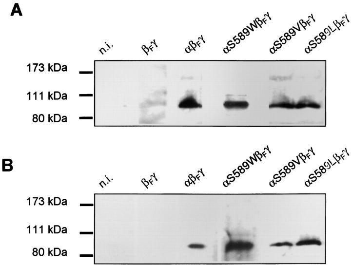
The absence of cell-surface expression of αS589V, αS589L, and αS589W mutants is not due to an impaired translation of the mutant subunit. After solubilization from oocytes expressing wt αβγ or the mutant αS589 subunits with wt βγ ENaC, wt and mutant α subunits could be detected on Western blots using an anti–rat αENaC antibody (May et al. 1997; Fig. 3 A), indicating that the mutant ENaC subunits are efficiently translated. The ability of the mutant α subunits to assemble with β subunits was tested by coimmunoprecipitation experiments in oocytes coexpressing the wt or mutant α subunits with β subunits that carry a FLAG epitope, i.e., αβFγ, αS589VβFγ, αS589LβFγ, αS589WβFγ, and as a negative control, βFγ (Firsov et al. 1996). The ENaC channel complexes tagged with the FLAG epitope on β ENaC were immunoprecipitated with the anti-FLAG antibody under nondenaturing conditions, subjected to SDS-PAGE, transferred to nitrocellulose membranes, and probed with an anti–rat αENaC antibody. An α ENaC-specific band was detected with αβFγ and αS589V/L/WβFγ but not βFγ (Fig. 3 B), indicating that the mutant subunits αS589V, αS589L, and αS589W retain their ability to assemble with β subunits. Thus, mutant α subunits with large amino acid substitutions at position α589 retain some ability to assemble with the β subunit but do not reach the cell surface.
Figure 3.
Analysis of translation of wt and mutant α subunits and of assembly with β subunits. Oocytes were not injected or injected with RNAs encoding ENaC carrying a FLAG epitope on the β subunit, αβFγ, αS589LβFγ, αS589VβFγ, αS589WβFγ, or βFγ, as indicated. (A) Western blot immunostaining of solubilized wt and mutant ENaC subunits. Solubilized proteins were subjected to SDS-PAGE, and αENaC was visualized on Western blots using an anti–rat αENaC antibody (May et al. 1997). Molecular markers are indicated. (B) Western blot immunostaining for coimmunoprecipitation of wt and mutant ENaC under nondenaturing conditions. Nondenaturing coimmunoprecipitation was performed with an anti-FLAG antibody. Immunoprecipitates were subjected to SDS-PAGE and αENaC was visualized on Western blots using an anti–rat αENaC antibody. Specificity of the anti–rat αENaC antibody is demonstrated by a control in which no αENaC RNA was injected (βFγ).
Permeability of ENaC Mutants to Inorganic Cations
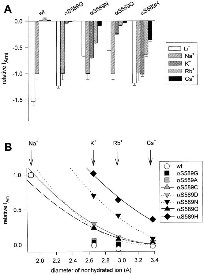
We have initially hypothesized that the increase in channel permeability to K+ of the αS589 mutants was due to changes in the pore diameter (Kellenberger et al. 1999a). To obtain further evidence for such alterations in pore geometry, we measured the permeability properties of the different channel mutants to alkali and organic cations of different sizes and shapes. Fig. 4 A illustrates the amiloride-sensitive currents measured in the presence of different alkali metal cations at −100 mV, normalized to the values obtained with Na+ as the charge carrier (IX/INa). When the main cation in the extracellular solution was K+, Rb+, or Cs+, wt ENaC showed small outward amiloride-sensitive currents at −100 mV (Fig. 4 A, upward bars) carried by intracellular Na+ ions. This indicates that ENaC wt is virtually impermeable to K+, Rb+, and Cs+. Among the mutants αS589G, αS589Q, αS589N, and αS589H, the ones with the highest permeability to K+ ions also show the highest conductance for Rb+ and Cs+. For all the mutants tested, the permeability sequence follows the size of the permeant ions, i.e., Na+ ≥ K+ > Rb+ > Cs+ (Table ). These results confirm the observation for amino acid substitutions of smaller sizes at position αS589, i.e., αS589A, αS589C, and αS589D (Kellenberger et al. 1999a) and further support the conclusion that αS589 mutations with larger side chain residues increase in pore diameter at the selectivity filter. For K+, Rb+, and Cs+ ions that are similar in size (Pauling diam, 2.7–3.4 Å), the permeability in αS589 mutants is inversely correlated with the size of the ion. This is demonstrated in Fig. 4 B, where the relationship between the relative permeability of the cation and its diameter is fitted by a simple equation that describes permeation based purely on molecular sieving (Dwyer et al. 1980; Kellenberger et al. 1999a; see Fig. 4 legend). However, the relative permeability of cations of very different sizes, as Na+ (1.9 Å) versus K+, Rb+, and Cs+, cannot be fitted by the above equation in most αS589 mutants (Fig. 4 B), indicating that mechanisms other than geometrical and frictional effects are critical for Na+ permeability. The fact that Na+ permeability is lower than expected for its diameter in the mutants αS589N and αS589H suggests that the selectivity filter becomes less favorable for permeation of Na+ ions when its diameter becomes larger. Such an effect could also contribute to the lower macroscopic INa expressed by the αS589 mutants with large amino acid substitutions (Table ).
Figure 4.
Selectivity of αS589 mutants to alkali metal cations. Two-electrode voltage-clamp recordings from Xenopus oocytes expressing ENaC wt or αS589 mutants. (A) Macroscopic amiloride-sensitive currents were measured at −100 mV in oocytes superfused with Li+, Na+, K+, Rb+, or Cs+ external solutions (materials and methods). Amiloride-sensitive currents are normalized to INa, and presented as mean ± SEM (n = 4–83 per condition). Positive current values correspond to outward currents. (B) Relationship between the size of the ion and permeability. The normalized amiloride-sensitive currents are plotted as a function of the diameter of the nonhydrated ion. Fits to the equation Ix/INa = f x [1 − (dS/2)/(dC/2)]2, where f is a scaling factor, d S is the diameter of permeating spheres, and d C is the diameter of the cylinder (the pore; Dwyer et al. 1980) are shown for the αS589 mutants that are permeable to at least K+ and Rb+. Data and fits of the previously studied mutants αS589A, C, and D are shown in gray. The values of d C obtained from the fit are 3.8 Å for αS589N, 3.5 Å for αS589Q, and 4.4 Å for αS589H.
Table 2.
Selectivity Sequences of Na+ and Larger Alkali Metal Cations and Organic Cations
| Mutant | Selectivity sequence | Cations not permeable |
|---|---|---|
| wt | Na+ | K+, Rb+, Cs+, NH4 +, MA, DMA, TriMA, guanidine |
| αS589C | Na+ > K+ = NH4 + > Rb+ > guanidine | Cs+, MA, DMA, TriMA |
| αS589D | Na+ > NH4 + = K+ > Rb+ > Cs+ > guanidine | MA, DMA, TriMA |
| αS589N | NH4 + > Na+ > K+ > Rb+ > Cs+ > guanidine > MA | DMA, TriMA |
| αS589Q | Na+ > NH4 + = K+ > DMA = Rb+ ≥ MA = guanidine = Cs+ = TriMa | |
| αS589H | NH4 + ≥ Na+ = K+ > Rb+ >guanidine > Cs+ > MA ≥ DMA >TriMA | |
| βG529D, C, S | Na+ > NH4 + ≥ K+ > Rb+ | MA |
The selectivity sequences are based on data of experiments shown in Fig. 4 and Fig. 6, Table , and Kellenberger et al. 1999a. “>” in the selectivity sequence means that the relative permeability between the two ions is significantly different (P < 0.05).
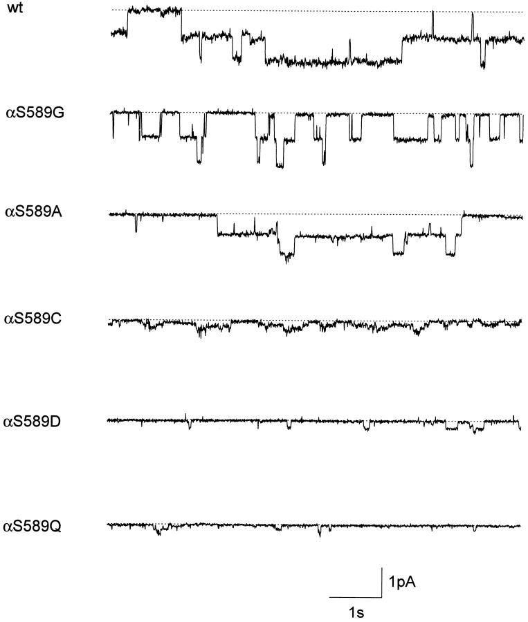
To test the possibility that the larger pore diameter makes it more difficult for small ions such as Na+ and Li+ to permeate the channel, we have performed single-channel recordings of different αS589 substitution mutants. Patch-clamp recordings of the functional αS589 mutants showed that the unitary conductance for Li+ and Na+ decreased with the increasing size of the substituent amino acid (Fig. 5 and Table ). For instance, the single-channel Li+ conductance was 9.0 pS for wt, 7.8 pS for αS589G, 1.5 pS for αS589C, and 1.9 pS for αS589Q, and <1 pS for the mutants αS589N and αS589H where single-channel openings could not be resolved. Thus, it appears that increasing the pore diameter at the selectivity filter with αS589 substitutions allows larger cations to permeate the channel to the prejudice of smaller ions such as Na+ or Li+.
Figure 5.
Single-channel records of ENaC wt and αS589 mutants. Traces are from outside-out patches from Xenopus oocytes at a holding potential of −100 mV in extracellular Li+ solution. The dotted lines indicate the baseline when all ENaC channels in the patch are closed. The outside-out configuration was chosen to verify amiloride-sensitivity of channel activity.
Permeability of ENaC Mutants to Organic Cations
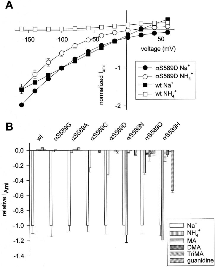
Organic cations such as ammonium and its methyl derivatives methyl-, dimethyl-, and trimethylammonium (MA, DMA, and TriMA) or guanidine have been used to estimate the minimum diameter of the pore of channels such as voltage-gated Na+, K+, and Ca2+ channels (Hille 1992). These organic cations are not completely symmetrical like the alkali metal cations. To determine the permeabilities of organic cations in ENaC wt and the functional αS589 mutants, the amiloride-sensitive current carried by organic cations was measured at −100 mV and normalized to INa. Fig. 6 A shows the current-voltage relationship of the amiloride-sensitive current carried by Na+ or NH4 + ions in oocytes expressing either ENaC wt or the αS589D mutant. Both, ENaC wt and the αS589 mutant show clear inward Na+ currents (Fig. 6 A, closed symbols). The αS589D Na+ current shows a strong inward rectification, which is due to the fact that the expression level of the mutant compared with the wt was low in this experiment and intracellular Na+ concentration was much lower in the mutant compared with the wt (Kellenberger et al. 1998). For ENaC wt, no NH4 + inward current could be measured in oocytes even at very negative potentials (Fig. 6 A, open squares), whereas a substantial NH4 + inward current was evident in the mutant αS589D (Fig. 6 A, open circles). All αS589 mutants are permeant to NH4 +, as shown in Fig. 6 B. The permeability ratios of the αS589 mutants for K+ or NH4 + ions are comparable, and as for K+ ions, the channel permeability to NH4 + increases with the size of the residue at position αS589 (Table ). Regarding ammonium derivatives, the smaller substitution mutants (αS589G/A/C/D) are not permeable to any of the ammonium derivatives. αS589N is only permeant to the smallest derivative, MA, and αS589Q and αS589H show significant but low permeability to all three derivatives tested (MA, DMA, and TriMA; Fig. 6 B). αS589C, D, N, Q, and H are permeable to guanidine.
Figure 6.
Selectivity of αS589 mutants to organic cations. (A) I-V relationship of amiloride-sensitive Na+ and NH4 + currents in Xenopus oocytes expressing either ENaC wt or the αS589D mutant. Currents were measured during 500- ms voltage-steps from a holding potential of −100 mV to test potentials of −160 to +60 mV in 20-mV increments. Currents measured in the presence of 5 μM amiloride were subtracted from currents measured in the absence of amiloride. Amiloride-sensitive currents were in each oocyte normalized to the amiloride-sensitive Na+ current at −100 mV. (B) Macroscopic amiloride-sensitive currents were measured at −100 mV in oocytes superfused with Na+, NH4 +, MA, DMA, TriMA, or guanidine external solution (materials and methods). Amiloride-sensitive currents are normalized to INa, and presented as mean ± SEM (n = 2–20 per condition). Positive current values correspond to outward currents.
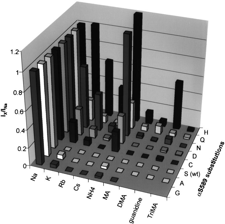
The permeability data obtained with organic and inorganic cations from all functional αS589 mutants are summarized in Fig. 7. In this representation, the ions are aligned according to their increasing size from left to right (Fig. 7 legend) and the mutants are shown with increasing volume of the substituting amino acid from front to back. Fig. 7 illustrates two aspects of organic and inorganic cation permeability of αS589 mutants. First, substitution of αS589 by large amino acid residues increases the relative permeability of large inorganic and organic cations, i.e., increasing the size of the amino acid at position α589 increases the pore diameter. Second, large cations have generally a lower permeability than small cations (Table ). NH4 + and guanidine carry higher currents than expected from their size. For the functional mutants with substituting residues larger than Ser, there is a significant correlation between the increase in volume of the amino acid and the relative permeability to K+ and NH4 +. Only for the αS589Q mutant, the K+ and NH4 + permeability relative to Na+ is lower than expected from its size. However, the αS589Q mutant still remains highly permeant to large cations such as K+ and NH4 +, which is consistent with the general conclusion that substitution of αS589 by large amino acids increases the pore diameter at the selectivity filer. The particularity of the αS589Q mutant indicates that the size of the residue at position α589 is not the only determinant of the pore diameter at the selectivity filter. Regarding the second observation addressed here, we found, in most mutants, a relative permeability for NH4 + and guanidine that was higher than expected from their size. NH4 + is somewhat flexible due to its ability to form hydrogen bonds (see discussion). Guanidine is of a similar size as TriMA, but is a planar molecule in contrast to the more spherical shape of ammonium derivatives. αS589C, D, N, Q, and H are permeable to guanidine. The highest guanidine permeability ratio was obtained for αS589H (Iguanidine/INa = 0.54 ± 0.03, n =10; Fig. 6 B). The higher guanidine permeability relative to ammonium derivatives in the αS589C, D, N, and H mutants suggests that the pore at the selectivity filter of these mutants is not completely symmetrical but has the shape of a slotlike rectangle.
Figure 7.
Graphical summary of the main results with inorganic and organic cations. Bar graph summarizing the amiloride-sensitive current ratios IX/INa for all ions in all αS589 mutants tested. On the axis “αS589 substitutions,” the different mutants are labeled by the single-letter code of the substituting amino acid. The ions are represented in the order of their size (diameter derived from Pauling radius for alkali metal cations and minimum diameter [Sun et al. 1997] for organic cations). The minimum diameter is as follows (in Å): 3.6 for NH4 +, 3.8 for MA, 4.6 for DMA, 6.0 for TriMA, and 5.8 for guanidine.
In addition to mutations of αS589, mutations of βG529 to Ser, Cys, and Asp (but not mutations to Ala and Arg) make ENaC permeable to K+. The IK/INa ratio was ∼0.2 for βG529D and βG529S and 0.06 for βG529C (Kellenberger et al. 1999b). Mutations of βG529 that increase the IK/INa ratio also increase the IRb/INa ratio, however, these mutant channels are not permeant to Cs+ (Kellenberger et al. 1999b). The INH4/INa ratio is 0.19 ± 0.09, 0.46 ± 0.07, and 0.17 ± 0.01 (n = 5) for βG529D, S, and D, respectively, and these three mutants are impermeable to MA (unpublished data). This is consistent with a mechanism similar to that of αS589 mutations in which these βG529 mutations change the ionic selectivity by enlarging the pore. The low permeability to large cations indicates that the βG529 mutations enlarge the pore by less than the αS589 mutations.
Coexpression of αS589N with βG529S and wt γ yielded a channel with reduced but still detectable current expression with an amiloride-sensitive Na+ current at −100 mV of 0.1 ± 0.05 μA. The current ratio IK/INa was 1.46 ± 0.23 (n = 4), showing a slightly higher selectivity to K+ ions than the single substitution mutant αS589N. Thus, the effects of the αS589 and βG529 mutations on channel permeability to K+ ions are additive, as expected for α and β subunits lining the channel pore and contributing to the selectivity filter.
Contributions of the Second Transmembrane Segment M2 and the Intracellular COOH Terminus to Ionic Selectivity
The increase in relative channel permeability to large cations in αS589 mutants indicates that αS589 is part of the selectivity filter. Adjacent residues not directly facing the pore lumen might also contribute to the ionic selectivity of the channel by holding the selectivity filter in place and at the correct diameter. In addition it is possible that other amino acids along the ion permeation pathway may contribute to ion selectivity. We have replaced systematically all residues from L591 to M610 in the putative M2 segment of the α subunit with Cys residues to investigate the role of the second membrane-spanning segment M2 in ion selectivity. We have tested the accessibility of these engineered Cys residues to extracellularly applied sulfhydryl reagents. The hydrophilic sulfhydryl reagent MTSEA attaches covalently and selectively a charged group to Cys residues. If a substituted Cys residue lies on the water-accessible surface of the external pore, it should react with MTSEA, which would affect ionic current. The ratio of the INa after a 2-min extracellular application of 2.5 mM MTSEA relative to the initial INa was 0.77 ± 0.33 for ENaC wt and was not significantly different for the mutants (unpublished data). This suggests that the Cys residues in the M2 segment of αENaC are not accessible to extracellular MTSEA, or that their modification does not change channel function, in contrast to residues in the preM2 segment of the ENaC subunits (Kosari et al. 1998; Snyder et al. 1999). When coexpressed with wt β and γ subunits, all mutants apart from αE598C produced functional channels. However, mutation αE598A was functional and was not different from wt with regard to ion selectivity and amiloride block. The mutants αL591C to αM610C exhibit a IK/INa in the range of 0.00–0.03 compared with 0.01 ± 0.00 of ENaC wt, indicating that the M2 segment of α ENaC does not contribute significantly to the ionic selectivity of the channel (Table ).
It was recently reported that truncation of the COOH terminus of rat αENaC at residue R613 changed the Li+ single-channel conductance and the Li+/K+ selectivity as determined from macroscopic currents (Jovov et al. 1999). In the context of this work, we have analyzed the properties of αR613Xβγ ENaC expressed in Xenopus oocytes. The unitary conductance, measured in outside-out patches was 4.7 ± 0.2 pS for Na+ and 8.0 ± 0.4 pS for Li+ (n = 5 patches each), directly compared with 4.6 ± 0.2 pS (Na+) and 8.8 ± 0.4 pS (Li+; n = 2 patches each) for ENaC wt. The αR613Xβγ mutant remained totally impermeant to K+ ions, as shown by the macroscopic current ratio IK/INa at −100 mV of zero as for ENaC wt. Thus, we have no indication that residues within the intracellular COOH terminus or M2 determine ion selectivity or unitary conductance. Therefore, we conclude that the selectivity filter is restricted to αG587–αS589 and the analogous residues in β and γ subunits.
Amiloride Sensitivity
Amiloride is a pore blocker and interacts with the permeating ions. We have addressed the possibility that in addition to changing ion selectivity αS589 mutations might also affect channel block by amiloride, since amiloride binds in the preM2 segment in the proximity of the selectivity filter (Fig. 1). We have analyzed the block by amiloride of currents generated by all αS589 mutants. In most mutants, amiloride block was not different from wt, except for αS589N, which marginally increased the IC50 and αS589H, which increased the IC50 for amiloride block by a factor of 16. With Na+ as the main extracellular cation, the IC50 for amiloride block was 0.44 ± 0.05 and 2.80 ± 0.65 μM for αS589N and αS589H, respectively, compared with 0.18 ± 0.08 μM for ENaC wt (n = 6–16). The important structural changes of the selectivity filter induced by the αS589H mutation that enlarges the channel pore allowing large cations to pass may extend to the nearby amiloride binding site and also impair channel block. We measured amiloride block of the NH4 + current for two mutants, αS589D and αS589N. The IC50 values for amiloride block of the NH4 + current were 0.10 ± 0.02 and 0.19 ± 0.06 μM for αS589D and αS589N, respectively (n = 6–7 each), indicating that amiloride blocks NH4 + currents with similar affinity as currents carried by Na+. The Cys substitution mutations in the M2 segment have no effect on amiloride block with an IC50 for the mutants αL591C to αM610C ranging from 0.06 to 0.33 μM (unpublished data).
DISCUSSION
It was initially found that mutations of two residues, αS589 and βG529, change Na+/K+ selectivity and make ENaC permeable to K+ and to a lesser extent to Rb+ and Cs+ (Kellenberger et al. 1999a,Kellenberger et al. 1999b). Subsequently, two additional mutations were shown to affect the channel K+/Na+ selectivity, αG587C at the position homologous to βG529 (Sheng et al. 1999) and γS542C at the position homologous to αS589 (Snyder et al. 1999). The present work provides evidence that the changes in ionic selectivity caused by αS589 and βG529 mutations are due to an enlargement of the pore at the selectivity filter. Substitutions of αS589 by a variety of amino acids of different volume and subsequent analysis of the permeability of these mutant channels to different organic and inorganic cations of varying sizes show that the larger the substituting amino acid at position α589, the easier it becomes for large organic and inorganic cations to permeate the channel. Enlargement of the pore at the selectivity filter not only allows large cations to permeate ENaC mutants, but also decreases channel permeability to small cations such as Na+ or Li+ ions. Thus, the pore size at the αS589 residue is critical for the selectivity properties of ENaC
Ion Selectivity in wt and Mutant ENaC
Ionic selectivity of ENaC wt allows Na+ and smaller cations such as Li+ and H+ to permeate the pore, whereas large cations such as K+, Rb+, and Cs+ are excluded (Palmer 1990). However, these monovalent nonpermeant cations block ENaC in a voltage-dependent manner, and as amiloride they are competitive blockers with regard to Na+ ions. These observations indicate that K+, Rb+, and Cs+ ions interact with Na+ ions in the outer part of the channel pore down to the site where amiloride binds (i.e., αS583 and the corresponding residues βG525 and γG537), but do not pass the selectivity filter. Thus, the outer pore of ENaC acts as a molecular sieve to discriminate among inorganic cations.
What are the consequences of enlarging the channel pore at the selectivity filter on the ion permeation properties of ENaC? Enlargement of the channel pore at the selectivity filter due to substitutions of αS589 by bulkier residues allows larger cations to pass through the selectivity filter (Kellenberger et al. 1999a). For all αS589 mutants, the permeability to K+, Rb+, and Cs+ still depends on the relative size of the ion and of the pore as supported by the fit to the molecular sieving model (Fig. 4 B). However, enlargement of the pore is also associated with a decrease in permeability to small ions such as Na+ and Li+. We observed that the permeability to Na+ relative to K+ ions of the αS589H and αS589N mutants is lower than expected for the size of Na+ ions from the fit to the hydrodynamic model (Fig. 4 B). But more importantly, we demonstrated a reduction in the single-channel conductance for Li+ and Na+ ions in αS589 ENaC mutants that is inversely related to the increase in channel permeability to K+, Rb+, and Cs+ ions. These two observations indicate that the higher the permeability of the αS589 mutant becomes to larger cations such as K+, Rb+, or Cs+, the more difficult it is for small ions such as Na+ and Li+ to permeate the channel. Thus, the geometry of the ENaC pore at the selectivity filter is critical for the relative permeability to Na+ over K+ and larger ions. A similar but more direct conclusion came from the solution of the 3-D structure of the K+ channel KcsA. All K+ channels show a selectivity sequence of K+ ≈ Rb+ > Cs+, whereas permeability for the smallest alkali metal cations Na+ and Li+ is immeasurably low. In the KcsA channel, the high selectivity for K+ over smaller ions (Li+ and Na+) is based on the tight interaction of the permeating K+ ion with the carbonyl oxygens of the backbone of the GYG sequence that lines the selectivity filter (Doyle et al. 1998). Permeant ions have to dehydrate to pass through the narrow channel pore. The ion–pore interaction is critical to compensate for the energetic cost of dehydration, and requires an optimal size ratio of the permeating ion and the selectivity filter. Na+ ions are too small to tightly fit in the K+ channel selectivity filter. As a consequence, the ion–pore interaction energy is too low to compensate for the cost of dehydration of Na+ ions, making Na+ permeation through K+ channels almost impossible. Similarly, as the ENaC pore becomes larger in the αS589 mutants, accommodation of a Na+ ion in the selectivity filter likely becomes energetically less favorable because of a lower interaction energy with the pore-lining residues, resulting in the observed decrease in unitary conductance.
Permeability to Organic Cations
NH4 + and its methyl derivatives, MA, DMA, and TriMA, all have the same basic geometry and differ only by the substitution of methyl groups for hydrogen atoms. Examples of channel proteins that select this series of cations entirely by molecular sieving are the nicotinic acetylcholine receptor (Adams et al. 1980; Dwyer et al. 1980) and voltage-gated Ca2+ channels in the absence of calcium (McCleskey and Almers 1985). In these channels, the permeability to NH4 +-methyl derivatives decreases in a monotonic fashion with size or volume, as expected for pure molecular sieving. The analysis of the permeability of αS589 ENaC mutants to NH4 + and its methyl derivatives did not show a graded decrease in permeability with increasing size of the ion (Fig. 6 and Fig. 7 and Table ). All αS589 mutants except for αS589G and αS589A have a high permeability to NH4 + and all of these mutants are not, or only slightly, permeable to the methyl derivatives of ammonium. This suggests that factors such as the shape of the ion and the interaction between the ion and the pore-lining residues codetermine ionic permeability in ENaC αS589 mutants. The magnitude of the current carried by NH4 + (INH4) is in many αS589 mutants similar to IK despite the fact that NH4 + is larger than K+, Rb+, and Cs+ ions (diam = 3.6 Å; Sun et al. 1997). The high relative NH4 +/Na+ permeability ratio stresses the importance of interactions between the permeating ion and the pore for ion discrimination. NH4 + ions can readily permeate relatively small openings by formation of hydrogen bonds with pore-lining oxygen-containing groups thereby reducing the N-O distances (Hille 1992). The effective radius of the NH3 + group when probed by an oxygen ligand (of the channel pore) was estimated to be 1.4 Å (Hille 1992). The high permeability of NH4 + in αS589 mutants suggests that NH4 + indeed does form hydrogen bonds in the ENaC pore. Due to this flexibility, NH4 + can adapt to different pore conformations, resulting in a higher permeability in αS589N or αS589H for NH4 + compared with Na+ or K+ ions. NH4 + can, in principle, form hydrogen bonds either with carbonyl oxygens of the peptide backbone of the residue at position α589 or in the case of the mutants αS589D, αS589Q, and αS589H with the oxygen of the side chain. High permeability to NH4 + was not only observed in the substitution mutants containing oxygens in the α589 side chain, but also in mutants without oxygens in the side chain at positions αS589 and βG529 (αS589C/H mutants as well as βG529C/S mutants). To explain the high NH4 + permeability of the latter mutants, the carbonyl oxygens have to be involved in hydrogen bonds with NH4 +, supporting the hypothesis that, as for the KcsA channel, the carbonyl residues of αS589 line the channel pore at the selectivity filter.
An Estimate of the Size of the ENaC Pore
An estimate of the minimum diameter of the wt and mutant ENaC selectivity filter can be obtained from the largest permeant and the smallest nonpermeant ion. It is assumed that ions permeating highly selective channels are completely dehydrated or carry at most one water molecule with them in the narrowest part of the pore (Doyle et al. 1998), and therefore the size of the nonhydrated ion gives a good estimate of the pore at this site. Ionic (Pauling) radii are established from contact distances between touching, nonbonding atoms in crystals (Hille 1992). The diameter of Na+ and K+ ions derived from their Pauling radius is 1.9 and 2.7 Å, respectively. Therefore, the minimum pore diameter of ENaC wt at its narrowest point is estimated to 1.9–2.7 Å. The pore diameters of αS589A and αS589G, which are permeant to K+ but not to Rb+, is estimated to be between 2.7 and 3.0 Å, and that of αS589C (permeant to Rb+ but not to Cs+) is between 3.0 and 3.4 Å. The fits to the relationship between the alkali cation permeability and the ion diameter in αS589 mutants that are permeant to Cs+ (Fig. 4 B) predict a pore radius in these mutants of the range of 3.5 (αS589D) to 4.4 Å (αS589H).
The organic cations that we have tested are not perfect spheres as the alkali metal cations. Judged from their minimum diameter, i.e., the smallest possible diameter of a circular pore through which the molecule would pass, all organic cations tested are larger than Cs+ (Sun et al. 1997). The permeability pattern of these asymmetric cations (Fig. 6 and Fig. 7) allows predictions about the shape of the ENaC pore at the selectivity filter. The observation that the planar guanidine can permeate the αS589C mutant which is not permeable to Cs+ (3.4 Å) and the fact that guanidine has a higher permeability than NH4 + derivatives in the αS589N, Q and H mutants indicate that the pore of mutant ENaC rather forms a slotlike rectangle. Voltage-gated Na+ channels are permeable to NH4 + (INH4/INa = ∼0.2), to guanidine and to some of its derivatives but not to methyl derivatives of ammonium (Hille 1992; Sun et al. 1997). From these permeation data, the minimum pore size of voltage-gated Na+ channels was estimated to a rectangle of 3.1 × 5.1 Å (Hille 1992). The permeability of K+ channels to organic cations is more restricted, and the largest molecule passing through K+ channels is NH4 + (Hille 1992). The minimum pore size of voltage-gated K+ channels was estimated to be a circle with a diameter of 3.3 Å (Hille 1992). The ENaC wt pore is narrower than that of either of these channel types and the shape of the cross-sectional area of ENaC αS589 mutants appears to be closer to that of voltage-gated Na+ than K+ channels.
Model of the ENaC Pore
There is good evidence that the preM2 segments of ENaC subunits line the outer part of the channel pore. The preM2 segments contain the binding site for the pore blocker amiloride, S583 in α and homologous residues in β and γ ENaC (Schild et al. 1997). Cys residues introduced at this position can bind extracellular zinc ions and can be modified by extracellular sulfhydryl reagents resulting in channel block (Schild et al. 1997; Firsov et al. 1998; Kosari et al. 1998; Snyder et al. 1999). The outer channel pore is relatively wide at the amiloride binding site (diam ∼5 Å; Kellenberger et al. 1999b) and gets narrower toward the selectivity filter. Mutations of the Gly and Ser residues at α587 and corresponding positions in β and γ subunits have dramatic effects on single channel conductance and ENaC affinity for Na+ and Li+ ions, indicating that these residues tightly interact with the permeating ions. The residue αS589 that determines the channel permeability cutoff is in the continuation of the amiloride binding site (position αS583) and the Na+/Li+ binding site (position αG587). The 583–589 sequence might represent a β sheet structure that precedes the putative transmembrane spanning M2 helix and lines the outer ion channel pore. It is quite understandable that introducing large residues such as His at position α589 induce steric changes in the pore with consequences on channel block by amiloride. All other functional αS589 mutants preserved the high affinity of ENaC wt for amiloride block, indicating that the selectivity filter does not overlap with the amiloride binding site.
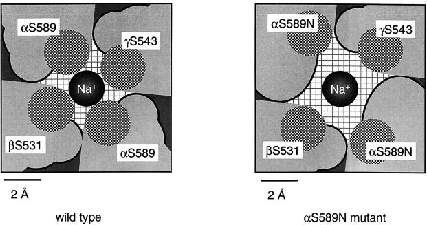
How do substitutions of αS589 by large residues increase the diameter of the pore and facilitate the passage of large organic cations such as ammonium or guanidine? This effect on the pore size is only possible if the side chain of residue αS589 points away from the pore lumen. Thus, most likely the carbonyl oxygens of the peptide backbone face the pore lumen to accommodate permeating cations. This orientation of the carbonyl oxygens is also suggested by the high permeability of NH4 + in αS589 mutants. This orientation of the polypeptide is similar to the KcsA channel in which the carbonyl oxygens are exposed to the pore lumen and the side chains of the amino acids point away from the pore. The side chain of αS589 and of the homologous Ser residues in β and γ subunits may point toward the interface between the subunits. Fig. 8 illustrates possible steric changes that may occur when αS589 is replaced by larger residues. This figure shows schematically a cross-section of the channel pore at residue αS589 in wt ENaC (Fig. 8, left) and the αS589N mutant (Fig. 8, right). The carbonyl oxygens of the polypeptide backbone, shown as hatched circles, face the pore lumen. In our model, the side chains of the residues at position α589 and the homologous positions in β and γ ENaC, identified by the dark border, are oriented toward the subunit interface. Increasing the size of the residue at position α589 pushes the backbone of the β and γ subunits away from the pore lumen, leading to an enlargement of the pore diameter (Fig. 8, right). Due to the α-β-α-γ subunit arrangement around the channel pore, large amino acid substitutions at the α-β and α-γ subunit interface may result in changes in the pore diameter predominantly in the direction of one axis (Firsov et al. 1998). Such a rectangular geometry of the ion-selective region of the pore could account for the higher channel permeability to the planar guanidine compared with bulky organic cations in αS589 mutants. We have shown that larger substituting residues of αS589 lead to a wider pore at the selectivity filter and that the enlargement depends on the size of the residue (Table ). The only exception from this rule is the αS589Q mutant that shows a lower permeability to large cations such as K+ and NH4 + than expected from its size, although this mutant is still highly permeant to K+ and NH4 +. This indicates that the size of the residue at position α589 is not the only determinant of the pore diameter. Other factors such as interactions between αS589 and adjacent residues may also control the diameter of the channel pore at the selectivity filter. These factors are not considered in our model. Substitution of αS589 by smaller residues such as Gly and Ala resulted in an increased K+ permeability. This observation suggests that other structures in the selectivity filter prevent the pore from collapsing when αS589 is replaced with smaller residues.
Figure 8.
Model illustrating the mechanism by which mutations of αS589 may enlarge the channel pore at the selectivity filter. Residue αS589 and analogues in the β and γ subunit arranged around the channel pore are shown in a cross-section of the pore seen from the top for wt ENaC (left) and the mutant αS589N (right). Each residue is represented by its backbone part in gray, with the carbonyl oxygen shown as a hatched circle pointing toward the pore lumen and the side chain identified by the black edge. The side chains are located at the interface between the subunits. Increasing the size of the side chain of the two αS589 residues adds extra volume at the subunit–subunit interface and makes the pore wider.
Mutations at position 529 of the β subunit resulted in a smaller pore enlargement compared with αS589 mutations. This can be explained by the fact that there is one β subunit in an ENaC tetramer compared with two α subunits (Firsov et al. 1998). Alternatively, the smaller effects of βG529 mutations on K+/Na+ selectivity may be a function of the position of this residue in the pore. βG529 is two residues more NH2-terminal in the sequence than αS589 (Fig. 1).
In this context, it is interesting to note that the substitution of αS589 by amino acids with a van der Waals volume ≥118 Å3 is not compatible with channel function at the cell surface (Table ). Given the steric effects of the αS589 mutations on the pore geometry it is possible that substitution by large amino acids prevents correct folding and assembly of subunits in the ER. Such changes in the ENaC biosynthetic process would direct the channel to degradation by a quality control in the ER. Mutation of the Ser homologous to αS589 has been shown to result in nonfunctional channels in other ENaC/DEG family members, DEG-1 (Garcia-Anoveros et al. 1995), MEC-4 (Hong and Driscoll 1994), and ASIC2a (Waldmann et al. 1996). Recently, a mutation of the homologous residue in human αENaC, S562, to leu was found in a patient with pseudohypoaldosteronism type 1, which is consistent with loss of channel function (Schaedel et al. 1999).
We use here a model of the channel pore that is based on the tetrameric stoichiometry of ENaC (Coscoy et al. 1998; Firsov et al. 1998; Kosari et al. 1998). An alternative ENaC subunit stoichiometry of eight or nine subunits also has been proposed (Snyder et al. 1998; Eskandari et al. 1999). The present work together with previous studies provides evidence that the carbonyl oxygens of all subunits that form the channel line the pore at its narrowest part (Kellenberger et al. 1999a,Kellenberger et al. 1999b). K+ channels are tetramers and form a selectivity filter that is slightly wider than that of wt ENaC. It seems very unlikely that nine subunits can be arranged around a unique ion permeation pathway with all nine subunits lining the pore to form a narrow pore of a diameter smaller than K+ channels.
It is not clear what the structures are that interact with the external channel pore to maintain the selectivity filter at the proper diameter and position. It is conceivable that mutations within these neighboring structures also affect channel ionic selectivity. The predicted transmembrane segment M2 could be part of this scaffold, or it might form the continuation of the pore. Our mutagenesis screen shows that M2 is not involved in ion discrimination. This transmembrane domain of the ENaC subunit is not accessible to extracellular sulfhydryl reagents. We extended our mutational analysis to the cytoplasmic COOH terminus of the α subunit because truncation of this region was recently claimed to influence ionic selectivity and single-channel conductance (Jovov et al. 1999). As for the truncation in the β subunit (Schild et al. 1996), the α COOH terminus truncation has no effect on the permeation properties of the channel. Together, these observations indicate that the ion-selective region of ENaC is restricted to a short sequence of three residues in the preM2 segment that represents the narrowest part of the external channel pore.
Acknowledgments
We thank J.-D. Horisberger and P. Greasely for critical reading of the manuscript.
This work was supported by a grant from the Swiss National Foundation for Scientific Research (to L. Schild, No. 3100-059217.99).
Footnotes
Abbreviations used in this paper: DEG, degenerin; DMA, dimethylammonium; ENaC, epithelial Na+ channel; ILi, amiloride-sensitive Li+ current; INa, amiloride-sensitive Na+ current; INH4, amiloride-sensitive NH4 + current; M2, second transmembrane domain; M2Ab, anti-FLAG M2 mouse mAb; pre-M2, segment NH2-terminal of M2; MA, methylammonium; MTSEA, aminoethyl methanethiosulfate; TriMA, trimethylammonium; wt, wild type.
References
- Adams D.J., Dwyer T.M., Hille B. The permeability of endplate channels to monovalent and divalent metal cations. J. Gen. Physiol. 1980;75:493–510. doi: 10.1085/jgp.75.5.493. [DOI] [PMC free article] [PubMed] [Google Scholar]
- Canessa C.M., Schild L., Buell G., Thorens B., Gautschi I., Horisberger J.-D., Rossier B.C. Amiloride-sensitive epithelial Na+ channel is made of three homologous subunits. Nature. 1994;367:463–467. doi: 10.1038/367463a0. [DOI] [PubMed] [Google Scholar]
- Coscoy S., Lingueglia E., Lazdunski M., Barbry P. The phe-met-arg-phe-amide-activated sodium channel is a tetramer. J. Biol. Chem. 1998;273:8317–8322. doi: 10.1074/jbc.273.14.8317. [DOI] [PubMed] [Google Scholar]
- Doyle D.A., Cabral J.M., Pfuetzner R.A., Kuo A.L., Gulbis J.M., Cohen S.L., Chait B.T., MacKinnon R. The structure of the potassium channelmolecular basis of K+ conduction and selectivity. Science. 1998;280:69–77. doi: 10.1126/science.280.5360.69. [DOI] [PubMed] [Google Scholar]
- Dwyer T.M., Adams D.J., Hille B. The permeability of the endplate channel to organic cations in frog muscle. J. Gen. Physiol. 1980;75:469–492. doi: 10.1085/jgp.75.5.469. [DOI] [PMC free article] [PubMed] [Google Scholar]
- Eskandari S., Snyder P.M., Kreman M., Zampighi G.A., Welsh M.J., Wright E.M. Number of subunits comprising the epithelial sodium channel. J. Biol. Chem. 1999;274:27281–27286. doi: 10.1074/jbc.274.38.27281. [DOI] [PubMed] [Google Scholar]
- Firsov D., Gautschi I., Merillat A.M., Rossier B.C., Schild L. The heterotetrameric architecture of the epithelial sodium channel (ENaC) EMBO J. 1998;17:344–352. doi: 10.1093/emboj/17.2.344. [DOI] [PMC free article] [PubMed] [Google Scholar]
- Firsov D., Schild L., Gautschi I., Merillat A.M., Schneeberger E., Rossier B.C. Cell surface expression of the epithelial Na channel and a mutant causing Liddle syndromea quantitative approach. Proc. Natl. Acad. Sci. USA. 1996;93:15370–15375. doi: 10.1073/pnas.93.26.15370. [DOI] [PMC free article] [PubMed] [Google Scholar]
- Garcia-Anoveros J., Ma C., Chalfie M. Regulation of Caenorhabditis elegans degenerin proteins by a putative extracellular domain. Curr. Biol. 1995;5:441–448. doi: 10.1016/s0960-9822(95)00085-6. [DOI] [PubMed] [Google Scholar]
- Garty H., Palmer L.G. Epithelial sodium channelsfunction, structure, and regulation. Physiol. Rev. 1997;77:359–396. doi: 10.1152/physrev.1997.77.2.359. [DOI] [PubMed] [Google Scholar]
- Geering K., Theulaz I., Verrey F. A role for the beta-subunit in the expression of functional Na+-K+-ATPase in Xenopus oocytes. Am. J. Physiol. 1989;257:C851–C858. doi: 10.1152/ajpcell.1989.257.5.C851. [DOI] [PubMed] [Google Scholar]
- Hille B. Ionic Channels of Excitable Membranes 2nd ed 1992. Sinauer Associates, Inc; Sunderland, MA: pp. 607 pp [Google Scholar]
- Hong K., Driscoll M. A transmembrane domain of the putative channel subunit MEC-4 influences mechanotransduction and neurodegeneration in C. elegans . Nature. 1994;367:470–473. doi: 10.1038/367470a0. [DOI] [PubMed] [Google Scholar]
- Jovov B., Tousson A., Ji H.L., Keeton D., Shlyonsky V., Ripoll P.J., Fuller C.M., Benos D.J. Regulation of epithelial Na+ channels by actin in planar lipid bilayers and in the Xenopus oocyte expression system. J. Biol. Chem. 1999;274:37845–37854. doi: 10.1074/jbc.274.53.37845. [DOI] [PubMed] [Google Scholar]
- Kellenberger S., Gautschi I., Rossier B.C., Schild L. Mutations causing Liddle syndrome reduce sodium-dependent downregulation of the epithelial sodium channel in the Xenopus oocyte expression system. J. Clin. Invest. 1998;101:2741–2750. doi: 10.1172/JCI2837. [DOI] [PMC free article] [PubMed] [Google Scholar]
- Kellenberger S., Gautschi I., Schild L. A single point mutation in the pore region of the epithelial Na+ channel changes ion selectivity by modifying molecular sieving Proc. Natl. Acad. Sci. USA. 96 1999. 4170 4175a [DOI] [PMC free article] [PubMed] [Google Scholar]
- Kellenberger S., Hoffmann-Pochon N., Gautschi I., Schneeberger E., Schild L. On the molecular basis of ion permeation in the epithelial Na+ channel J. Gen. Physiol. 114 1999. 13 30b [DOI] [PMC free article] [PubMed] [Google Scholar]
- Kosari F., Sheng S.H., Li J.Q., Mak D.D., Foskett J.K., Kleyman T.R. Subunit stoichiometry of the epithelial sodium channel. J. Biol. Chem. 1998;273:13469–13474. doi: 10.1074/jbc.273.22.13469. [DOI] [PubMed] [Google Scholar]
- Mano I., Driscoll M. DEG/ENaC channelsa touchy superfamily that watches its salt. Bioessays. 1999;21:568–578. doi: 10.1002/(SICI)1521-1878(199907)21:7<568::AID-BIES5>3.0.CO;2-L. [DOI] [PubMed] [Google Scholar]
- May A., Puoti A., Gaeggeler H.P., Horisberger J.D., Rossier B.C. Early effect of aldosterone on the rate of synthesis of the epithelial sodium channel alpha subunit in A6 renal cells. J. Am. Soc. Nephrol. 1997;8:1813–1822. doi: 10.1681/ASN.V8121813. [DOI] [PubMed] [Google Scholar]
- McCleskey E.W., Almers W. The Ca channel in skeletal muscle is a large pore. Proc. Natl. Acad. Sci. USA. 1985;82:7149–7153. doi: 10.1073/pnas.82.20.7149. [DOI] [PMC free article] [PubMed] [Google Scholar]
- Palmer L.G. Epithelial Na channelsthe nature of the conducting pore. Renal Physiol. Biochem. 1990;13:51–58. doi: 10.1159/000173347. [DOI] [PubMed] [Google Scholar]
- Schaedel C., Marthinsen L., Kristoffersson A.C., Kornfalt R., Nilsson K.O., Orlenius B., Holmberg L. Lung symptoms in pseudohypoaldosteronism type 1 are associated with deficiency of the alpha-subunit of the epithelial sodium channel. J. Pediatr. 1999;135:739–745. doi: 10.1016/s0022-3476(99)70094-6. [DOI] [PubMed] [Google Scholar]
- Schild L., Lu Y., Gautschi I., Schneeberger E., Lifton R.P., Rossier B.C. Identification of a PY motif in the epithelial Na channel subunits as a target sequence for mutations causing channel activation found in Liddle syndrome. EMBO J. 1996;15:2381–2387. [PMC free article] [PubMed] [Google Scholar]
- Schild L., Schneeberger E., Gautschi I., Firsov D. Identification of amino acid residues in the α, β, γ subunits of the epithelial sodium channel (ENaC) involved in amiloride block and ion permeation. J. Gen. Physiol. 1997;109:15–26. doi: 10.1085/jgp.109.1.15. [DOI] [PMC free article] [PubMed] [Google Scholar]
- Sheng S.H., Li J.Q., McNulty K.A., Kieber-Emmons T., Kleyman T.R. Epithelial sodium channel pore regionstructure and role in gating. J. Biol. Chem. 1999;276:1326–1334. doi: 10.1074/jbc.M008117200. [DOI] [PubMed] [Google Scholar]
- Snyder P.M., Cheng C., Prince L.S., Rogers J.C., Welsh M.J. Electrophysiological and biochemical evidence that DEG/ENaC cation channels are composed of nine subunits. J. Biol. Chem. 1998;273:681–684. doi: 10.1074/jbc.273.2.681. [DOI] [PubMed] [Google Scholar]
- Snyder P.M., Olson D.R., Bucher D.B. A pore segment in DEG/ENaC Na+ channels. J. Biol. Chem. 1999;274:28484–28490. doi: 10.1074/jbc.274.40.28484. [DOI] [PubMed] [Google Scholar]
- Sun Y.M., Favre I., Schild L., Moczydlowski E. On the structural basis for size-selective permeation of organic cations through the voltage-gated sodium channeleffect of alanine mutations at the DEKA locus on selectivity, inhibition by Ca2+ and H+, and molecular sieving. J. Gen. Physiol. 1997;110:693–715. doi: 10.1085/jgp.110.6.693. [DOI] [PMC free article] [PubMed] [Google Scholar]
- Waldmann R., Champigny G., Voilley N., Lauritzen I., Lazdunski M. The mammalian degenerin MDEG, an amiloride-sensitive cation channel activated by mutations causing neurodegeneration in Caenorhabditis elegans . J. Biol. Chem. 1996;271:10433–10436. doi: 10.1074/jbc.271.18.10433. [DOI] [PubMed] [Google Scholar]


