Abstract
With the advent of thoracoscopy, anterior release procedures in adolescent idiopathic scoliosis (AIS) have come into more frequent use, however, the indication criteria for an anterior release in thoracic AIS are still controversial in the literature. To date, few studies have assessed the influence on spinal flexibility and no study has so far been able to show a beneficial effect on the correction rate as compared to a single posterior procedure. The objective of this study was to evaluate the influence of thoracic disc excision on coronal spinal flexibility. Six patients (5 females, 1 male) with AIS and a mean age of 15.6 years (range 13–20 years) underwent an open anterior thoracic release prior to posterior instrumentation. Cotrel dynamic traction along with radiographs of the whole spine including traction films were conducted pre- and postoperatively and were evaluated retrospectively. The mean preoperative thoracic curve was 89.7° ± 15.4° (range 65°–110°). The flexibility rate in Cotrel traction was 22.8 ± 8.1%. After performance of the anterior release the thoracic curve showed a mean increase of coronal correction by 5.5° ± 5.0° as assessed by traction radiographs. The flexibility index changed by 6.2 ± 5.6%. After posterior instrumentation the thoracic curve was corrected to a mean of 36.5° ± 10.1° (correction rate 59.6%). Disc excision in idiopathic thoracic scoliosis only slightly increased spinal flexibility as assessed by traction films. In our view a posterior release with osteotomy of the concave ribs (concave thoracoplasty, CTP) is more effective in increasing spinal flexibility. According to our clinical experience, an anterior release prior to posterior instrumentation in AIS should only be considered in hyperkyphosis, coronal imbalance or massive curves.
Keywords: Adolescent idiopathic scoliosis, Thoracic anterior release, Spinal flexibility, Traction films, Correction rate
Introduction
The indication for an anterior release prior to posterior instrumentation is well established in thoracic hyperkyphosis like in Scheuermann`s disease. With the advent of thoracoscopic procedures, the anterior release has become increasingly popular even in adolescent idiopathic scoliosis (AIS) [1, 2, 5, 11–13, 18–25]. The aim of the disc excision is to increase spinal flexibility and to improve subsequent deformity correction by posterior instrumentation. The efficacy of a preceding anterior release in AIS in terms of leading to a better curve correction as compared to a single posterior approach has not been proven and therefore the indication for the combined approach is still controversial. But the current literature focuses on the technical advantages of the thoracoscopic approach as compared to the open procedure. Therefore the question arises whether the indication for thoracoscopic release has currently expanded to cases which could be sufficiently corrected posteriorly alone. In the literature there is only sparse data available assessing the effect of an anterior release on coronal flexibility in AIS because the combined approach was mainly performed as a one stage procedure. Only one recent paper showed an improvement of spinal flexibility after thoracoscopic anterior release using bending radiographs [5]. In the authors department, Cotrel traction in AIS is routinely performed to determine the most suitable type of instrumentation and the fusion levels utilizing traction radiographs. This enabled us to assess the effect of the anterior release and to the best of our knowledge this is the first study to investigate the influence of an anterior release on coronal spinal flexibility in AIS by traction radiographs.
Materials and methods
Six patients (5 females, 1 male) with a mean age of 15.6 years (range 13–20 years) with AIS underwent a staged procedure with an open anterior release prior to posterior instrumentation. Preoperatively all patients had an extensive diagnostic evaluation including X-rays of the whole spine in two planes, ap bending and traction films as well as MRI. Congenital abnormalities of the spinal canal were not found in any patients, none of them had previous spinal surgery.
Cotrel dynamic traction
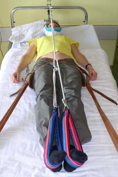
All patients underwent Cotrel dynamic traction before and after the anterior release procedure. Tension was applied to the head halter with an occipital piece and a chin strap (Fig. 1). A counterforce is applied with two sets of straps, one on each side of the pelvis fixated with a triangular trochanter pad. The straps are attached to the foot of the bed. The third component of the traction consists of two foot pieces that connects to the head halter via a pulley system. The patients were supervised in Cotrel traction by physiotherapists and the system was adjusted to create the highest tolerable force when the patient extends the legs. Patients performed traction six times a day for 20 min and stayed in traction for 1 week.
Fig. 1.
Patient performing Cotrel traction. Traction radiographs were made before and after performance of the anterior release
Traction radiographs
Traction radiographs of the whole spine in two planes were performed after 1 week of traction. The indication for a combined procedure was made and the levels to be anteriorly released were determined. Postoperatively patients again performed cotrel traction and further traction films were made to assess curve flexibility and coronal balance. According to these X-rays, fusion levels for posterior instrumentation were determined. Traction radiographs were retrospectively evaluated and the flexibility rate was assessed [(ap−bending)/ap × 100] [15].
Surgical procedure
Open thoracotomy and rib resection was performed in all patients. The anterior longitudinal ligament was resected and the discs completely excised. After resection of the endplates the intervertebral space was filled with rib grafts. After removal of the chest drain patients again started Cotrel traction before the second operation. Before posterior instrumentation and correction, a posterior release by facettectomy and concave thoracoplasty (CTP) with osteotomy of the ribs was performed [17].
Statistics
The Wilcoxon-signed rank non-parametric test for comparison of paired groups was used for statistical analysis. Differences were considered significant with a P-value of less than 0.05.
Results
The mean preoperative thoracic curve was 89.7° ± 15.4° (range 65°–110°), the mean kyphosis was 30.5° ± 9°, hyperkyphosis was not found in any patients. Rotation of the thoracic apical vertebra was grade III in three patients and grade II in three patients. The flexibility rate for side bending was 20.5 ± 10.1%, the results for curve correction in Cotrel traction are shown in Table 1. The mean lumbar curve was 47.7° ± 20.4° and corrected to a mean of 19.3° ± 14.8° in side bending.
Table 1.
Preoperative results
| Subject | Age (years) | Lenke type | Thoracic curve (°) | Side bending (°) | Cotrel traction (°) | Flexibility rate traction (%) |
|---|---|---|---|---|---|---|
| 1 | 16 | 1BN | 100 | 80 | 75 | 25 |
| 2 | 15 | 4CN | 93 | 88 | 86 | 8 |
| 3 | 21 | 2BN | 86 | 62 | 60 | 30 |
| 4 | 15 | 2AN | 84 | 74 | 68 | 19 |
| 5 | 13 | 1BN | 65 | 45 | 47 | 28 |
| 6 | 14 | 3BN | 110 | 80 | 84 | 24 |
| Mean (SD) | 15.6 | 89.7 (15.4) | 71.5 (15.6) | 70 (14.9) | 22.2 (8.1) |
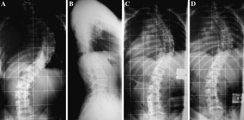
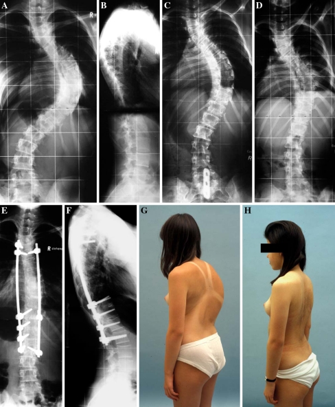
All patients underwent an open thoracic release; levels of disc excision are shown in Table 2. The average number of thoracic discs excised was 5.8. In one patient the most caudad vertebra was L1 (Table 2, subject 4) and one patient also had a lumbar release down to L4 (Table 2, subject 6). Postoperative results are shown in Table 2. After performance of the anterior release the thoracic curve showed a mean increase of correction by 5.5° ± 5.0° as shown in traction radiographs (Figs. 2, 3). The flexibility index changed significantly by 6.2 ± 5.6% (P = 0.028). After posterior correction and instrumentation the curve was corrected to a mean of 36.5° ± 10.1°.
Table 2.
Postoperative results
| Subject | Levels released | Cotrel traction (°) | Flexibility rate traction (%) | Thoracic curve (after post instrument) (°) | Correction rate (%) |
|---|---|---|---|---|---|
| 1 | Th5–Th12 | 65 | 35 | 40 | 60 |
| 2 | Th6–Th12 | 83 | 11 | 46 | 50,5 |
| 3 | Th5–Th12 | 55 | 36 | 39 | 55 |
| 4 | Th6–L1 | 55 | 35 | 23 | 73 |
| 5 | Th4–Th11 | 46 | 29 | 25 | 62 |
| 6 | Th3–L4 | 83 | 25 | 46 | 58 |
| Mean (SD) | 64.5 (15.5) | 28.3 (9.7) | 36.5 (10.1) | 59.6 (7.5) |
Fig. 2.
Subject 3 preoperative ap and lateral radiographs showing right convex thoracic scoliosis of 86° (a, b). The preoperative ap traction radiograph shows scoliosis correction to 60° (c) and traction films after anterior release from Th5–Th12 shows correction to 55° (d)
Fig. 3.
Subject 4 preoperative ap and lateral radiographs showing right convex thoracic scoliosis of 84° (a, b). The preoperative ap traction radiograph shows scoliosis correction to 68° (c) and traction films after anterior release from Th6–L1 shows correction to 55° (d). Postoperative pictures showing correction to 23° (e, f). Clinical photographs showing the preoperative deformity with a significant rib hump (g) and the postoperative result (h)
Discussion
With the advent of thoracoscopic techniques for spinal surgery, numerous studies have been published concerning anterior release in AIS [1, 2, 11–13, 18–26]. Excision of the intervertebral discs prior to posterior instrumentation is believed to impart additional flexibility and it has been presumed that disc excision improves the correction rate [12, 19, 20]. But the extent of improvement has only been roughly estimated [20, 24] and the benefit of the release as compared to the single posterior procedure is largely undocumented. In most publications, the effect of the anterior release could not be addressed separately because anterior release and posterior instrumentation were performed as a one-stage procedure. To our knowledge, only two papers separately assessed the results after the anterior release in AIS patients: Wang et al. [27] concluded that the anterior release alone has only little effect on severe scoliosis whereas Cheung et al. [5] stated that thoracoscopic release improves spinal flexibility as shown by fulcrum bending radiographs (four patients). The current literature mainly focuses on technical descriptions of thoracoscopy and its advantages as compared to the open procedure, i.e. the less traumatic approach. But the important question is not whether the release should be performed open or thoracoscopically, the question is whether it is necessary at all since indication criteria for thoracic release in AIS patients remain controversial.
Preoperative Cotrel traction along with traction radiographs is a routine procedure in AIS patients at the author’s institution (Figs. 1, 2, 3). Additional to bending films this gives information about flexibility together with coronal balancing of the spine. All patients in this study had a staged procedure and traction radiographs were made before and after performance of the anterior release. This enabled us to assess the effect of the anterior release on spinal flexibility by comparing the traction radiographs pre- and postoperatively. The mean preoperative thoracic curve was 89.7° with a mean flexibility rate of 22.2% in traction. The flexibility rate changed by a mean of 6.1% after performance of the anterior release (mean improvement of the Cobb angle 5.5°). Although this change was statistically significant, the achieved amount of correction was little and averaged approximately 1° per segment why we consider it to be clinically not relevant. The question is of whether this change as seen in the traction films justifies the performance of an anterior release and whether this improves the correction rate after posterior instrumentation. Presuming that traction radiographs can predict the postoperative correction to some degree [6, 7] especially in curves larger than 65° [10], the change attributable to anterior release is only marginal in our study. But it should be noted that the study group is small and definition of curvature criteria for anterior release would require a case control study. The optimal indication for an anterior release in AIS remains unclear: it has been recommended for curves greater than 60° [20], 70° [24], curves greater than 100° [4] and curves greater than 40° in fulcrum bending [6]. In a meta-analysis it has been shown that in most series using thoracoscopic anterior release, the Cobb angle averaged 65° [2] and even patients with a scoliosis of 42° have been anteriorly released [1]: we are convinced that the additional morbidity by the anterior release is not justified in such cases. Recent paper showed that there is no need for an anterior release even for curves in the 70°–90° range [3] and that in curves up to 100° a single posterior approach with pedicle screw constructs leads to the same coronal correction rate [14]. Lenke [12] has published the most precise indication criteria, which includes curves with a magnitude greater than 75°, a diminished curve flexibility with thoracic bending greater than 50° or sagittal malalignement with severe thoracic lordosis or kyphosis. All but one of the presented patients fulfilled these criteria. Nevertheless, at least in four of these, in retrospect the anterior release did not sufficiently increase spinal flexibility as had been intended.
In kyphotic deformity, the anterior release improves sagittal correction. However, most patients with AIS have a hypokyphosis and the results of this study indicate that the increase in flexibility by disc resection is probably less than it had been suspected by several authors [20, 24]. In a cadaver model it has been shown that disc excision alone does not enable a significant increase in motion when unaccompanied by rib head resection [8]. In our experience the rigidity of the rib cage impedes a sufficient correction. Accordingly Halsall et al. [9] found that “...only rib resection on the scoliotic concavity significantly increased spinal flexibility.” In our view, the single posterior approach with posterior release by opening of the facet joints and osteotomy of the concave ribs is more efficient in the increase of spinal flexibility than excision of intervertebral discs [16, 17]. This is a routine procedure in the authors clinic and CTP has been performed in more than 800 patients. This procedure has improved the correction rates as well as the cosmetic result. According to our clinical experience as well as the literature, a combined approach in AIS is only necessary in concomitant kyphosis, in coronal imbalance and massive curves beyond 100° [14].
To conclude, indication criteria for the combined approach in AIS with the aim of achieving an optimal correction remains to be determined. In our view, with the progress of thoracoscopic procedures the indication for an anterior release in AIS has probably been expanded to curves which could have been sufficiently corrected with a single posterior procedure. Despite the small patient collective, our results indicate that whilst thoracoscopic release is coming into more frequently use in AIS, the indication criteria still require some further clarification. “Just because a procedure can be done doesn’t mean that it should be done” [22].
Acknowledgment
No benefits in any form have been received or will be received from a commercial party related directly or indirectly to the subject of this article. The authors thank M. Daniels, Heidelberg, for the help in editing the manuscript.
References
- 1.Al-Sayyad MJ, Crawford AH, Wolf RK (2004) Early experiences with video-assisted thoracoscopic surgery: our first 70 cases. Spine 29:1945–1951; discussion 1952 [DOI] [PubMed]
- 2.Arlet V. Anterior thoracoscopic spine release in deformity surgery: a meat-analysis and review. Eur Spine J. 2000;9:17–23. doi: 10.1007/s005860000186. [DOI] [PMC free article] [PubMed] [Google Scholar]
- 3.Arlet V, Jiang Ouellet L J. Is there a need for anterior release for 70–90° thoracic curves in adolescent scoliosis ? Eur Spine J. 2005;13:740–745. doi: 10.1007/s00586-004-0729-x. [DOI] [PMC free article] [PubMed] [Google Scholar]
- 4.Bridwell K. Surgical treatment of idiopathic adolescent scoliosis. Spine. 1999;24:2607–2616. doi: 10.1097/00007632-199912150-00008. [DOI] [PubMed] [Google Scholar]
- 5.Cheung Luk KM KD. Prediction of correction of scoliosis with use of the fulcrum-bending radiograph. J Bone Joint Surg Am. 1997;79:1144–1150. doi: 10.2106/00004623-199708000-00005. [DOI] [PubMed] [Google Scholar]
- 6.Cheung KM, Lu DS, Zhang H, et al. In-vivo demonstration of the effectiveness of thoracoscopic anterior release using the fulcrum-bending radiograph: a report of five cases. Eur Spine J. 2005;21:1–5. doi: 10.1007/s00586-005-0027-2. [DOI] [PMC free article] [PubMed] [Google Scholar]
- 7.Davis BJ, Gadgil A, Trivedi J, et al. Traction radiography performed under general anesthetic: a new technique for assessing idiopathic scoliosis curves. Spine. 2004;29:2466–2470. doi: 10.1097/01.brs.0000143109.45744.12. [DOI] [PubMed] [Google Scholar]
- 8.Feiertag M, Horton W, Norman J, et al. The effect on different surgical releases on thoracic spinal motion: a cadaveric study. Spine. 1995;20:1604–1611. doi: 10.1097/00007632-199507150-00009. [DOI] [PubMed] [Google Scholar]
- 9.Halsall A, James D, Kostuik J, et al. An experimental evaluation of spinal flexibility with respect to scoliosis surgery. Spine. 1983;8:482–488. doi: 10.1097/00007632-198307000-00005. [DOI] [PubMed] [Google Scholar]
- 10.Hamzaoglu A, Talu U, Tezer M, et al. Assessment of curve flexibility in adolescent idiopathic scoliosis. Spine. 2005;30:1637–1642. doi: 10.1097/01.brs.0000170580.92177.d2. [DOI] [PubMed] [Google Scholar]
- 11.Krasna M, Jiao X, Eslami A, et al. Thoracoscopic approach for spine deformities. J Am Coll Surg. 2003;197:777–779. doi: 10.1016/S1072-7515(03)00755-5. [DOI] [PubMed] [Google Scholar]
- 12.Lenke LG. Anterior endoscopic discectomy and fusion for adolescent idiopathic scoliosis. Spine. 2003;28:S36–S43. doi: 10.1097/00007632-200308011-00007. [DOI] [PubMed] [Google Scholar]
- 13.Liljenqvist U, Steinbeck J, Niemeyer T, et al. Thoracoscopic interventions in deformities of the thoracic spine. Z Orthop Ihre Grenzgeb. 1999;137:496–502. doi: 10.1055/s-2008-1039378. [DOI] [PubMed] [Google Scholar]
- 14.Luhmann S, Lenke L, Kim Y, et al. Thoracic adolescent idiopathic scoliosis curves between 70 degrees and 100 degrees: is anterior release necessary? Spine. 2005;15:2061–2067. doi: 10.1097/01.brs.0000179299.78791.96. [DOI] [PubMed] [Google Scholar]
- 15.Mann D, Nash C, Wilham M. Evaluation of the role of concave rib osteotomies in the correction of thoracic Scoliosis. Spine. 1989;14:491–495. doi: 10.1097/00007632-198905000-00003. [DOI] [PubMed] [Google Scholar]
- 16.Metz-Stavenhagen P, Hempfing A, Völpel H et al (2002) Concave thoracoplasty (CTP) and posterior instrumentation for correction of rigid thoracic scoliosis: results at 4–6 years. In: 37th annual meeting of the Scoliosis Research Society, Seattle
- 17.Metz-Stavenhagen P, Morgenstern W (2003) Concave thoracoplasty for stiff thoracic scoliosis. In: Haher TR, Merola AA (eds) Surgical techniques for the spine. New York, pp 128–130
- 18.Newton PO, Shea Granlund KG KF. Defining the pediatric spinal thoracoscopy learning curve: sixty-five consecutive cases. Spine. 2000;25:1028–1035. doi: 10.1097/00007632-200004150-00019. [DOI] [PubMed] [Google Scholar]
- 19.Newton PO, Wenger DR, Mubarak SJ, et al. Anterior release and fusion in pediatric spinal deformity. A comparison of early outcome and cost of thoracoscopic and open thoracotomy approaches. Spine. 1997;22:1398–1406. doi: 10.1097/00007632-199706150-00020. [DOI] [PubMed] [Google Scholar]
- 20.Niemeyer T, Freeman BJ, Grevitt MP, et al. Anterior thoracoscopic surgery followed by posterior instrumentation and fusion in spinal deformity. Eur Spine J. 2000;9:499–504. doi: 10.1007/s005860000181. [DOI] [PMC free article] [PubMed] [Google Scholar]
- 21.Pollock M, O’Neal K, Picetti G, et al. The results of video-assisted exposure of the anterior thoracic spine in idiopathic scoliosis. Video-assisted scoliosis repair Ann Thorac Surg 199662818–823. 10.1016/S0003-4975(96)00505-X8815822 [DOI] [Google Scholar]
- 22.Regan JJ, Guyer RD. Endoscopic techniques in spinal surgery. Clin Orthop. 1997;335:122–139. [PubMed] [Google Scholar]
- 23.Sucato Elerson DJ E. A comparison between the prone and lateral position for performing a thoracoscopic anterior release and fusion for pediatric spinal deformity. Spine. 2003;28:2176–2180. doi: 10.1097/01.BRS.0000084641.96288.8D. [DOI] [PubMed] [Google Scholar]
- 24.Waisman M, Saute M. Thoracoscopic spine release before posterior instrumentation in scoliosis. Clin Orthop. 1997;336:130–136. doi: 10.1097/00003086-199703000-00018. [DOI] [PubMed] [Google Scholar]
- 25.Wall E, Al-Sayyad M (2003) Early experience with video assisted thoracoscopic surgery (VATS): the first hundred cases. In: 38th annual meeting of the Scoliosis Research Society, Quebec City
- 26.Wall EJ, Bylski-Austrow DI, Shelton FS et al (1998) Endoscopic discectomy increases thoracic spine flexibility as effectively as open discectomy. A mechanical study in a porcine model. Spine 23:9–15; discussion 15–16 [DOI] [PubMed]
- 27.Wang YP, Xu HG, Qiu GX, et al. The effect of anterior spinal release on severe adolescent idiopathic scoliosis. Zhonghua Wai Ke Za Zhi. 2004;42:77–80. [PubMed] [Google Scholar]


