Abstract
We studied 79 patients with unilateral injury to the anterior cruciate ligament (ACL). The patients were randomly allocated to reconstruction with autologous patellar bone-tendon-bone (BTB) grafts (49 knees) or hamstring tendon (ST) grafts (30 knees). We measured anterior tibial translation (ATT) during isokinetic concentric contraction exercise 18–20 months after surgery using a computerized electrogoniometer. In both groups the highest ATT during exercise was observed at a knee flexion of about 20° and was 13.5±3.0 mm in the BTB group and 13.9±3.4 mm in the ST group. There was no difference in the ATT between the reconstructed and healthy knees. For a range of knee flexion between 30 and 50° the ATT in the ST group was significantly higher on the reconstructed side than on the healthy side. In the BTB group, the mean ATT in the reconstructed group was similar to that on the healthy side at a knee flexion angle between 0 and 90°.
Résumé
Nous avons étudié 79 malades avec une rupture unilatérale du ligament croiseur antérieur (ACL). Les malades étaient randomisés pour avoir une reconstruction avec greffe autologue os-tendon-os rotulien (BTB; 49 genoux) ou une greffe des tendons ischiojambiers (ST; 30 genoux). Nous avons mesuré la translation tibiale antérieure (ATT) pendant la contraction concentrique isocinétique, 18–20 mois après la chirurgie en utilisant un électrogoniomètre informatisé. Dans les deux groupes le plus haut ATT pendant l'exercice a été observé à une flexion du genou d'approximativement 20° et était de 13,5±3,0 mm dans le groupe BTB et 13,9±3,4 mm dans le groupe ST. Il n'y avait aucune différence dans la translation entre les genoux reconstruit et les genoux sains. Pour une flexion du genou entre 30 et 50°, l'ATT dans le groupe ST était significativement plus haut du côté reconstruit que du côté sain. Dans le groupe BTB, l'ATT dans le groupe reconstruit était semblable à celui sur le côté sain à un angle de la flexion du genou entre 0 et 90°.
Introduction
Recently, Beynnon et al. [3] performed a prospective randomized study of anterior cruciate ligament (ACL) reconstruction using bone-tendon-bone (BTB) grafts or hamstring tendon (ST) grafts, and reported that the knee joint stability and the knee flexion muscle performance were slightly better with the BTB method than ST, but that there were no differences in the subjective satisfaction and the postoperative activity level. Several cohort studies demonstrated that the clinical outcome and the knee stability were good with both methods, with no differences between them [1, 6–8, 15]. It has been reported that recovery of knee extension is delayed in patients with anterior knee pain after ACL reconstruction using BTB [23]. This suggests that reconstruction using BTB is not always superior to reconstruction using ST.
Knee joint stability is generally measured under static conditions without muscular activity using a knee arthrometer, such as the KT-1000 [5, 25]. However, since the instability of the knee joint with ACL injury is induced by movement, it is important to evaluate the stability during movement of the knee joint. Recently, some researchers drew attention to this by trying to evaluate stability during isokinetic concentric contraction exercises [13, 14] and analysis of kinematics using a 3D optoelectronic system [10]. In particular, the tension of the ACL is considered high during isokinetic concentric contraction [12], and evaluation of the anterior tibial translation (ATT) under such conditions may lead to evaluation of the changes in knee joint kinematics after ACL reconstruction.
The purpose of this study was to examine ATT during isokinetic concentric contraction exercises in patients with ACL reconstruction using either BTB grafts or ST grafts, and evaluate the differences in biomechanical characteristics between the two procedures.
Material and methods
Seventy-nine patients (79 knees) with unilateral ACL injury who underwent ACL-reconstruction between 1998 and 2000 were randomly allocated into two groups according to the graft to be used in the surgical reconstruction. In the BTB group the middle one-third of the patellar tendon with bone on both ends was collected, and fixed using interference screws arthroscopically [11]. In the ST group the semitendinosus tendon was harvested, and a four-stranded tendon graft was prepared. Following Rosenberg's method, the graft was inserted into the bone foramen using the inside-out technique, and fixed with an end-button on the femoral side and to a post-screw on the tibial side [24].
There were 49 patients, 21 men and 28 women, mean age 25.4 (range 16–42) years in the BTB group and the biomechanical examination was performed a mean of 20 (14–29) months after reconstruction. In the ST group, there were 30 patients, 17 men and 13 women, mean age 27.2 (range 15–44) years and the examination was performed a mean of 18 (15–23) months after surgery. The same method of rehabilitation after ACL reconstruction was used in the two groups. At the time of examination, the knee joint stability in the Lachman test was negative in 45 patients in the BTB group, and a hard end point was observed in the remaining four patients showing a positive Lachman test. In the ST group, 26 patients had a negative Lachman test, and a hard end point was observed in the remaining four patients with a positive Lachman test.
The Biodex exercise dynamometer system (Biodex, Shirley, NY, USA) was used for the isokinetic concentric contraction exercise. Both system and methods were well explained, and immediately before measurement, warming-up exercise using an aero-bike was performed for about 5 min. Each patient was seated in the Biodex dynamometer and positioned to align the flexion-extension axis of the knee with the rotational axis of the leg attachment. The patient was then strapped in the seat and the resistance pad on the lower leg was fixed just above the ankle joint. This distal pad position was selected because the applied loads provoke higher joint forces and displacement, than with a more proximal application at similar muscle torques. Patients were tested at a velocity of 30°/s along a 90° motion range, knowing that the largest tibial sagittal translation in isokinetic exercises was noted at a speed of 30°/s with various velocities. After several submaximal cycles, the participants were asked to perform three times at maximum thigh action.
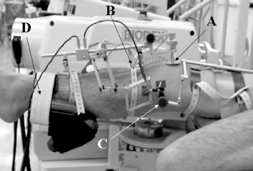
To measure ATT, a computerized goniometer linkage (CA-4000, OSI, Hayward, CA, USA) was fixed to the knee with broad elastic bands (Fig. 1). The reproducibility of this system has been reported previously [20] and it has been employed by several investigators to study characteristics of knee motion [9, 17, 19, 21]. It is composed of femoral and tibial frames and three goniometers in a rotation module to measure the relative rotation between the femur and the tibia. The potentiometer for sagittal motion mounted in the tibial frame registers the difference in position between a spring-loaded patellar pad and the fixation point of the tibial tuberosity during knee motion. The sagittal plane direction is perpendicular to the tibial frame. The linear accuracy of the sagittal parallelogram linkage was 0.1 mm and the angular accuracy for the potentiometers was 0.125°. The application of the CA-4000 followed the manufacturer's instructions. The system was zeroed with the participant lying relaxed at full knee extension and neutral knee rotation. Following previous reports [9, 20, 21], the alignment of the potentiometers was checked repeatedly and carefully in the zeroing screen of the computer during exercise. The protocol was repeated with fresh zeroing if values were different from the original. Only the sagittal plane translation (mm) and the change in flexion angles (degrees) were studied during the two different measurements (isokinetic and passive motion) assisted by the Biodex machine. For comparison the same procedure was repeated on the unaffected side.
Fig. 1.
The electrogoniometer system (CA-4000) fitted to a lower limb of a patient sitting on a Biodex seat. The removal arm is padded on the patella (A) and the tibial tuberosity (B). The potentiometer (C) is used to measure the knee joint angle and is aligned with the knee joint center. The resistance pad (D) of the Biodex is placed on the tibia just above the ankle joint
Analysis of electrogoniometric data
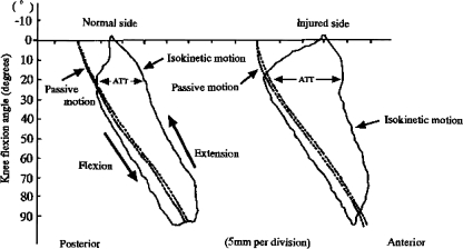
The CA-4000 system recorded two vertically oriented curves throughout the range of motion in each exercise. A graphical display of sagittal plane translation during a test cycle of the two different exercises was used. During a passive knee motion cycle, the curve representing tibial sagittal translation from 90 to 0° demonstrated a gradually increasing posterior translation towards extension, both during the extension and the flexion phase. During isokinetic motion, in contrast, an obvious difference in tibial sagittal translation was apparent between the extension and the flexion phase from 90 to 0°. It is generally considered that the slope of this line in exercise represents the relative motion between the measuring arm and the patella during the range of motion [9, 20, 21]. In this study, ATT was measured in terms of the difference with isokinetic exercise compared to the value for passive extension motion (Fig. 2) [17, 19]. From 90 to 0° of knee flexion in the extension phase, differences in anterior sagittal displacement between passive and isokinetic motion were measured at every 10° position with the help of a computer (IBM PC/AT compatible EVEREX computer, Fremont, CA, USA) equipped for the CA-4000. Data from the second cycle of each test were used for calculation, as in previous reports [19–21]
Fig. 2.
Typical graphical display (CA-4000 system) of sagittal plane knee translation during passive and isokinetic test cycles. Anterior tibial translation (ATT), in terms of the difference with isokinetic extension exercise compared with the value for passive extension motion with the Biodex system, was measured at every 10° position with the help of a computer. X-axis, sagittal plane translation; Y-axis, knee flexion angle
Statistics
Commercially available software (Statistica; Stat Soft, Tulsa, OK, USA) was used. Using the same test in the same patient to compare the normal and the injured knee, the Student's t test was employed with the significance level set at p<0.05. The coefficients of variation were calculated from the actual tests, and correlation analysis was also performed, with a significance level of 1%.
Results
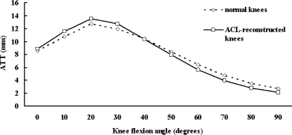
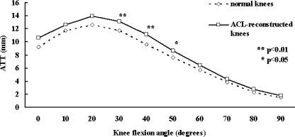
The ATT during isokinetic concentric contraction exercise increased in proportion to the extension angle of the knee by between 0 and 20°, and reached the maximum of 20° in both BTB and ST groups. At a knee flexion angle of 0°, the ATT was 8.5±1.8 mm on the healthy side and 8.9±2.1 mm on the reconstructed side in the BTB group, while in the ST group, the ATT was 9.2±2.0 mm on the healthy side and 10.6±2.2 mm on the reconstructed side. In both groups, there were no differences in the ATT between the healthy and reconstructed sides. At a knee flexion angle of 20°, the maximal ATT was 12.7±2.8 mm on the healthy side and 13.5±3.0 mm on the reconstructed side in the BTB group, showing no difference between the healthy and reconstructed sides. In the ST group, the maximal ATT was 12.6±3.2 mm on the healthy side and 13.9±3.4 mm on the reconstructed side, the difference not being significant (Figs. 3, 4). In the BTB group, the mean ATT on the reconstructed side was similar to that on the healthy side at a knee flexion angle between 0 and 90°, and the stability of the knee during isokinetic concentric contraction exercise was good (Fig. 3). In the ST group, the mean ATT on the reconstructed side was higher than that on the healthy side at a knee flexion angle between 0 and 90°, and the differences in the mean ATT between the reconstructed and healthy sides were significant at a knee flexion angle between 30 and 50° (Fig. 4).
Fig. 3.
Comparison of ATT between the anterior cruciate ligament (ACL) reconstructed with bone-tendon-bone (BTB) grafts and normal knees for isokinetic extension exercise. No significant difference was seen with any testing flexion angles. ATT was measured as described in Fig. 2
Fig. 4.
Comparison of ATT between the ACL reconstructed with hamstring tendon (ST) grafts and normal knees for isokinetic extension exercise. A significant difference was seen within a range of flexion between 30 and 50°. ATT measured as described in Fig. 2
Discussion
Currently, BTB or ST grafts are most commonly used for ACL reconstruction. Reconstruction with BTB grafts is considered to be the gold standard because the mechanical strength of the graft is high, the stability of the graft fixed with interference screws is initially high and good outcomes are reported [4]. However, anterior knee pain causing delay in the recovery of the knee extensors [23] has been reported. In the mid-1990s the ST method was therefore introduced. Recently, studies on randomized controlled trials of these methods have been published [1, 6–8, 15]. There is, however, no consensus as to which method is best.
“Giving way” and an unstable feeling in the knee joint are the major subjective symptoms of ACL injury. Such symptoms are induced by exercise, especially landing from a jump and performing a cutting motion. Since it is difficult to evaluate the stability of joints during such activities, static measurement using an arthrometer, such as the KT-1000, is generally performed to evaluate the degree of improvement after surgery [5, 25]. Recently, we have reported the stability of knee joints with ACL injury and of those after ACL reconstruction during isokinetic concentric contraction exercise [13, 14]. Based on these studies, the ATT during isokinetic concentric contraction exercise was prospectively examined in knee joints that had been reconstructed by either the BTB method or ST method. In both methods, the ATT of the knee joints during isokinetic concentric contraction exercise was maximal at a knee flexion angle of about 20°. The same result was obtained on the healthy knee. There was no difference in the maximal ATT between the knees on the affected and the healthy sides, nor was there a difference between the BTB and ST methods. As in studies on the evaluation of the stability of knee joints after ACL reconstruction under static conditions, our results indicated that the instability in the ATT during isokinetic concentric contraction exercise was also improved. However, the ATT was higher in the knees reconstructed by the ST method than in the control knees between 30 and 50° of flexion. This may be the main biomechanical difference between the BTB and ST methods of reconstruction. It is difficult to identify the cause of this difference, but harvest site morbidity due to the collection of ST grafts may be involved. It is generally considered that harvest site morbidity is smaller with ST than with BTB [2, 22]. On the other hand, it has been reported that reduction in the strength of the knee flexor muscles was more marked with the ST method than with the BTB method [1, 3, 8, 22]. The peak torque of knee flexion during isokinetic concentric contraction exercise is observed at 35–40° [18], and it was interesting to note that the ATT changed at around these angles. Among the functions of the hamstring tendon, its role as a dynamic stabilizer of ATT is important [16]. This suggests that the harvesting of the hamstring tendon was a cause of the increase in the ATT during isokinetic concentric contraction exercise between 30 and 50° with the ST method.
In conclusion, the maximal ATT during isokinetic concentric contraction exercise was restored to a level within the normal range by the BTB and ST methods. With the BTB method, no articular instability was observed during isokinetic concentric contraction exercise in the knee between 0 and 90°, while with the ST method, joint instability was observed during isokinetic concentric contraction exercise between 30 and 50°.
References
- 1.Aune AK, Holm I, Risberg MA, Jensen HK, Steen H (2001) Four-strand hamstring tendon autograft compared with patellar tendon-bone autograft for anterior cruciate ligament reconstruction: a randomized study with two-year follow-up. Am J Sports Med 29:722–728 [DOI] [PubMed]
- 2.Aglietti P, Buzzi R, Zaccherotti G, De Biase P (1994) Patellar tendon versus doubled semitendinosus and gracillis tendons for anterior cruciate ligament reconstruction. Am J Sports Med 22:211–218 [DOI] [PubMed]
- 3.Beynnon BD, Johnson RJ, Fleming BC (2002) Anterior cruciate ligament replacement: comparison of bone-patellar tendon-bone grafts with two-strand hamstring grafts: a prospective, randomized study. J Bone Joint Surg Am 84:1503–1513 [DOI] [PubMed]
- 4.Clancy WG Jr, Nelson DA, Reider B (1982) Anterior cruciate ligament reconstruction using one-third of the patellar ligament, augmented by extra-articular tendon transfers. J Bone Joint Surg Am 64:352–359 [PubMed]
- 5.Daniel DM, Stone ML, Sachs R, Malcom L (1985) Instrumented measurement of anterior knee laxity in patients with acute anterior cruciate ligament disruption. Am J Sports Med 13:401–407 [DOI] [PubMed]
- 6.Ejerhed L, Kartus J, Sernert N, Kohler K, Karisson J (2003) Patellar tendon or semitendinosus tendon autografts for anterior cruciate ligament reconstruction? A prospective randomized study with a two-year follow-up. Am J Sports Med 31:19–25 [DOI] [PubMed]
- 7.Eriksson K, Anderberg P, Hamberg P (2001) A comparison of quadruple semitendinosus and patellar tendon grafts in reconstruction of the anterior cruciate ligament. J Bone Joint Surg Br 83:348–354 [DOI] [PubMed]
- 8.Feller JA, Webster KE (2003) A randomized comparison of patellar tendon and hamstring tendon anterior cruciate ligament reconstruction. Am J Sports Med 31:564–573 [DOI] [PubMed]
- 9.Gillquist J, Messner K (1995) Instrumented analysis of the pivot shift phenomenon after reconstruction of the anterior cruciate ligament. Int J Sports Med 16:484–488 [DOI] [PubMed]
- 10.Hagemeister N, Long R, Yahia L, Duval N, de Guise JA (2002) Quantitative comparison of three different types of anterior cruciate ligament reconstruction methods: laxity and 3-D kinematic measurements. Biomed Mater Eng 12:47–57 [PubMed]
- 11.Hardin GT, Bach BR Jr, Bush-Joseph CA, Farr J (1992) Endoscopic single-incision anterior cruciate ligament reconstruction using patellar tendon autograft. Surgical technique. Am J Knee Surg 5:144–155 [PubMed]
- 12.Henning CE, Lynch MA, Glick KR (1985) An in vivo strain gage study of elongation of the anterior cruciate ligament. Am J Sports Med 13:22–26 [DOI] [PubMed]
- 13.Higuchi H, Terauchi M, Kimura M, Shirakura K, Takagishi K (2002) Characteristics of anterior tibial translation with active and isokinetic knee extension exercise before and after ACL reconstruction. J Orthop Sci 7:341–347 [DOI] [PubMed]
- 14.Higuchi H, Terauchi M, Kimura M, Watanabe H, Takagishi K (2003) The relation between static and dynamic knee stability after ACL reconstruction. Acta Orthop Belg 69:257–266 [PubMed]
- 15.Jannsson KA, Linko E, Sandelin J, Harilainen A (2003) A prospective randomized study of patellar versus hamstring tendon autografts for anterior cruciate ligament reconstruction. Am J Sports Med 31:12–18 [DOI] [PubMed]
- 16.Jenkins WL (1997) A measurement of anterior tibial displacement in the closed and open kinetic chain. J Orthop Sports Phys 25:49–56 [DOI] [PubMed]
- 17.Kanai H (1993) Dynamic analysis in the knees with chronic anterior cruciate ligament insufficiency. An evaluation of antero-posterior instability, leg rotation and ground reaction force. J Jpn Orthop Assoc 67:617–630 [PubMed]
- 18.Kannus P, Beynnon B (1993) Peak torque occurrence in the range of motion during isokinetic extension and flexion of the knee. Int J Sports Med 14:422–426 [DOI] [PubMed]
- 19.Kizuki S, Shirakura K, Kimura M, Fukasawa N, Udagawa E (1995) Dynamic analysis of anterior tibial translation during isokinetic quadriceps femoris muscle concentric contraction exercise. Knee 2:151–155 [DOI]
- 20.Lysholm M, Goertzen D, Messner K (1994) Reproducibilty of sagittal plane knee translation during isokinetic exercises. Isokinet Exerc Sci 4:16–21
- 21.Lysholm M, Messner K (1995) Sagittal plane translation of the tibia in anterior cruciate ligament-deficient knees during commonly used rehabilitation exercises. Scand J Med Sci Sports 5:49–56 [DOI] [PubMed]
- 22.Marder RA, Raskin JR, Carroll M (1991) Prospective evaluation of arthroscopically assisted anterior cruciate ligament reconstruction: patellar tendon versus semitendinous and gracilis tendon. Am J Sports Med 19:478–484 [DOI] [PubMed]
- 23.Natri A (1996) Isokinetic muscle performance after anterior cruciate ligament surgery. Int J Sports Med 17:223–228 [DOI] [PubMed]
- 24.Rosenberg TD, Graf B (1994) Techniques for ACL reconstruction with multi-trac drill guide. Acufex Microsurgical, Mansfield
- 25.Staubli HU (1990) Stress radiography. Measurements of knee motion limits. In: Daniel DM, Akeson WH, O'Connor JJ (eds) Knee ligaments. Structure, function, injury, and repair. Raven, New York, pp 449–459


