

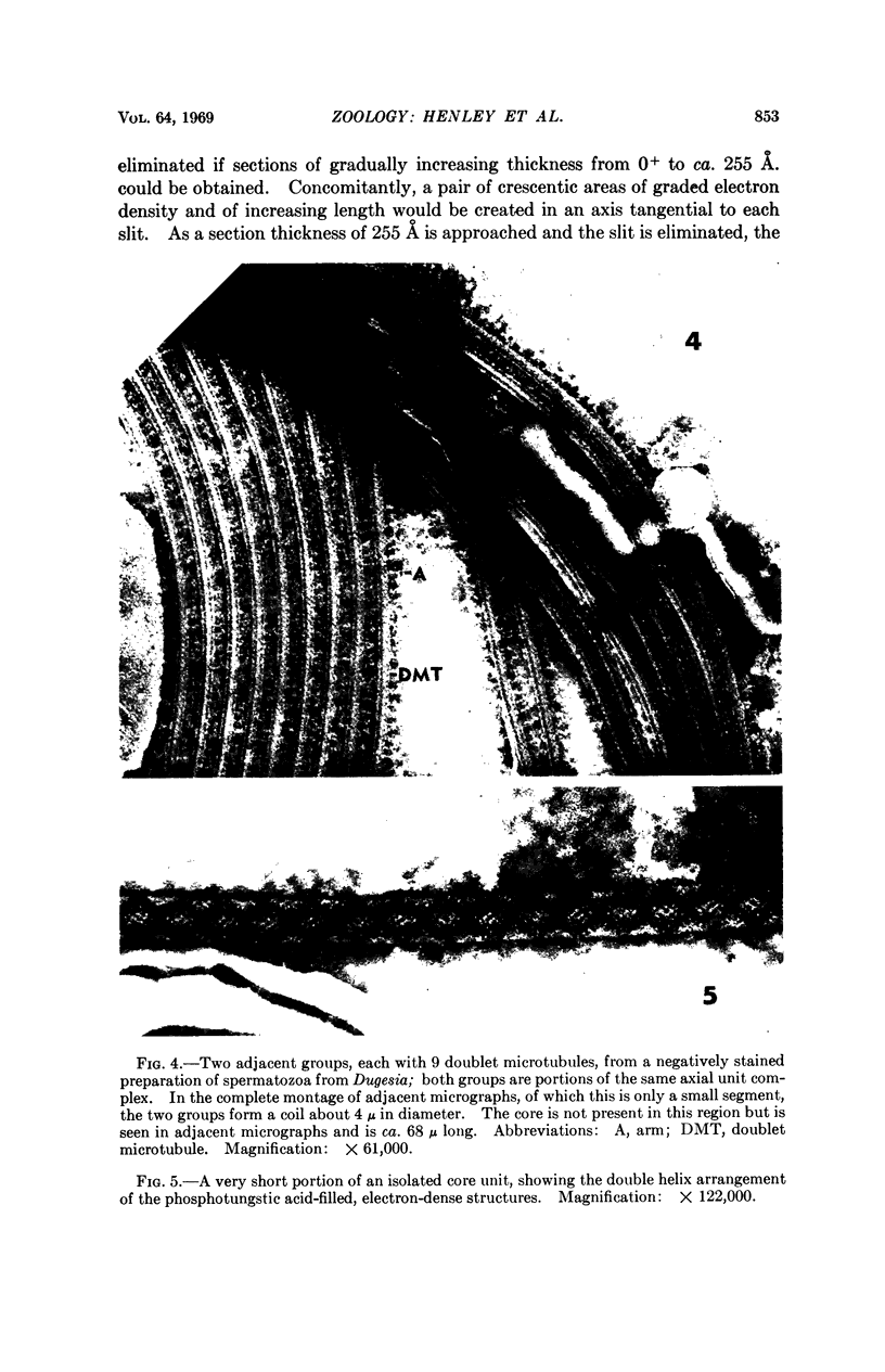
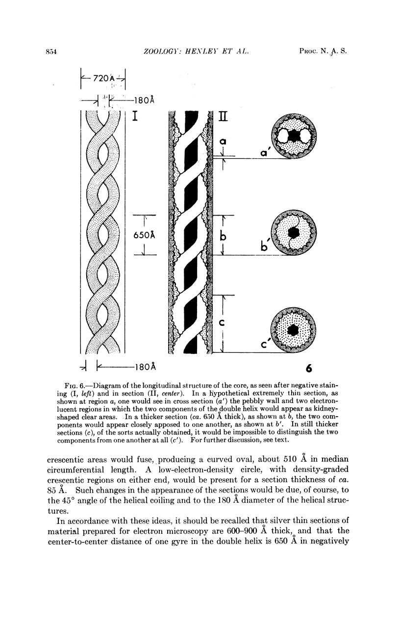
Abstract
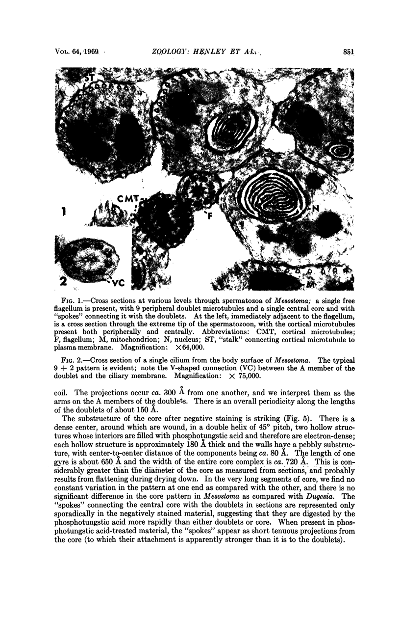
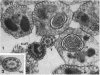
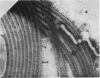
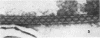
The living spermatozoa of the flatworm Mesostoma georgianum have a sperm body about 100 μ long and 0.5 μ wide, and two motile free flagella, ca. 200 μ long. In sections examined with the electron microscope, these flagella have the usual nine pairs of peripheral doublet microtubules and have a single central core unit which is connected to the A members of the doublets by spokelike structures. There are also short connections between the doublets and the flagellar membrane. In material negatively stained with phosphotungstic acid, the doublet microtubules seem to have very different elastic properties than the core; they tend to fall on the copper grids in coils of rather uniform diameter (2-4 μ), while the core is much more rigid and is often found extending alone, along a relatively straight course, for very long distances (up to 73 μ). After negative staining, the core has a striking appearance with a dense center around which are wound two hollow structures in a double helix of 45° pitch. The center-to-center distance of each gyre is approximately 650 Å, and the hollow structures are ca. 180 Å in diameter.
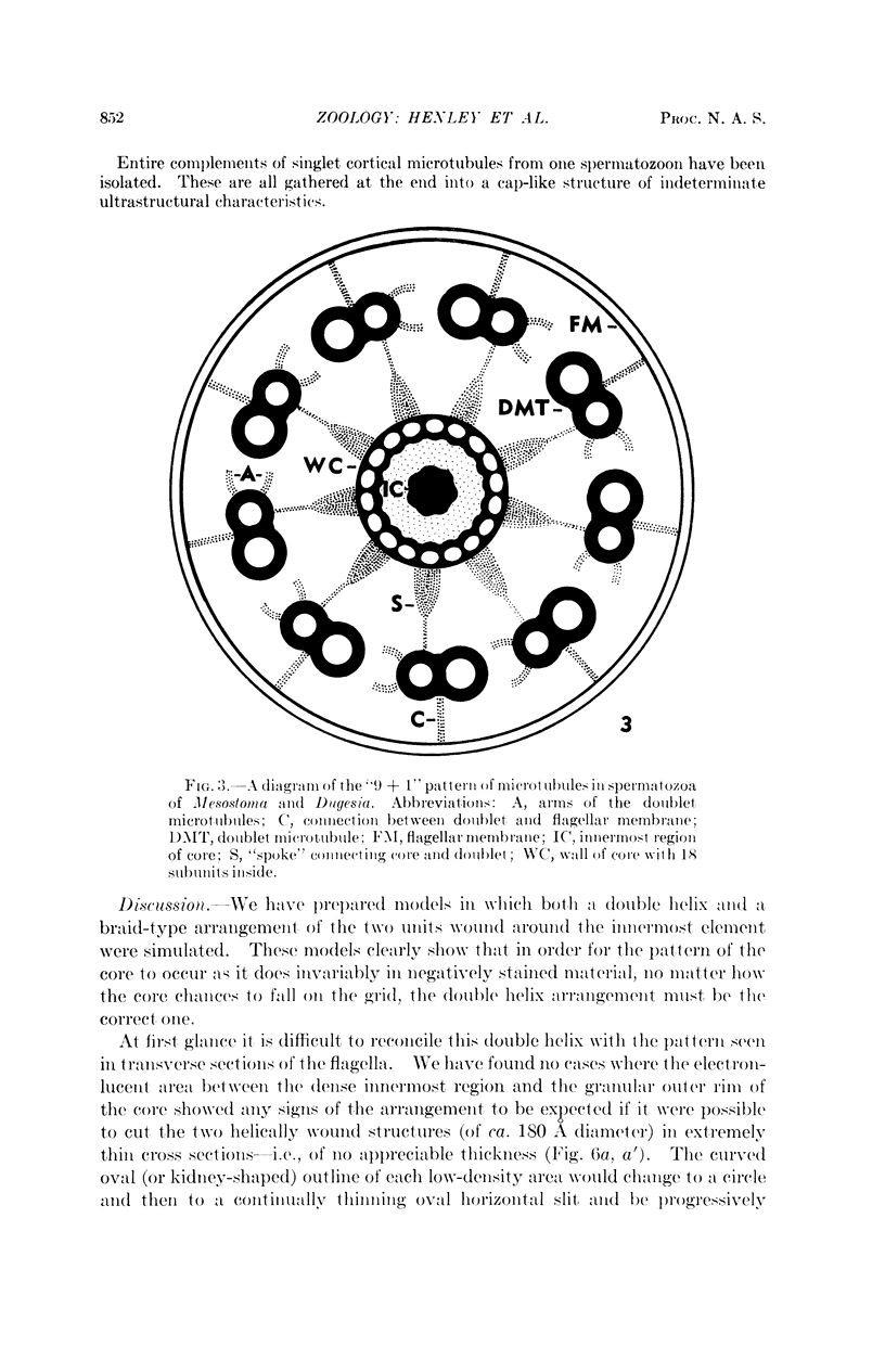
Full text
PDF
Images in this article
Selected References
These references are in PubMed. This may not be the complete list of references from this article.
- Burton P. R. Fine structure of the unique central region of the axial unit of lung-fluke spermatozoa. J Ultrastruct Res. 1967 Jul;19(1):166–172. doi: 10.1016/s0022-5320(67)80066-2. [DOI] [PubMed] [Google Scholar]
- Costello D. P., Henley C., Ault C. R. Microtubules in spermatozoa of childia (turbellaria, acoela) revealed by negative staining. Science. 1969 Feb 14;163(3868):678–679. doi: 10.1126/science.163.3868.678. [DOI] [PubMed] [Google Scholar]
- Hershenov B. R., Tulloch G. S., Johnson A. D. The fine structure of trematode sperm-tails. Trans Am Microsc Soc. 1966 Jul;85(3):480–483. [PubMed] [Google Scholar]
- LUMSDEN R. D. MACROMOLECULAR STRUCTURE OF GLYCOGEN IN SOME CYCLOPHYLLIDEAN AND TRYPANORHYNCH CESTODES. J Parasitol. 1965 Aug;51:501–515. [PubMed] [Google Scholar]
- Morseth D. J. Spermtail finestructure of Echinococcus granulosus and Dicrocoelium dendriticum. Exp Parasitol. 1969 Feb;24(1):47–53. doi: 10.1016/0014-4894(69)90220-3. [DOI] [PubMed] [Google Scholar]
- ROSARIO B. AN ELECTRON MICROSCOPE STUDY OF SPERMATOGENESIS IN CESTODES. J Ultrastruct Res. 1964 Dec;11:412–427. doi: 10.1016/s0022-5320(64)80073-3. [DOI] [PubMed] [Google Scholar]
- Silveira M. Ultrastructural studies on a "nine plus one" flagellum 1. J Ultrastruct Res. 1969 Feb;26(3):274–288. doi: 10.1016/s0022-5320(69)80007-9. [DOI] [PubMed] [Google Scholar]
- Tulloch G. S., Hershenov B. R. Fine structure of platyhelminth sperm tails. Nature. 1967 Jan 21;213(5073):299–300. doi: 10.1038/213299a0. [DOI] [PubMed] [Google Scholar]