Abstract
We have measured the stability and stoichiometry of variants of the human p53 tetramerization domain to assess the effects of mutation on homo- and hetero-oligomerization. The residues chosen for mutation were those in the hydrophobic core that we had previously found to be critical for its stability but are not conserved in human p73 or p51 or in p53-related proteins from invertebrates or vertebrates. The mutations introduced were either single natural mutations or combinations of mutations present in p53-like proteins from different species. Most of the mutations were substantially destabilizing when introduced singly. The introduction of multiple mutations led to two opposite effects: some combinations of mutations that have occurred during the evolution of the hydrophobic core of the domain in p53-like proteins had additive destabilizing effects, whereas other naturally occurring combinations of mutations had little or no net effect on the stability, there being mutually compensating effects of up to 9.5 kcal/mol of tetramer. The triple mutant L332V/F341L/L344I, whose hydrophobic core represents that of the chicken p53 domain, was nearly as stable as the human domain but had impaired hetero-oligomerization with it. Thus, engineering of a functional p53 variant with a reduced capacity to hetero-oligomerize with wild-type human p53 can be achieved without any impairment in the stability and subunit affinity of the engineered homo-oligomer.
Keywords: negative dominance, p51, p73, protein engineering, stability
During molecular evolution, potentially deleterious mutations may be fixed in the population if additional mutations occur that compensate for the negative phenotypic effect of the former (1). Multiple amino acid substitutions in a protein generally act in an additive fashion in that their combined effect equals the sum of the effects of each mutation separately (2–5). However, close to neutral “suppressor” mutations may revert the negative effect of other mutations in the same protein. “Mutually compensatory” mutations involve two or more potentially detrimental replacements able to compensate for each other’s effect, with no or little net variation in protein stability or function. Such nonadditive effects may arise if the residues involved interact with each other, either directly or indirectly (6). Partially compensatory effects on thermodynamic stability have been observed in proteins designed or selected in vitro for repacking of the hydrophobic core (4, 7–14). Such compensatory effects appeared to be infrequent, and incomplete exploration of the sequence space may have restricted even more their occurrence during natural evolution. Even so, a few examples of natural mutations with nonadditive effects on stability have been documented (15, 16). Evaluation of the quantitative limits to such compensatory effects, and of their frequency and general significance in nature, is important to understand the molecular evolution of proteins (1, 17).
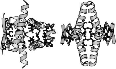
The multifunctional transcription factor p53 is a protein of biomedical importance because of its role as a natural tumor suppressor in humans (18–20). p53 is fully functional only as a tetramer, and its ability to oligomerize resides in a small domain (p53tet, residues 326–355). Inactive p53 with a mutated DNA-binding domain, but with an intact tet domain, is frequently found in cancer cells (21). These mutants may exert a negative dominant effect through hetero-oligomerization with nonmutated p53 (22–24). Thus, the design of a fully functional, tetrameric p53 containing an altered tet domain unable to hetero-oligomerize with the inactive p53 found in tumor cells may be relevant for the development of an anticancer gene therapy based on p53 (25). p53tet can be considered a dimer of dimers, with each primary dimer formed by an antiparallel β-sheet and two antiparallel α-helices, and the dimers associated through the helices in a quasi-orthogonal way (refs. 26–28; Fig. 1). Folding and tetramerization of this domain are coupled processes (29). The side chains found critical for thermodynamic stability and oligomerization were identified by alanine-scan mutagenesis as being most of those involved in the hydrophobic core (30). These residues were essentially conserved in mammalian p53, but not in human p53 homologues (p73 or p51; refs. 31–33) or in other p53-like proteins from more than 20 species of vertebrates and invertebrates (ref. 34 and references therein and ref. 35; see also Table 1).
Figure 1.
Ribbon model of the human p53tet structure. Two different orientations are shown on the left and right sides. The 1sak coordinates (28) for the minimum tet domain (p53 residues 326–356), obtained from the Protein Data Bank, and the program molscript (43) were used. The residues whose side chains were found critical for stability form most of the hydrophobic core and have been depicted as ball-and-stick models.
Table 1.
Key residues within the hydrophobic core of the oligomerization domain of human p53 and alignment with p53 and p53-like proteins from different species
| Species | Hydrophobic core/critical side chains*
|
|||||||
|---|---|---|---|---|---|---|---|---|
| 328 | 330 | 332 | 338 | 340 | 341 | 344 | 348 | |
| Human p53/mammals† | F/Y* | L | I | F/Y‡ | M | F | L | L |
| Xenopus laevis | I | |||||||
| Rainbow trout | L | F | ||||||
| Zebrafish | V | I | L | |||||
| Human p73 | V | I | L | |||||
| Squid | V | I | L | M | ||||
| Chicken | V | L | I | |||||
| Clam | V | L | I | |||||
| Human p51 | L | V | L | I | ||||
| Rat KET | L | V | L | I | ||||
| Japanese fish | F | V | F | L | I | |||
The side chains of these residues are those found to contribute most to the thermodynamic stability of human p53tet (30). They correspond very closely with the side chains involved in the hydrophobic core (Fig. 1). The side chain of Arg-337 was found also critical for p53tet stability. However, the effect appeared to be due mainly to the contribution of its hydrophobic part; the few natural substitutions found at this position (Lys in trout, Thr in humans p51, and Asn in human p73, clam and squid) may not affect these hydrophobic interactions. Amino acid residue numbering is for human p53. For all other proteins, only amino acids that differ from those of human/mammalian p53tet at the equivalent positions are indicated.
See ref. 34 and the Internet address http://perso.curie.fr/Thierry.Soussi/for references. The “mammalian” sequence corresponds to that of the following species: human, african green monkey, rhesus monkey, dog, cat, hamster, chinese hamster, rat, mouse, sheep, and bovine.
At position 328, Tyr was found in human p73 and trout, whereas Phe was found in all other sequences; at position 338, Phe was found in all mammalian sequences except dog, bovine, and human p51, and Tyr was found in all other sequences. Molecular modeling suggested that replacement of Phe by Tyr at either position would not entail any alteration of the hydrophobic core of p53tet; the hydroxyl group of Tyr could be accommodated on the protein surface without disrupting any interaction.
The dual aim of this work has been (i) to assess the contribution of compensatory mutations to the natural evolution of the hydrophobic core of a protein and (ii) to use the information gathered to engineer a stable p53tet domain with an impaired hetero-oligomerization ability. We have introduced in the core of human p53tet several natural single mutations and combinations of mutations occurring in p53-related proteins and quantitated their effect on the thermodynamic stability of p53tet. The results indicate that mutations able to mutually compensate for drastic destabilization have occurred during the evolution of this domain. A triple mutant that was as stable as the human domain showed impaired hetero-oligomerization with the latter.
MATERIALS AND METHODS
Site-Directed Mutagenesis and Protein Expression and Purification.
Subcloning of human p53tet, both a long version with a histidine tag (tetL, p53 residues 303–366) and a shorter version with no tag (tetS, residues 311–367), has been described (30). The construction of single and multiple tetS mutants was carried out by using the QuickChange site-directed mutagenesis kit (Stratagene) and pairs of 26-mer to 35-mer oligonucleotides incorporating the desired mutations. tetL, tetS, and all mutants were sequenced, expressed to high levels, and purified to apparent homogeneity essentially as described (30).
Analytical Size-Exclusion Chromatography.
A calibrated Superdex 75 HR 10/30 FPLC (fast protein liquid chromatography) column (Pharmacia) was used. The protein was eluted in 25 mM phosphate buffer (pH 7) and 200 mM NaCl (30).
Equilibrium Denaturation.
Equilibrium unfolding of tetrameric tetS and tetP variants was analyzed in 25 mM phosphate buffer (pH 7) by far-UV CD spectroscopy in a Jasco-720 spectropolarimeter by using either guanidinium chloride (GdmCl) at 25°C or temperature as denaturant, and the data were fitted to a two-state transition from folded tetramers to denatured monomers as described (method I in ref. 30).
Hetero-Oligomerization Assays.
The first procedure was essentially that of Davison et al. (36). Equimolar amounts of histidine-tagged human p53tetL and of tetS (either wild-type or mutants) were mixed (8 nmol of each tetramer in a volume of 400 μl) and incubated at 37°C for 20 h to allow for complete subunit exchange (26). A 10-μl aliquot was taken for analysis and the remaining solution was loaded into a column containing 200 μl of Qiagen (Chatsworth, CA) Ni-NTA agarose beads. The nontagged protein was eluted with 3 ml of 40 mM Tris⋅HCl (pH 7.4), 500 mM NaCl, and 100 mM imidazole (buffer A), and the histidine-tagged protein was eluted with 2 ml of 40 mM Tris⋅HCl (pH 7.4), 500 mM NaCl, and 500 mM imidazole (buffer B). Fractions (300 μl) were collected and analyzed by SDS/PAGE and Coomassie Blue staining. In the second method, 2 nmol of His-tagged tetL were mixed with different amounts (up to 4 nmol) of nontagged tetS or mutants in a volume of 100 μl and incubated at 42°C for 24 h. The mixtures were added to 50 μl of washed Ni-NTA resin and briefly incubated. The nontagged protein was removed by washing the gel three times with 1 ml of buffer A, followed by centrifugation and removal of the supernatant. Finally, the tagged protein was eluted with 100 μl of buffer B. The samples were centrifuged and the supernatants analyzed by SDS/PAGE as above.
Molecular Modeling.
A personal computer, a Silicon Graphics workstation, and the programs rasmol (37), swiss-pdbviewer (38), and insightii (Biosym Technologies, San Diego) were used.
RESULTS
Effect of Naturally Occurring Single Mutations on the Thermodynamic Stability of p53tet.
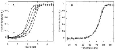
A previous thermodynamic analysis of human p53tet (residues 326–353) showed that the side chains critical for stability were those of residues Leu-330, Ile-332, Phe-341, Leu-344, and Leu-348 and, to a lesser extent, those of Phe-328, Arg-337, Phe-338, and Met-340 (30). All these residues comprise most of the hydrophobic core (Fig. 1). Table 1 specifies the amino acid differences at these key positions between human p53tet and the equivalent region of p53-like proteins whose sequences have been published. We have determined the individual effect of most of these natural amino acid differences on the oligomerization status and the thermodynamic stability of isolated p53tet. At the concentration tested (40 μM monomer), all single mutants tested gave a far-UV CD spectrum very similar to that of wild-type tetS. In addition, they behaved as tetramers in size-exclusion chromatography (data not shown). Thermodynamic stability was quantitated by using that same protein concentration in GdmCl denaturation experiments (Fig. 3A). The values determined for the relevant thermodynamic parameters were compared with those of the parental tetS and refer to unfolding of tetramer (Table 2). Ile substitutions of the partially buried Met-340 or the buried Leu-344 caused only small effects in stability. In contrast, all other natural replacements tested individually led to substantial, although not dramatic, destabilization (from 3.3 to 6.9 kcal/mol of tetramer). These latter substitutions involved hydrophobic replacements of the partially buried Phe-328 or of the buried critical residues Leu-330, Ile-332, Phe-341, or Leu-344 (Fig. 1).
Figure 3.
(A) GdmCl denaturation (at 25°C and pH 7) of human p53tetS and some variants with natural mutations. The fraction of protein denatured is represented as a function of GdmCl concentration for wild-type tetS (□) and mutants L332V (▵), F341L (▿), L344I (⋄), and L332V/F341L/L344I (○). The protein (monomer) concentration was 40 μM. (B) Thermal denaturation (at pH 7) of human p53tetS (□) and the triple mutant L332V/341L/344I (○). The fraction of protein denatured is shown as a function of the temperature. The protein (monomer) concentration was 10 μM. For clarity, only 1 in every 10 experimental points is represented. The fitting of the experimental values to two-state transition curves by using the program Microsoft excel (method II in ref. 30) is indicated by solid lines.
Table 2.
Equilibrium unfolding of p53tet with natural mutations in the hydrophobic core
| Mutant | Hydrophobic core represented* | m, kcal/(M.mol) | [D]50%, M | ΔGuH2O, kcal/mol) | ΔΔGuH2O, kcal/mol | Expected additive ΔΔGuH2O, kcal/mol |
|---|---|---|---|---|---|---|
| wt tetS | Human p53, mammals | 5.1 ± 0.2 | 2.78 ± 0.01 | 32.5 ± 0.5 | ||
| F328L | 7.5 ± 0.2 | 1.41 ± 0.01 | 29.0 ± 0.3 | 3.5 ± 0.6 | ||
| L330F | 7.4 ± 1.0 | 1.19 ± 0.03 | 27.3 ± 1.2 | 5.2 ± 1.3 | ||
| I332V | 4.2 ± 0.3 | 2.25 ± 0.04 | 27.5 ± 0.9 | 5.0 ± 1.0 | ||
| M340I | 6.9 ± 0.4 | 2.14 ± 0.02 | 33.2 ± 0.9 | −0.7 ± 1.0 | ||
| F341L | 5.4 ± 0.3 | 2.05 ± 0.02 | 29.2 ± 0.6 | 3.3 ± 0.8 | ||
| F341I | Xenopus | 6.0 ± 0.2 | 1.21 ± 0.01 | 25.6 ± 0.2 | 6.9 ± 0.6 | |
| L344F | 5.0 ± 0.2 | 1.60 ± 0.01 | 26.4 ± 0.3 | 6.1 ± 0.6 | ||
| L344I | 5.3 ± 0.1 | 2.42 ± 0.01 | 31.3 ± 0.3 | 1.2 ± 0.6 | ||
| F341L/L344F | Trout | 4.7 ± 0.4 | 1.20 ± 0.02 | 24.1 ± 0.5 | 8.4 ± 0.7 | 9.4 ± 0.7 |
| L332V/F341L | 6.3 ± 0.4 | 1.88 ± 0.02 | 30.3 ± 0.7 | 2.2 ± 0.8 | 8.3 ± 1.1 | |
| L332V/M340I/F341L | Human p73, zebrafish | 6.2 ± 0.1 | 1.76 ± 0.01 | 29.4 ± 0.2 | 3.1 ± 0.5 | 7.6 ± 1.4 |
| F341L/L344I | 4.9 ± 0.0 | 2.62 ± 0.00 | 31.2 ± 0.1 | 1.3 ± 0.5 | 4.5 ± 0.7 | |
| L332V/F341L/L344I | Chicken, clam | 5.5 ± 0.1 | 2.55 ± 0.01 | 32.4 ± 0.3 | 0.1 ± 0.6 | 9.5 ± 1.1 |
| F328L/L332V/F341L/L344I | Human p51, rat KET | 6.9 ± 0.5 | 1.20 ± 0.02 | 26.8 ± 0.5 | 5.7 ± 0.7 | 13.0 ± 1.3 |
m, Variation in the free energy of unfolding, ΔGu, with the GdmCl concentration; [D]50%, concentration of GdmCl at which the transition is half-completed by using 40 μM protein (monomer) concentration. ΔGuH2O, ΔGu extrapolated to absence of denaturant and given for a standard 1 M protein concentration; ΔΔGuH2O difference in ΔGu between wild-type p53tetS and any mutant at zero denaturant concentration. Expected additive ΔΔGuH2O values for the multiple mutants were obtained by addition of the experimental values found for the corresponding single mutants. The standard errors of fitting and the propagated errors are indicated. The m values obtained for some mutants were somewhat different than that obtained for wt tetS, possibly due to the nature of the mutation(s) introduced. For these mutants, ΔΔGu values differed somewhat depending on the denaturant concentration at which they were calculated. ΔΔGu values are thus given at zero denaturant concentration, because this corresponds to more physiological conditions and because the required extrapolation of the experimental data was relatively short, so that the fitting errors were not large. The values obtained for a 2 M GdmCl concentration (data not shown) led to essentially the same conclusions discussed in the text.
For the mutants indicated, all residues at the thermodynamically most critical positions of human p53tet (residues 330, 332, 340, 341, 344, and 348; ref. 30) correspond to those of p53 or p53-like protein specified. These six residues form the central hydrophobic minicores of each primary dimer and the critical, central part of the interdimer interface. See also text, Fig. 1, and Table 1.
Additive and Compensatory Effects of Combinations of Naturally Occurring Mutations on the Stability of p53tet.
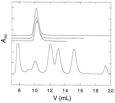
Relative to human p53, human p73 and p51 and nonmammalian p53-like proteins involve different combinations of the amino acid differences that were individually analyzed above (Table 1). Multiple mutants of p53tetS were constructed to mimic the key hydrophobic core sequences from most evolutionarily related p53-like proteins sequenced to date (compare Tables 1 and 2). These chimeric proteins contained, in essence, the surface residues found in human p53 and the hydrophobic core of different p53-like proteins. Again, the far-UV CD spectra obtained were similar to that of the parental p53tetS, and all multiple mutants behaved as tetramers in size-exclusion chromatography analysis (Fig. 2 and data not shown). The values of the thermodynamic parameters obtained for these mutants in GdmCl denaturation experiments (Fig. 3A) are given in Table 2. The multiple mutants analyzed showed, as a group, a destabilization smaller than the single mutants. Some multiple mutants were only moderately destabilized relative to the wild type (3 kcal/mol of tetramer or less). Remarkably, the triple mutant L332V/F341L/L344I was as stable as the human domain (compare Table 2). This was confirmed by thermal denaturation experiments (Fig. 3B). The Tm values (transition temperature) obtained at 10 μM (monomer) concentration were 73.5°C and 74.3°C for human tetS and the mutant 332V/341L/344I, respectively. The ΔHuTm values (variation in the enthalpy of unfolding at the transition temperature) were 165 and 166 kcal/mol of tetramer, respectively.
Figure 2.
Analytical size-exclusion chromatography of human p53tetS and multiple mutants with the hydrophobic cores of human p51 and p73. The column was calibrated by using a set of molecular weight markers as shown in the lower part of the figure. The peaks correspond to thyroglobulin (660 kDa, V0), BSA (67 kDa), ovalbumin (43 kDa), chymotrypsin (25 kDa), ribonuclease A (13.7 kDa), aprotinin (6.5 kDa), and acetone (Vt). p53tet chromatograms are shown on the upper part and have been offset for clarity. They correspond (from top to bottom) to human p53tetS and mutants L332V/M340I/F341L (core from p73), and F328L/L332V/F341L/l344I (core from p51). The initial (monomer) concentration was 40 μM.
For each combination of mutations analyzed, the destabilization expected if the individual effects were additive is also given in Table 2. Comparison with the experimental values revealed a few near-additive effects. For example, the double mutant F341L/L344F, which represents the hydrophobic core of trout p53tet, was destabilized by 8.4 kcal/mol of tetramer, which is close to the expected additive value of 9.4 kcal/mol. However, in most cases nonadditivity was clearly occurring to dramatic extents (Table 2). The double substitution L332V/F341L, found in human p73 and p51 and in most nonmammalian p53-like proteins, was destabilized only by 2.2 kcal/mol of tetramer, compared with an expected additive value of 8.4 kcal/mol. The double mutation F341L/L344I, found in human p51 and other p53-like proteins, also behaved nonadditively (1.3 kcal/mol of tetramer instead of 4.5 kcal/mol). The most extreme example is that of the triple mutant L332V/F341L/L344I, which combines the above substitutions and represents the hydrophobic core of chicken p53 and clam p53-like protein. As described above, this triple mutant was not destabilized relative to the parental p53tet, whereas the expected additive destabilizing effect of the three substitutions together amounted to 9.5 kcal/mol (Table 2 and Fig. 3).
Impaired Hetero-Oligomerization Between Human p53tet and a Mutant with Compensatory Core Mutations and Wild-Type Stability.
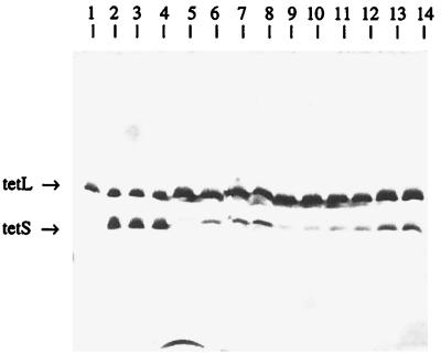
Although the mutant homo-oligomer L332V/F341L/L344I was not destabilized relative to human p53tet, the 12 core residues substituted in the tetramer could lead to impaired hetero-oligomerization with nonmutated human p53tet (see Discussion). We have tested the hetero-oligomerization ability of mutants L332V/F341L/L344I and L332V/M340I/F341L with human p53tet (Fig. 4) in subunit-exchange assays (refs. 26 and 36; see Materials and Methods). Human His-tetL and either human tetS or the mutant L332V/M340I/F341L did quantitatively hetero-oligomerize. In contrast, the mutant L332V/F341L/L344I showed clearly impaired hetero-oligomerization with human p53tet (Fig. 4). A rough estimate suggested that about 5–10 times more protein from this mutant was required to achieve hetero-oligomerization to the same extent as that achieved with the wild type. Although hetero-oligomerization was not completely abolished, this result shows that careful engineering of p53tet can lead to decreased hetero-oligomerization with wild-type p53 without undesirable effects on the thermodynamic stability and oligomerization affinity of the homo-oligomer.
Figure 4.
Analysis of hetero-oligomerization between p53tet mutants and wild-type human p53tet. The second procedure described in Materials and Methods was followed. A total of 2 nmol of His-tagged tetL were incubated either alone (lane 1) or with nontagged wild-type tetS (lane 2), mutant L332V/F341L/L344I (lane 3), or L332V/M340I/F341L (lane 4). The His-tagged homo-oligomers and the hetero-oligomers formed were separated from the remaining nontagged homo-oligomers by specific binding to Ni-NTA beads followed by elution with imidazole. Lanes 5–14, eluates containing His-tagged homo-oligomers and hetero-oligomers (if applicable) from tetL alone (lane 5) or mixtures of tetL with wild-type tetS (lanes 6–8), L332V/F341L/L344I (lanes 9–11), or L332V/M340I/F341L (lanes 12–14). TetL:tetS (wild-type or mutants) molar ratios in the corresponding incubation mixtures were 1:0.5 (lanes 6, 9, and 12), 1:1 (lanes 7, 10, and 13), or 1:2 (lanes 2–4, 8, 11, and 14). The conditions for the experiment were set up by using the alternative procedure described in Materials and Methods. In control samples with individual proteins, no His-tagged tetL was found in the washing fractions, and no untagged wild-type or mutant tet were found in the fractions eluted. Similar results consistent with those shown here were obtained in repeated experiments.
DISCUSSION
Are p53-Like Proteins Tetrameric?
It is still unclear whether human p73 and p51 and other p53-like proteins are tetramers. High sequence identity with human p53 (higher than 35%) is consistent with an ability to oligomerize, and there is experimental evidence which suggests that Xenopus p53 and human p73 are oligomers (31, 39). The chimeric tet domains analyzed here included the residues found in p53-like proteins at positions equivalent in human p53 to those critical for stability and located at the interfaces. Thus, the tetrameric status and stability of these chimeric domains favor the possibility of p51 and p73 and most p53-like proteins sequenced to date being also tetrameric. However, definitive proof must await direct analysis of the unmodified natural proteins.
Naturally Occurring “Size-Swap” Mutations Do Not Preserve p53tet Stability.
Comparison of the hydrophobic cores of human and trout p53tet provides an extreme example derived from nature which emphasizes the importance of structural context and core packing complementarity in modulating the effect of amino acid substitutions on protein stability. Despite preserving core size and hydrophobicity, the eight central core residues replaced in the tetrameric variant F341L/L344F were strongly destabilizing. Although these side chains contacted each other (Fig. 1), the replacements acted additively. Molecular modeling suggested that good packing would be difficult to achieve. The hydrophobic cores of trout and Xenopus p53tet were found substantially destabilized, but those of zebrafish and clam appeared to be little destabilized. Thus, core stability has not been kept invariant during p53tet evolution, but no evolutionary trend toward improved stability was apparent. It should be noted, however, that the effect of the natural differences found in surface-exposed residues was not analyzed, and their combined effect might be important in modulating the overall stability. Individual truncation to Ala of most surface-exposed side chains in human p53tet had a relatively minor, but not negligible, effect on stability (30).
Compensatory Effects During Evolution of the p53tet Hydrophobic Core.
It has been proposed that the occurrence of compensatory mutations plays a role in the molecular evolution of proteins (1, 17). Statistical sequence analyses suggest that covariation of mutations within proteins may be a relatively common, if not widespread, occurrence (40, 41). The present study provides a dramatic example of mutually compensatory effects during the natural evolution of the hydrophobic core of a protein domain. The side chains found most critical for human p53tet stability were those of Leu-330, Ile-332, and Phe-341, which formed the central minicores within each of the primary dimers in the tetramer (Fig. 1). These residues were conserved in all 12 mammalian p53 analyzed. In contrast, human p73 and p51, rat KET, and eight nonmammalian p53-like proteins (all those sequenced except Xenopus and trout p53) had Val and Leu at positions 332 and 341, respectively. Sequence comparisons suggested that human p73 and p51, and p53-like proteins from lower eukaryotes, may be more closely related to a putative ancestral protein, whereas p53 from upper vertebrates may have arisen by gene duplication and divergent evolution (31, 35). Thus, it is tempting to speculate that a compensatory double mutation V332I/L341F occurred late in evolution and gave rise to mammalian p53 with the same or even improved stability but with a different core packing when compared with ancestral proteins more related to p73 and p51. Other natural differences like those at residue 344 may have modulated the above effects. In summary, variation within the tet core of p53-related proteins appears to be centered on the most critical positions regarding oligomerization and stability, namely 332, 341, and 344. Evolution toward mammalian p53tet with a different core packing may have entailed compensatory substitutions at these positions which, instead of being detrimental for stability, may have preserved or even improved stability.
Design of a Mutant p53 with Impaired Hetero-Oligomerization.
An engineered human p53 incorporating the chimeric tet domain L332V/F341L/L344I described here may show impaired hetero-oligomerization with inactive p53 from tumor cells and provide a step in the development of anticancer gene therapy based on p53. Our rationale was to hypothesize, from the modeling of natural amino acid differences found at key positions within the tet core, that some multiple mutations during evolution of p53tet might have preserved stability, but not hydrophobic core packing specificity. The results indicate that both assumptions were correct in particular instances, especially with L332V/F341L/L344I. Potential problems with this engineered domain are that hetero-oligomerization, though clearly reduced, was not drastically impaired. This could still allow sequestration of the engineered p53 by high levels of inactive p53 within cancer cells. Also, the tet core sequence is a step closer to that of human p51 and p73. Thus, the engineered p53 might display a somewhat increased hetero-oligomerization with those proteins, whose function is unclear. Evaluation of such possibilities must await experiments with full-length p53 in biological assays. The advantages of our design relative to approaches based on the use of an engineered p53 with either an entirely different oligomerization domain (42) or drastic substitutions at the tet interfaces are that (i), the engineered tet sequence is very close to the human sequence, and the surface residues identical; this may be most important if the tet domain participates in p53 functions other than oligomerization, like interdomain or intermolecular recognition; and (ii) the stability and homo-oligomerization ability of the triple mutant are not reduced at all relative to human p53. Xenopus p53 also showed poor ability to hetero-oligomerize with human p53 (39), but this may be just a consequence of reduced stability and homo-oligomerization ability of Xenopus p53tet compared with the human domain (see Results; ref. 39). Our design may be also considered a first stage to try and further decrease p53 hetero-oligomerization by either a rational approach or random mutation and selection.
Acknowledgments
We thank Drs. M. Bycroft and J. L. Neira for scientific discussions. This work was supported by the Cancer Research Campaign of the United Kingdom. M.G.M. was supported by a grant from the European Union.
References
- 1.Kimura M. J Genet. 1985;64:7–19. [Google Scholar]
- 2.Wells J A. Biochemistry. 1990;37:8509–8517. doi: 10.1021/bi00489a001. [DOI] [PubMed] [Google Scholar]
- 3.Sandberg W S, Terwilliger T C. Proc Natl Acad Sci USA. 1991;88:1706–1710. doi: 10.1073/pnas.88.5.1706. [DOI] [PMC free article] [PubMed] [Google Scholar]
- 4.Gregoret L M, Sauer R T. Proc Natl Acad Sci USA. 1993;90:4246–4250. doi: 10.1073/pnas.90.9.4246. [DOI] [PMC free article] [PubMed] [Google Scholar]
- 5.Serrano L, Day A G, Fersht A R. J Mol Biol. 1993;233:305–312. doi: 10.1006/jmbi.1993.1508. [DOI] [PubMed] [Google Scholar]
- 6.Horovitz A, Serrano L, Avron B, Bycroft M, Fersht A. J Mol Biol. 1990;216:1031–1044. doi: 10.1016/S0022-2836(99)80018-7. [DOI] [PubMed] [Google Scholar]
- 7.Lim W A, Sauer R T. Nature (London) 1989;339:31–36. doi: 10.1038/339031a0. [DOI] [PubMed] [Google Scholar]
- 8.Lim W A, Sauer R T. J Mol Biol. 1991;219:359–376. doi: 10.1016/0022-2836(91)90570-v. [DOI] [PubMed] [Google Scholar]
- 9.Daopin S, Alber T, Baase W A, Wozniak J A, Matthews B W. J Mol Biol. 1991;221:647–667. doi: 10.1016/0022-2836(91)80079-a. [DOI] [PubMed] [Google Scholar]
- 10.Hurley J H, Baase W A, Matthews B W. J Mol Biol. 1992;224:1143–1159. doi: 10.1016/0022-2836(92)90475-y. [DOI] [PubMed] [Google Scholar]
- 11.Lim W A, Farruggio D C, Sauer R T. Biochemistry. 1992;31:4324–4333. doi: 10.1021/bi00132a025. [DOI] [PubMed] [Google Scholar]
- 12.Green S M, Shortle D. Biochemistry. 1993;32:10131–10139. doi: 10.1021/bi00089a032. [DOI] [PubMed] [Google Scholar]
- 13.Baldwin E P, Hajiseyedjavadi O, Baase W A, Matthews B W. Science. 1993;262:1715–1718. doi: 10.1126/science.8259514. [DOI] [PubMed] [Google Scholar]
- 14.Baldwin E, Xu J, Hajiseyedjavadi O, Baase W A, Matthews B W. J Mol Biol. 1996;259:542–559. doi: 10.1006/jmbi.1996.0338. [DOI] [PubMed] [Google Scholar]
- 15.Malcolm B A, Wilson K P, Matthews B W, Kirsch J F, Wilson A C. Nature (London) 1990;345:86–89. doi: 10.1038/345086a0. [DOI] [PubMed] [Google Scholar]
- 16.Wilson K P, Malcolm B A, Matthews B W. J Biol Chem. 1992;267:10842–10849. [PubMed] [Google Scholar]
- 17.Kimura M. Proc Natl Acad Sci USA. 1991;88:5969–5973. doi: 10.1073/pnas.88.14.5969. [DOI] [PMC free article] [PubMed] [Google Scholar]
- 18.Picksley S M, Lane D P. Curr Opin Cell Biol. 1994;6:853–858. doi: 10.1016/0955-0674(94)90056-6. [DOI] [PubMed] [Google Scholar]
- 19.Arrowsmith C H, Morin P. Oncogene. 1996;12:1379–1385. [PubMed] [Google Scholar]
- 20.Levine A J. Cell. 1997;88:323–331. doi: 10.1016/s0092-8674(00)81871-1. [DOI] [PubMed] [Google Scholar]
- 21.Hollstein M, Sidransy D, Vogelstein B, Harris C C. Science. 1991;253:49–53. doi: 10.1126/science.1905840. [DOI] [PubMed] [Google Scholar]
- 22.Shaulian E, Zauberman A, Ginsberg D, Oren M. Mol Cell Biol. 1992;12:5581–5592. doi: 10.1128/mcb.12.12.5581. [DOI] [PMC free article] [PubMed] [Google Scholar]
- 23.Harvey M, Vogel H, Morris D, Bradley A, Bernstein A, Donehower L A. Nat Genet. 1995;9:305–311. doi: 10.1038/ng0395-305. [DOI] [PubMed] [Google Scholar]
- 24.Hann B C, Lane D P. Nat Genet. 1995;9:221–222. doi: 10.1038/ng0395-221. [DOI] [PubMed] [Google Scholar]
- 25.Beaudry G A, Bertelsen A H, Sherman M I. Curr Opin Biotechnol. 1996;7:592–600. doi: 10.1016/s0958-1669(96)80069-3. [DOI] [PubMed] [Google Scholar]
- 26.Lee W, Harvey T S, Yin Y, Yau P, Litchfield D, Arrowsmith C H. Nat Struct Biol. 1994;1:877–890. doi: 10.1038/nsb1294-877. [DOI] [PubMed] [Google Scholar]
- 27.Jeffrey P D, Gorina S, Pavletich N P. Science. 1995;267:1498–1502. doi: 10.1126/science.7878469. [DOI] [PubMed] [Google Scholar]
- 28.Clore G M, Ernst J, Clubb R, Omichinski J G, Kennedy W M P, Sakaguchi K, Appella E, Gronenborg A. Nat Struct Biol. 1995;2:321–333. doi: 10.1038/nsb0495-321. [DOI] [PubMed] [Google Scholar]
- 29.Johnson C R, Morin P E, Arrowsmith C H, Freire E. Biochemistry. 1995;34:5309–5316. doi: 10.1021/bi00016a002. [DOI] [PubMed] [Google Scholar]
- 30.Mateu M G, Fersht A R. EMBO J. 1998;17:2748–2758. doi: 10.1093/emboj/17.10.2748. [DOI] [PMC free article] [PubMed] [Google Scholar]
- 31.Kaghad M, Bonnet H, Yang A, Creancier L, Biscan J-C, Valent A, Minty A, Chalon P, Lelias J-M, Dumont X, et al. Cell. 1997;90:809–819. doi: 10.1016/s0092-8674(00)80540-1. [DOI] [PubMed] [Google Scholar]
- 32.Osada M, Ohba M, Kawahara C, Ishioka C, Kanamaru R, Katoh I, Ikawa Y. Nat Med. 1998;4:839–843. doi: 10.1038/nm0798-839. [DOI] [PubMed] [Google Scholar]
- 33.Trink B, Okami K, Wu L, Sriuranpong V, Jen J, Sidransky D. Nat Med. 1998;4:747–748. doi: 10.1038/nm0798-747. [DOI] [PubMed] [Google Scholar]
- 34.Soussi T, May P. J Mol Biol. 1996;260:623–637. doi: 10.1006/jmbi.1996.0425. [DOI] [PubMed] [Google Scholar]
- 35.Schmale H, Bamberger C. Oncogene. 1997;15:1363–1367. doi: 10.1038/sj.onc.1201500. [DOI] [PubMed] [Google Scholar]
- 36.Davison T S, Yin P, Nie E, Kay C, Arrowsmith C H. Oncogene. 1998;17:651–656. doi: 10.1038/sj.onc.1202062. [DOI] [PubMed] [Google Scholar]
- 37.Sayle R A, Milner-White E J. Trends Biochem Sci. 1995;20:374–376. doi: 10.1016/s0968-0004(00)89080-5. [DOI] [PubMed] [Google Scholar]
- 38.Guex N, Peitsch M C. Electrophoresis. 1997;18:2714–2723. doi: 10.1002/elps.1150181505. [DOI] [PubMed] [Google Scholar]
- 39.Wang Y, Farmer G, Soussi T, Prives C. Oncogene. 1995;10:779–784. [PubMed] [Google Scholar]
- 40.Neher E. Proc Natl Acad Sci USA. 1994;91:98–102. doi: 10.1073/pnas.91.1.98. [DOI] [PMC free article] [PubMed] [Google Scholar]
- 41.Chelvanayagam G, Eggenschwiler A, Knecht L, Gonnet G H, Benner S A. Protein Eng. 1997;10:307–316. doi: 10.1093/protein/10.4.307. [DOI] [PubMed] [Google Scholar]
- 42.Waterman J F, Waterman J L F, Halazonetis T D. Cancer Res. 1996;56:158–163. [PubMed] [Google Scholar]
- 43.Kraulis P J. J Appl Crystallogr. 1991;24:946–950. [Google Scholar]


