Abstract
Inositol polyphosphate 4-phosphatase (4-phosphatase) is an enzyme that catalyses the hydrolysis of the 4-position phosphate from phosphatidylinositol 3,4-bisphosphate [PtdIns(3,4)P2]. In human platelets the formation of this phosphatidylinositol, by the actions of phosphatidylinositol 3-kinase (PI 3-kinase), correlates with irreversible platelet aggregation. We have shown previously that a phosphatidylinositol 3,4,5-trisphosphate 5-phosphatase forms a complex with the p85 subunit of PI 3-kinase. In this study we investigated whether PI 3-kinase also forms a complex with the 4-phosphatase in human platelets. Immunoprecipitates of the p85 subunit of PI 3-kinase from human platelet cytosol contained 4-phosphatase enzyme activity and a 104-kDa polypeptide recognized by specific 4-phosphatase antibodies. Similarly, immunoprecipitates made using 4-phosphatase-specific antibodies contained PI 3-kinase enzyme activity and an 85-kDa polypeptide recognized by antibodies to the p85 adapter subunit of PI 3-kinase. After thrombin activation, the 4-phosphatase translocated to the actin cytoskeleton along with PI 3-kinase in an integrin- and aggregation-dependent manner. The majority of the PI 3-kinase/4-phosphatase complex (75%) remained in the cytosolic fraction. We propose that the complex formed between the two enzymes serves to localize the 4-phosphatase to sites of PtdIns(3,4)P2 production.
Following agonist-induced cellular activation, two distinct pathways of phosphatidylinositol (PtdIns) metabolism have been well characterized. Phosphatidylinositol 4,5-bisphosphate [PtdIns(4,5)P2] hydrolysis by phospholipase C produces soluble inositol phosphates and diacylglycerol (1, 2). Phosphatidylinositol 3-kinase (PI 3-kinase) phosphorylates phosphatidylinositol in the D3 position of the inositol ring, forming phosphatidylinositol 3-phosphate (PtdIns3-P), phosphatidylinositol 3,4-bisphosphate [PtdIns(3,4)P2], and phosphatidylinositol 3,4,5-trisphosphate [PtdIns(3,4,5)P3], respectively (reviewed in refs. 3 and 4). Downstream targets of PtdIns(3,4,5)P3 and PtdIns(3,4)P2 include various isoforms of protein kinase C and the serine/threonine kinase Akt (5, 6). The activation of Akt via these 3-position polyphosphatidylinositols serves to regulate apoptosis and, thereby, cell survival (7, 8).
Thrombin stimulation of platelets results in a rapid transient formation of PtdIns(3,4,5)P3 and PtdIns(3,4)P2 (9–12). After platelet aggregation a sustained, delayed accumulation of PtdIns(3,4)P2 occurs, which is mediated by wortmannin-inhibitable generation of PtdIns3-P, followed by phosphorylation by a specific PtdIns3-P 4-kinase (13–15). This pathway requires activation of the calcium-dependent protease calpain. The production of PtdIns(3,4,5)P3 and PtdIns(3,4)P2 correlates with the translocation of PI 3-kinase to the platelet actin cytoskeleton and an increase in PI 3-kinase enzyme activity (12, 16).
Inositol polyphosphate 4-phosphatase (4-phosphatase) is a 105-kDa Mg2+-independent enzyme that hydrolyzes the 4-position phosphate from the inositol ring of inositol 1,3,4- trisphosphate [Ins(1,3,4)P3], inositol 3,4-bisphosphate [Ins(3,4)P2], and PtdIns(3,4)P2 (17, 18). Kinetic analysis suggests that the phospholipid is the preferred substrate. Two cDNAs encoding 4-phosphatases types I and II have been cloned that predict proteins with 37% amino acid identity (19, 20). The enzymatic properties of the isoforms are similar, and both 4-phosphatases are widely expressed. A role for calpain-mediated inactivation of the 4-phosphatase in the calcium- and aggregation-dependent accumulation of PtdIns(3,4)P2 in thrombin-activated platelets has been demonstrated recently (21).
It was shown previously that an inositol polyphosphate 5-phosphatase forms a complex with the p85 subunit of PI 3-kinase (22). The p85-associated 5-phosphatase has a unique substrate specificity, in that the enzyme only hydrolyses PtdIns(3,4,5)P3 to form PtdIns(3,4)P2. Liu et al. (23) have also demonstrated a similar 5-phosphatase, constitutively associated with the p85 subunit of PI 3-kinase, which forms a complex with the IL-3 receptor (23). In this report, we show that analogous to the p85-associated 5-phosphatase, the 4-phosphatase I also associates with the p85 subunit of the PI 3-kinase in vivo in human platelet cytosol and, thereby, may enable localization of the 4-phosphatase to sites of PtdIns(3,4)P2 production.
EXPERIMENTAL PROCEDURES
Platelet Subcellular Fractionation.
Human platelets were obtained and washed, and subcellular fractions were isolated as described (24). In experiments determining Ins(1,3,4)P3 4-phosphatase activity in the cytoplasmic actin cytoskeleton, the washed cytoskeletal pellet was resuspended in 20 mM Tris, pH 7.2/150 mM NaCl.
Affinity Capture of PI 3-Kinase.
The peptide (Y751) CDESVDYPVPML (where Yp represents phosphotyrosine), which corresponds to the binding site on the platelet-derived growth factor β receptor for the p85 subunit of PI 3-kinase, was coupled to Actigel Superflow resin (Sterogen, Arcadia, CA), as described (25). Sixty microliters of the Y751 peptide-coupled resin was mixed with 600 μl of platelet cytosol overnight at 4°C. The resin was pelleted and washed either three times with 1 ml of low-stringency buffer (20 mM Tris, pH 7.2/5 mM MgCl2/1 mM EDTA) followed by 1 ml × three times with kinase assay buffer (20 mM Hepes, pH 7.4/1 mM EGTA/5 mM MgCl2) or three times with 1 ml of high-stringency buffer (10 mM Tris, pH 7.2/0.5 M NaCl/1 mM EDTA/1% NP-40/0.1% SDS), followed by three times with 1 ml of kinase assay buffer.
Antibodies.
The C-terminal 4-phosphatase antipeptide antibody was developed as described (19). The N-terminal 4-phosphatase antipeptide antibody was raised against amino acids 1–16 MTAREHSPRHGARAR in the type I 4-phosphatase. The peptide was coupled to keyhole limpet hemocyanin with N-hydroxysuccinimide ester, and antigen was injected into rabbits by Pocono Rabbit Farm (Canadensis, PA).
Immunoprecipitation of Inositol Polyphosphate 4-Phosphatase.
Five microliters of polyclonal antisera directed to either an N-terminal or C-terminal type I 4-phosphatase peptide was incubated overnight at 4°C with 600 μl of human platelet cytosol (5 mg/ml) and 60 μl of a 50% slurry of protein A-Sepharose. Pelleted immunoprecipitates were washed with 1 ml × 6 of 20 mM Tris, pH 7.4/150 mM NaCl.
Immunoprecipitation of the p85 Subunit of the PI 3-Kinase.
The p85 subunit of PI 3-kinase was immunoprecipitated from human platelet cytosol by using 5 μl of polyclonal antibody to the p85 subunit (Upstate Biotechnology, Lake Placid, NY). The conditions for immunoprecipitation were as those for the 4-phosphatase.
Inositol Polyphosphate 4-Phosphatase Assay.
The immunoprecipitates or immunodepleted samples (2, 3, 5, or 10 μl) were incubated with [3H]Ins(1,3,4)P3 (16 μM, 16,680 cpm/nmol) in 100 μl 50 mM Mes, pH 6.5/5 mM EDTA at 37°C for 15 min, and products of the reaction were analyzed as described (18).
PI 3-Kinase Assay.
Platelet PI 3-kinase captured on Actigel Superflow resin or protein A-Sepharose pellets with associated anti-p85 or anti-4-phosphatase immune complexes were washed three times with ice-cold 20 mM Tris, pH 7.4/150 mM NaCl/1 mM sodium orthovanadate/250 μg/ml phenylmethylsulfonyl fluoride, followed by three washes with ice-cold kinase assay buffer (20 mM Hepes, pH 7.4/1 mM EGTA/5 mM MgCl2). Immunoprecipitates were resuspended in kinase assay buffer and mixed with either sonicated PtdIns (200 μM) and PtdSer (300 μM), or PtdIns 4-P (200 μM) and PtdSer (300 μM), and [γ-32P]ATP (50 μM, 1 μCi/nmol) to a final reaction volume of 100 μl. The reactions were stopped after 20 min at room temperature, lipid products were analyzed by TLC (22), and the identity of deacylated lipid products was confirmed as described (26).
RESULTS AND DISCUSSION
PtdIns(3,4)P2 4-Phosphatase Associates with PI 3-Kinase in Human Platelet Cytosol.
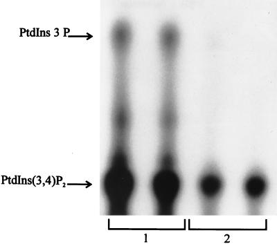
We have shown previously that a PtdIns(3,4,5)P3 5-phosphatase forms a stable complex with the p85 subunit of PI 3-kinase (22). We investigated whether the 4-phosphatase, which dephosphorylates the 4-position phosphate from PtdIns (3,4)P2, forming PtdIns3-P, also associates with the p85 subunit of PI-3-kinase. The phosphorylated peptide (CDESVDYPVPML), which represents the p85-binding domain of the platelet-derived growth factor β receptor, was coupled to Actagel Superflow resin, and PI 3-kinase was affinity-captured from platelet cytosol. PI 3-kinase assays were performed with affinity-captured enzyme, using PtdIns 4-P as substrate (Fig. 1). After washing of the resin with low-stringency buffer (20 mM Tris, pH 7.4/150 mM NaCl), two lipid reaction products, PtdIns (3,4)P2 and PtdIns3-P, were observed. In this assay, PtdIns(3,4)P2 is synthesized by the affinity-captured PI 3-kinase and subsequently is hydrolyzed to PtdIns3-P by a PtdIns(3,4)P2 4-phosphatase, which complexes with the kinase. Disassociation of the PtdIns(3,4)P2 4-phosphatase/PI 3-kinase complex was achieved after washing the peptide affinity resin with 10 mM Tris, pH 7.2/0.5 M NaCl/1 mM EDTA/1% NP-40/0.1% SDS. PI 3-kinase activity still was demonstrated by the observed production of PtdIns(3,4)P2, but no PtdIns(3,4)P2 4-phosphatase activity was detected. These studies suggest PtdIns(3,4)P2 4-phosphatase activity is not intrinsic to PI 3-kinase, but may complex with either the p85 adapter or p110 catalytic subunit of PI 3-kinase, or with another protein that, in turn, binds to p85 or p110.
Figure 1.
PI 3-kinase was affinity-purified from platelet cytosol. A peptide (Y751) corresponding to the p85-binding site on the platelet-derived growth factor receptor was coupled to Actigel Superflow resin. Six hundred microliters of human platelet cytosol (12 mg/ml) was incubated with 60 μl of a 50% slurry of the peptide-coupled resin. The resin was pelleted and washed with either (1) low-stringency or (2) high-stringency buffer. PI 3-kinase assays were performed on washed pellets in duplicate by using PtdIns 4-P as substrate.
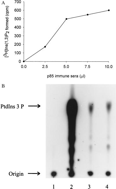
Two isoforms of 4-phosphatases (types I and II) have been identified (19, 20). Both isoforms hydrolyze Ins(1,3,4)P3, Ins(3, 4)P2, and PtdIns(3,4)P2, forming inositol 1,3-bisphosphate [Ins(1,3)P2], inositol 3-phosphate, and PtdIns3-P, respectively. To further characterize the PtdIns(3,4)P2 4-phosphatase that complexes with PI-3 kinase, increasing amounts of immune serum to the p85 subunit of the PI-3-kinase were incubated with platelet cytosol, and immune complexes were captured on protein A-Sepharose. Ins(1,3,4)P3 4-phosphatase activity was determined in both the supernatant and pellet of the immunoprecipitations. A dose-dependent increase in Ins(1,3,4)P3 4-phosphatase activity was observed in protein A-Sepharose pellets, with increasing amounts of antiserum to the p85 subunit of PI 3-kinase (Fig. 2A). Loss of Ins(1,3,4)P3 4-phosphatase activity was observed in the supernatant of immunoprecipitation reactions (results not shown). These studies demonstrate that the PtdIns(3,4)P2 4-phosphatase, which complexes with PI 3-kinase, also hydrolyses Ins(1,3,4)P3 and, therefore, may represent a previously characterized 4-phosphatase.
Figure 2.
(A) Four hundred microliters of platelet cytosol (9 mg/ml) was incubated with the indicated volume of p85 antibody overnight at 4°C. Each immunoprecipitation was made to contain a total of 10 μl of serum using nonimmune serum. Immunoprecipitates were captured on protein A-Sepharose and washed. 4-Phosphatase enzyme activity was determined by measuring the hydrolysis of [3H]Ins(1,3,4)P3 to form [3H]Ins(1,3)P2. (B) One milliliter of platelet cytosol was immunoprecipitated with (1) 5 μl nonimmune serum, (2) 5 μl of p85 antiserum, (3) 5 μl of antiserum to the N terminus of the 4-phosphatase, or (4) 5 μl of the antiserum to the C terminus of the 4-phosphatase. Washed immunoprecipitates were assayed for PI 3-kinase activity by using PtdIns as a substrate.
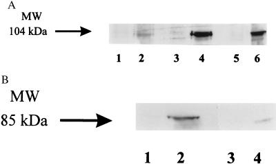
To support this contention, PI 3-kinase enzyme activity was determined in 4-phosphatase immunoprecipitates. Antipeptide antibodies to the N-terminal region of the type I 4-phosphatase recognize only the type I 4-phosphatase, whereas antibodies to the C-terminal peptide of the type I 4-phosphatase recognize both the type I and II isoenzymes (19). Similar PI 3-kinase enzyme activity was detected by using both N- and C-terminal 4-phosphatase antipeptide antibodies (Fig. 2B). That the N-terminal antibody is specific for the type I 4-phosphatase implies it is the predominant 4-phosphatase isoform associating with the PI 3-kinase. Immunoprecipitation of PtdIns 3-kinase activity was much less (1.14%, SD ± 0.12%, n = 5) in 4-phosphatase compared with p85 immunoprecipitations. However, we cannot exclude that the 4-phosphatase antibody sterically inhibits PI 3-kinase enzyme activity under the assay conditions used or that complexing with 4-phosphatase reduces its activity. Western blot analysis of p85 or 4-phosphatase immunoprecipitates was undertaken by using antibodies to either the phosphatase or kinase, respectively. Immunoblot analysis of p85 immunoprecipitates demonstrated a 104-kDa polypeptide detected by 4-phosphatase antibodies, consistent with the presence of the type I 4-phosphatase in a complex with the p85 subunit of the PI 3-kinase (Fig. 3A). The 4-phosphatase immunoprecipitated by p85 antibodies represented approximately 10% of that observed in 4-phosphatase immunoprecipitates, as determined by densitometric analysis. Western blot analysis of 4-phosphatase immunoprecipitates using p85 antiserum demonstrated the presence of an 85-kDa polypeptide, which comigrated with immunoprecipitated PI 3-kinase (Fig. 3B). Densitometric analysis of 4-phosphatase immunoprecipitates revealed that the p85 detected represented 13% of that observed in p85 immunoprecipitations.
Figure 3.
(A) Six hundred microliters of platelet cytosol (9 mg/ml) was immunoprecipitated with 5 μl of nonimmune serum (lanes 1, 3, and 5), 5 μl of p85 polyclonal antibody (lane 2), 5 μl of C-terminal 4-phosphatase antipeptide antibody (lane 4), or 5 μl of N-terminal 4-phosphatase antibody (lane 6) and immunoblotted with 4-phosphatase antiserum (C-terminal antibody). (B) Six hundred microliters of platelet cytosol (9 mg/ml) was immunoprecipitated with 5 μl of nonimmune sera (lanes 1 and 3), 5 μl of p85 antiserum (lane 2), or 5 μl of antiserum to the N-terminal 4-phosphatase and immunoblotted by using p85 antiserum.
Translocation of 4-Phosphatase to the Thrombin-Activated Cytoskeleton.
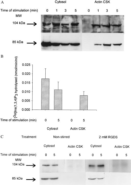
Thrombin stimulation of human platelets results in a relocation of the p85 subunit of PI 3-kinase to the cytoskeleton, which correlates with the formation of PtdIns(3,4)P2 and irreversible platelet aggregation (9, 10, 12, 16). We investigated whether the 4-phosphatase translocated to the actin cytoskeleton of thrombin-activated platelets. No 4-phosphatase was present in the actin cytoskeleton of resting platelets. However, a time-dependent translocation of 4-phosphatase to the cytoskeleton of thrombin-stimulated platelets was observed, which temporally correlated with the translocation of the p85 subunit of PI 3-kinase (Fig. 4A). Densitometric analysis of the intensity of the immunoreactive 104-kDa 4-phosphatase in the cytosol of nonstimulated platelets compared with the actin cytoskeleton after 5-min thrombin activation demonstrated that 15% (SD ± 2.5%, n = 5) of the total cytosolic 4-phosphatase translocated. Similar analysis of p85 translocation under the same conditions revealed that 36% (SD ± 8.6%, n = 8) of the enzyme accumulated in the thrombin-activated cytoskeleton, consistent with previous reports (12). Cytoskeletal translocation of the 4-phosphatase also was shown by Ins(1,3,4)P3 4-phosphatase activity in the cytoskeleton of thrombin-activated, but not resting, platelets (Fig. 4B). The specific activity of cytoskeletal vs. cytosolic 4-phosphatase was determined by densitometric analysis of immunoreactive 4-phosphatase in subcellular fractions per unit of enzyme activity and showed the enzyme was activated 3-fold in the cytoskeletal fraction, as shown in Table 1. Translocation of the 4-phosphatase to the actin cytoskeleton depended on both platelet aggregation and integrin engagement, because 4-phosphatase relocation was not observed when the platelets were not stirred or were preincubated with RGDS peptide (Fig. 4C).
Figure 4.
(A) Washed platelets were stimulated with thrombin (1 unit/ml) for the indicated times, and cytosol and actin cytoskeleton (actin CSK) fractions were isolated. Thirty microliters of each fraction was analyzed by SDS/PAGE and immunoblot analysis using either antibodies to the 4-phosphatase (Upper) or anti-p85 antibodies (Lower). (B) Platelet cytosol or actin CSK (1, 2.5, and 5 μl) was assayed for 4-phosphatase enzyme activity, the hydrolysis of [3H]Ins(1,3,4)P3 to form [3H]Ins(1, 3)P2. (C) Platelets were stimulated with thrombin (1 unit/ml) with or without stirring. As indicated, platelets were incubated with 2 mM RGDS for 10 min before thrombin stimulation and then analyzed as for A.
Table 1.
Subcellular localization of 4-phosphatase
| Nonstimulated
|
Thrombin-stimulated
|
|||
|---|---|---|---|---|
| Cytosol | CSK | Cytosol | CSK | |
| Enzyme activity, [3H]Ins(1,3,4)P3 hydrolyzed pmol/min per ml | 17 ± 6 (n = 5) | 0 | 11 ± 4 (n = 5) | 8 ± 2 (n = 3) |
| Densitometry, arbitrary units | 1 | 0 | 0.76 ± 0.015 (n = 3) | 0.15 ± 0.025 (n = 5) |
| Specific activity, enzyme activity per unit densitometry | 17 | 0 | 14 | 53 |
4-Phosphatase activity was determined in the cytosol and cytoskeleton (CSK) of nonstimulated platelets and platelets thrombin-stimulated for 5 min. Densitometric analysis was performed on 4-phosphatase immunoblots of the same platelet fractions, and the specific 4-phosphatase enzyme activity per unit protein was determined.
A 36% (SD ± 6.8, n = 5) decrease in cytosolic Ins(1,3,4)P3 4-phosphatase activity was routinely observed after thrombin stimulation. However, this decrease in enzyme activity does not result only from 4-phosphatase translocation to the actin cytoskeleton. Rather, previous studies have shown platelet activation with calcium ionophore or thrombin results in calpain-mediated proteolysis of 4-phosphatase (21).
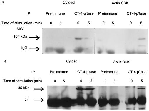
We have observed previously that the PtdIns(3,4,5)P3 5-phosphatase that forms a complex with the p85 subunit of PI 3-kinase remains in the cytosolic fraction after thrombin stimulation, whereas the p85 subunit translocates to the activated actin cytoskeleton (22). Complex formation between the 4-phosphatase and PI 3-kinase was determined in the cytosol and actin cytoskeleton of resting and thrombin-stimulated platelets. Platelet subcellular fractions were immunoprecipitated by using antibodies to the 4-phosphatase, captured on protein A-Sepharose, and immunoblotted using PI 3-kinase antiserum. The PI 3-kinase/4-phosphatase complex remained stable in the cytosolic fraction of thrombin-activated platelets (Fig. 5). A small decrease (23%, n = 4) in the amount of complex between the two enzymes was determined by densitometric analysis of p85 immunoblots of 4-phosphatase immunoprecipitates in the cytosol after thrombin stimulation, compared with resting cells. In the actin cytoskeleton of unstimulated platelets, no PI 3-kinase/4-phosphatase complex was detected. However, after thrombin activation, the PI 3-kinase/4-phosphatase complex was observed in the thrombin-activated actin cytoskeleton.
Figure 5.
One milliliter of cytosol or actin cytoskeleton (CSK) from resting or thrombin-stimulated platelets (1 × 10⁹/ml platelets) was immunoprecipitated with 5 μl of nonimmune serum or C-terminal 4-phosphatase antibody and immunoblotted with 4-phosphatase antiserum (A) or p85 antiserum (B).
In human platelets there is a rapid, early formation of PtdIns(3,4,5)P3 and PtdIns(3,4)P2, but not PtdIns3-P, after thrombin stimulation, which probably is generated by PI 3-kinase phosphorylation of PtdIns(4,5)P2 and PtdIns 4-P. Dephosphorylation of PtdIns(3,4,5)P3 by inositol polyphosphate 5-phosphatases also can form PtdIns(3,4)P2 (2, 9, 11, 12). Aggregation-dependent formation of PtdIns(3,4)P2 recently has been described that is generated by the action of PtdIns3-P-4-kinase (13–15). Phosphatidylinositol phosphate 5-kinases (types I and II) phosphorylate PtdIns3-P in the 4-position, forming PtdIns(3,4)P2 (27). Calpain-mediated proteolysis of 4-phosphatase also is responsible for calcium- and aggregation-dependent accumulation of PtdIns(3,4)P2 in thrombin-stimulated platelets (21).
In this study we have demonstrated that 4-phosphatase is associated with PI 3-kinase in the cytosolic fraction of nonstimulated platelets and in the thrombin-activated cytoskeleton. We propose that this association serves to localize the phosphatase to sites of PtdIns(3,4)P2 production. The mechanism mediating association between the 4-phosphatase and PI 3-kinase has yet to be determined. A complex between the kinase and phosphatase may be mediated by the p85 SH3 domain associating with proline-rich motifs present in the 4-phosphatase and is the subject of current investigation in the laboratory.
Using densitometry of the amount of 4-phosphatase in p85 immunoprecipitates compared with 4-phosphatase immunoprecipitates, approximately 10% of the cytosolic 4-phosphatase is in complex with the p85 subunit of the PI 3-kinase. This association appears functionally significant, because we always demonstrate hydrolysis of the 4-position phosphate of PtdIns(3,4)P2 in affinity-purified PI 3-kinase preparations or in PI 3-kinase derived from platelet-cytosolic p85 immunoprecipitations.
After thrombin-stimulated platelet activation, both the PI 3-kinase and the 4-phosphatase translocate to the actin cytoskeleton, which requires both integrin engagement and platelet aggregation. The accumulation of 4-phosphatase in the cytoskeleton is associated with enzyme activation. The 3-fold activation of the 4-phosphatase correlates with p85 enzyme activation in the thrombin-activated cytoskeleton. Previous studies have shown that 29% of the cytosolic p85 translocates to the activated cytoskeleton, accompanied by a significant increase in PI 3-kinase activity, PtdIns(3,4)P2 production, cytoskeletal accumulation of pp60c-src, p125FAK, and platelet aggregation (12). The concomitant translocation and activation of 4-phosphatase to the thrombin-activated actin cytoskeleton provides a mechanism for increased hydrolysis of PtdIns(3,4)P2 after its synthesis by the activated PI 3-kinase.
Recent studies have shown that the inositol polyphosphate 4-phosphatases and 5-phosphatases function to negatively regulate signals transmitted by phosphatidylinositols. We have shown that both 4-phosphatase and 5-phosphatase can form a complex with PI 3-kinase. The recruitment of the kinase and phosphatase into a signaling network provides a means for the localized amplification and degradation of phosphatidylinositol signals at critical sites after cellular activation.
Acknowledgments
We thank Dr. Michael Berndt for platelet preparations, Dr. Rudiger Woscholski for helpful suggestions, Dr. Harshal Nandurkur and Cindy O’Malley for venesecting platelet donors, and all blood donors for their contribution. This work was funded by a grant from the National Health and Medical Research Council of Australia (9936645). This research also was supported by National Institutes of Health Grants HL 16634, HL 07088, and HL 55672.
ABBREVIATIONS
- 4-phosphatase
inositol polyphosphate 4-phosphatase
- PtdIns(3
4)P2, phosphatidylinositol 3,4-bisphosphate
- PtdIns3-P
phosphatidylinositol 3-phosphate
- PI 3-kinase
phosphatidylinositol 3-kinase
- Ins(1
3,4)P3, inositol 1,3,4-trisphosphate
- Ins(1
4)P2, inositol 1,4-bisphosphate
- Ins(3
4)P2, inositol 3,4-bisphosphate
References
- 1.Berridge M J. Nature (London) 1993;361:315–325. doi: 10.1038/361315a0. [DOI] [PubMed] [Google Scholar]
- 2.Zhang X, Majerus P W. Semin Cell Dev Biol. 1998;9:153–160. doi: 10.1006/scdb.1997.0220. [DOI] [PubMed] [Google Scholar]
- 3.Fruman D A, Meyers R E, Cantley L C. Annu Rev Biochem. 1998;67:481–507. doi: 10.1146/annurev.biochem.67.1.481. [DOI] [PubMed] [Google Scholar]
- 4.Vanhaesebroek B, Leevers S J, Panayotou G, Waterfield M D. Trends Biochem Sci. 1997;22:267–272. doi: 10.1016/s0968-0004(97)01061-x. [DOI] [PubMed] [Google Scholar]
- 5.Downward J. Curr Opin Cell Biol. 1998;10:262–267. doi: 10.1016/s0955-0674(98)80149-x. [DOI] [PubMed] [Google Scholar]
- 6.Franke T F, Kaplan D R, Cantley L C, Toker A. Science. 1997;275:665–668. doi: 10.1126/science.275.5300.665. [DOI] [PubMed] [Google Scholar]
- 7.Downward J. Science. 1998;279:673–674. doi: 10.1126/science.279.5351.673. [DOI] [PubMed] [Google Scholar]
- 8.Marte B M, Downward J. Trends Biochem Sci. 1997;22:355–358. doi: 10.1016/s0968-0004(97)01097-9. [DOI] [PubMed] [Google Scholar]
- 9.Cunningham T W, Lips D L, Bansal V S, Caldwell K K, Mitchell C A, Majerus P W. J Biol Chem. 1990;265:21676–21683. [PubMed] [Google Scholar]
- 10.Hawkins P J, Jackson T R, Stephens L R. Nature (London) 1992;358:157–159. doi: 10.1038/358157a0. [DOI] [PubMed] [Google Scholar]
- 11.Rittenhouse S E. Blood. 1996;88:4401–4414. [PubMed] [Google Scholar]
- 12.Guinebault C, Payrastre B, Racaudsultan C, Mazarguil H, Breton M, Mauco G, Plantavid M, Chap H. J Cell Biol. 1995;129:831–842. doi: 10.1083/jcb.129.3.831. [DOI] [PMC free article] [PubMed] [Google Scholar]
- 13.Banfic H, Tang X, Batty I H, Downes C P, Chen C, Rittenhouse S E. J Biol Chem. 1998;273:13–16. doi: 10.1074/jbc.273.1.13. [DOI] [PubMed] [Google Scholar]
- 14.Banfic H, Downes C P, Rittenhouse S E. J Biol Chem. 1998;273:11630–11637. doi: 10.1074/jbc.273.19.11630. [DOI] [PubMed] [Google Scholar]
- 15.Zhang J, Banfic H, Straforini F, Tosi L, Volina S, Rittenhouse S E. J Biol Chem. 1998;273:14081–14084. doi: 10.1074/jbc.273.23.14081. [DOI] [PubMed] [Google Scholar]
- 16.Kovacsovics T J, Bachelot C, Toker A, Vlahos C J, Duckworth B, Cantley L C, Hartwig J H. J Biol Chem. 1995;270:11358–11366. doi: 10.1074/jbc.270.19.11358. [DOI] [PubMed] [Google Scholar]
- 17.Bansal V S, Caldwell K K, Majerus P W. J Biol Chem. 1990;265:1806–1811. [PubMed] [Google Scholar]
- 18.Norris F A, Majerus P W. J Biol Chem. 1994;269:8716–8720. [PubMed] [Google Scholar]
- 19.Norris F A, Auethavekiat V, Majerus P W. J Biol Chem. 1995;270:16128–16133. doi: 10.1074/jbc.270.27.16128. [DOI] [PubMed] [Google Scholar]
- 20.Norris F A, Atkins R C, Majerus P W. J Biol Chem. 1997;272:23859–23864. doi: 10.1074/jbc.272.38.23859. [DOI] [PubMed] [Google Scholar]
- 21.Norris F A, Auethavekiat V, Majerus P W. J Biol Chem. 1997;272:10987–10989. doi: 10.1074/jbc.272.17.10987. [DOI] [PubMed] [Google Scholar]
- 22.Jackson S P, Schoenwaelder S M, Matzaris M, Brown S, Mitchell C A. EMBO J. 1995;14:4490–4500. doi: 10.1002/j.1460-2075.1995.tb00128.x. [DOI] [PMC free article] [PubMed] [Google Scholar]
- 23.Liu L, Jefferson A B, Zhang X, Norris F A, Majerus P W, Krystal G. J Biol Chem. 1996;271:29729–29733. doi: 10.1074/jbc.271.47.29729. [DOI] [PubMed] [Google Scholar]
- 24.Zhang J, Fry M J, Waterfield M D, Jaken S, Liao L, Fox J E B, Rittenhouse S E. J Biol Chem. 1992;267:4686–4692. [PubMed] [Google Scholar]
- 25.Fry M J, Panayotou G, Dhand R, Ruiz-Larrea F, Gout I, Nguyen O, Courtneidge S A, Waterfield M D. Biochem J. 1992;288:383–393. doi: 10.1042/bj2880383. [DOI] [PMC free article] [PubMed] [Google Scholar]
- 26.Stephens L R, Hughes K T, Irvine R F. Nature (London) 1991;351:33–39. doi: 10.1038/351033a0. [DOI] [PubMed] [Google Scholar]
- 27.Zhang X, Loijens J C, Boronenkov I V, Parker G J, Norris F A, Chen J, Thum O, Prestwich G D, Majerus P W, Anderson R A. J Biol Chem. 1997;272:17756–17761. doi: 10.1074/jbc.272.28.17756. [DOI] [PubMed] [Google Scholar]


