Abstract
Background
Floating Knee injuries are complex injuries. The type of fractures, soft tissue and associated injuries make this a challenging problem to manage. We present the outcome of these injuries after surgical management.
Methods
29 patients with floating knee injuries were managed over a 3 year period. This was a prospective study were both fractures of the floating knee injury were surgically fixed using different modalities. The associated injuries were managed appropriately. Assessment of the end result was done by the Karlstrom criteria after bony union.
Results
The mechanism of injury was road traffic accident in 27/29 patients. There were 38 associated injuries. 20/29 patients had intramedullary nailing for both fractures. The complications were knee stiffness, foot drop, delayed union of tibia and superficial infection. The bony union time ranged from 15 – 22.5 weeks for femur fractures and 17 – 28 weeks for the tibia. According to the Karlstrom criteria the end results were Excellent – 15, Good – 11, Acceptable – 1 and Poor – 3.
Conclusion
The associated injuries and the type of fracture (open, intra-articular, comminution) are prognostic indicators in the Floating knee. Appropriate management of the associated injuries, intramedullary nailing of both the fractures and post operative rehabilitation are necessary for good final outcome.
Background
The incidence of fractures resulting from motor vehicle accidents is on the rise. As a by-product of the horsepower race, high velocity accidents are now more common. Such accidents produce violent and complex injuries. Frequently, multiple fractures are produced in the same extremity, adding new dimensions to the problems of their management.
Floating Knee is the term applied to the flail knee joint segment resulting from a fracture of the shaft or adjacent metaphysis of the ipsilateral femur and tibia [1]. The fractures range from simple diaphyseal to complex articular types. This complex injury has increased in proportion to population growth, number of motor vehicles on the road, and high speed traffic. Although the exact incidence of the floating knee is not known, it is an uncommon injury. The largest series reported in the literature was of 222 patients over 11 years [2]. This injury is generally caused by high-energy trauma and the trauma to the soft tissues is often extensive. There also may be life-threatening injuries to the head, chest or abdomen and a high incidence of fat embolism. Management of this injury has been variously described in the literature [3-8].
The purpose of our study was to determine the outcome of patients after surgical management of the Floating Knee and identify prognostic factors for this injury.
Methods
This was a prospective study conducted at a tertiary care trauma centre after approval of the Research and Ethics committee over a 3 year period (1998 – 2001). All floating knee injuries that were managed surgically during the study period were included. Children and floating knee injuries managed conservatively were excluded from the study.
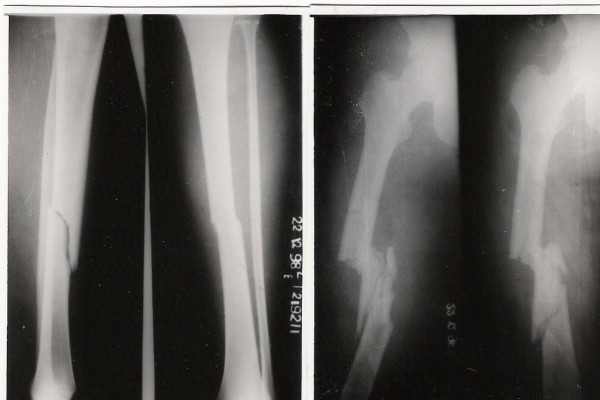
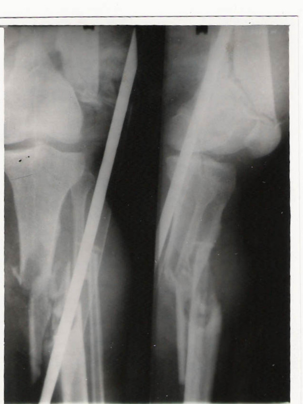
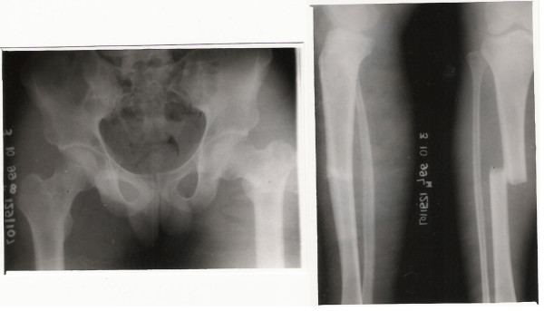
Our study included 29 patients with 29 floating knee injuries, 27 males and 2 females [Table 1]. Initial management involved resuscitation and haemodynamic stabilisation of the patient, splinting of the affected limb in a Thomas splint and a thorough secondary survey to identify other injuries. Radiographs of the chest, pelvis, affected lower limb including all its joints and other suspected bony injuries were done. Open fractures were classified according to Gustilo & Anderson's classification [9]. Initial wound toilet, tetanus immunisation and antibiotic therapy was initiated for open fractures. The floating knee injury was classified according to Blake & McBryde's Classification [10] [Table 2] [Figure 1, 2 &3].
Table 1.
Summary of patient profiles, treatment methods & final outcomes
| Patient No: | Age | Sex | Side | Type | Treatment | Complications | Final Outcome (Karlstrom criteria) |
| 1 | 40 | M | R | 2B | Other | None | Excellent |
| 2 | 19 | M | L | 2B | Other | Infection | Poor |
| 3 | 56 | M | R | 2B | Other | None | Excellent |
| 4 | 27 | M | R | 2A | Other | Delayed union | Poor |
| 5 | 24 | M | R | 1 | Both IM nail | None | Excellent |
| 6 | 42 | M | L | 2A | Other | None | Good |
| 7 | 27 | M | L | 1 | Both IM nail | Foot drop | Acceptable |
| 8 | 24 | M | R | 1 | Both IM nail | None | Excellent |
| 9 | 21 | M | R | 1 | Both IM nail | None | Excellent |
| 10 | 32 | F | L | 2B | Other | Knee stiff | Good |
| 11 | 30 | M | R | 2B | Both IM nail | None | Excellent |
| 12 | 21 | M | L | 1 | Other | Knee stiff | Good |
| 13 | 35 | M | R | 1 | Both IM nail | None | Excellent |
| 14 | 22 | F | R | 1 | Both IM nail | None | Good |
| 15 | 20 | M | L | 1 | Both IM nail | Delayed union | Poor |
| 16 | 22 | M | R | 1 | Both IM nail | Infection | Good |
| 17 | 35 | M | R | 1 | Both IM nail | None | Excellent |
| 18 | 19 | M | R | 1 | Both IM nail | None | Excellent |
| 19 | 22 | M | R | 0 | Both IM nail | None | Excellent |
| 20 | 40 | M | L | 1 | Both IM nail | None | Good |
| 21 | 22 | M | R | 1 | Both IM nail | None | Excellent |
| 22 | 35 | M | L | 2A | Other | Knee stiff | Acceptable |
| 23 | 24 | M | R | 1 | Both IM nail | None | Good |
| 24 | 25 | M | R | 1 | Both IM nail | None | Excellent |
| 25 | 18 | M | R | 2B | Both IM nail | None | Excellent |
| 26 | 28 | M | L | 1 | Other | Knee stiff | Good |
| 27 | 22 | M | L | 1 | Both IM nail | None | Excellent |
| 28 | 31 | M | R | 1 | Both IM nail | None | Excellent |
| 29 | 26 | M | R | 1 | Both IM nail | None | Good |
Table 2.
Blake and McBryde classification for Floating Knee injuries
| Type 1 – True Floating Knee | The knee joint is isolated completely and not involved, with either shaft fractured. |
| Type 2 – Variant Floating knee | Involves one or more joints with either shaft fractured. |
| Type 2A | The knee joint alone is involved |
| Type 2B | Involves the hip or ankle joints |
Figure 1.
Type 1 Floating knee (Blake & McBryde classification).
Figure 2.
Type 2A Floating knee showing involvement of distal femur and proximal tibia.
Figure 3.
Type 2B Floating Knee showing involvement of the hip joint.
Patients were observed closely to detect development of fat embolism. (Tachypnoea, confusion, tachycardia). If fat embolism was diagnosed, patients were managed in the surgical intensive care and surgical fixation of the fractures was postponed. Patients with associated chest or head injuries were managed appropriately prior to surgical stabilisation of the fractures. We inserted chest drains in patients with suspected haemothorax or pneumothorax. All patients with fluctuating conscious levels had a CT scan of the brain. If an intracranial haematoma or bleed was diagnosed, these patients were referred to the neurosurgery unit for further management. Surgical stabilisation of the fractures was delayed till the head injury is dealt with. Detection of abdominal injuries was by clinical assessment and ultrasonography. If there was a suspicion of intra-abdominal injury, an urgent CT scan was indicated. If significant abdominal injuries were detected, these took priority over surgical stabilisation of the fractures. In our study, we did not have any patients with significant abdominal trauma, intracranial haematoma or bleed.
Surgical management of both the fractures were done once patients were hemodynamically stable and fit to undergo surgery. The femur fracture was fixed prior to the tibia fracture. Intramedullary nailing of both fractures was the commonest method. Both the femur and tibia nails were inserted antegrade. When external fixation was used in open tibia fractures, this was the definitive management. Associated injuries that needed surgery were treated under the same anaesthesia. Knee ligament injuries were diagnosed by clinical assessment by the surgeon after surgical stabilisation of the fractures. Lachman's test and posterior drawer's test were used to clinically assess the anterior and posterior cruciate ligaments respectively. If a knee ligament injury was suspected, a diagnostic arthroscopy was performed under the same anaesthesia and primary ligament reconstruction done. Patellar bone-tendon-bone grafts were used for reconstruction of the torn cruciate ligaments.
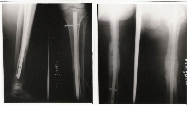
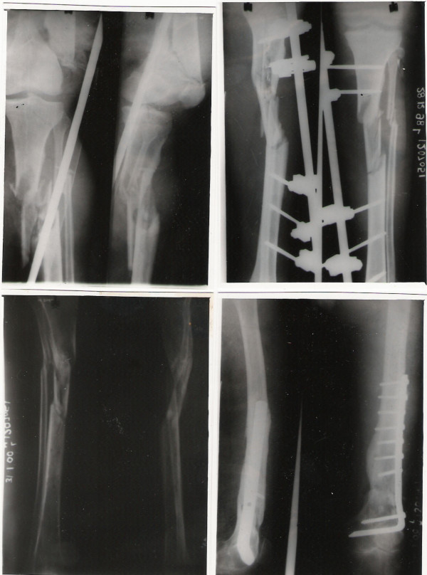
Thromboprophylaxis was initiated in all patients in the post-operative period. Physiotherapy and mobilisation was started as soon as possible after surgery. Patients were followed up regularly till bony union (clinical and radiological). Functional assessment and final outcome was measured using the Karlstrom's criteria [11] after bony union [Table 3] [Figure 4 &5].
Table 3.
Karlstrom criteria for functional assessment after management of floating knee injuries
| CRITERION | EXCELLENT | GOOD | ACCEPTABLE | POOR |
| Subjective symptoms from thigh or leg | none | Intermittent slight symptoms | More severe symptom impairing function | Considerable functional impairment: pain at rest |
| Subjective symptoms from knee or ankle joint | none | Same as above | Same as above | Same as above |
| Walking ability | Unimpaired | Same as above | Walking distance restricted | Uses cane, crutch or other support |
| Work and sports | Same as before the accident | Given up some sport; work same as before accident | Change to less strenuous work | Permanent disability |
| Angulation, rotational deformity or both | 0 | <10 degrees | 10 – 20 degrees | > 20 degrees |
| Shortening | 0 | < 1 centimetre | 1 – 3 centimetres | > 3 centimetres |
| Restricted joint mobility | 0 | <10 degrees at ankle; <20 degrees at hip, knee or both | 10 – 20 degrees at ankle; 20 – 40 at hip, knee or both | >20 degrees at ankle; >40 degrees at hip, knee or both |
Figure 4.
Fractures treated by intramedullary nailing showing union.
Figure 5.
Complex Floating Knee injury treated with a Dynamic Condylar screw for the distal femur and external fixation for open comminuted tibia fracture at bony union.
Results
The mean age of the study group was 28 years (18 – 56).27 patients were involved in motor vehicle accidents while 2 patients sustained the injury by fall from height. The right side was involved in 19 and left side in 10 patients. There were 20 Type 1, 3 Type 2A and 6 Type 2B floating knee injuries (Blake & McBryde classification) [Table 2] [Figure 1, 2 &3]. There were 6 open fractures of the tibia – 4 – Type 3B and 2 – Type 2. The average time from admission to surgery was 2 days (Range 1–11). Surgery was delayed in 6 patients due to head injury and fat embolism. Intramedullary nailing for both fractures was performed in 20 patients. Combination of Dynamic hip screw, dynamic condylar screw, external fixation for tibia fracture and buttress plating for tibia plateau fractures were performed in the remaining 9 patients. The bony union times for the femoral and tibia fractures with different fixation methods were as detailed in Table 4.
Table 4.
Bony union times for the femoral and tibial fractures with different fixation methods
| Fracture fixation methods | Number of patients | Bony union time |
| Intramedullary nailing – Diaphyseal femur | 20 | 19.1 weeks |
| Dynamic Hip screw – Proximal femur | 5 | 15 weeks |
| Dynamic condylar screw – Distal femur | 4 | 22.5 weeks |
| Intramedullary nailing – Diaphyseal tibia | 20 | 20.8 weeks |
| Buttress plating – Tibial plateau | 3 | 17 weeks |
| External Fixation – Open tibial fracture | 6 | 28 weeks |
38 associated injuries were noted in the 29 patients. These ranged from head injury to metatarsal fractures [Table 5]. 7 patients had ipsilateral knee injuries (3 patellar fractures, 2 anterior cruciate ligament tears, 1 posterior cruciate ligament tear and a bucket handle medial meniscal tear). 2 patients had haemopneumothoraces that needed tube thoracostomies. These patients underwent surgical stabilisation of the fractures without any delay. The chest drains were kept till the haemothorax was drained as monitored by serial chest radiographs. There was no delay in recovery or rehabilitation in these patients.
Table 5.
Associated injuries with Floating knee and their management
| Associated injury | Patients | Intervention |
| Patellar fractures | 3 | Open reduction internal fixation |
| Knee ligament injuries – Anterior cruciate, Posterior cruciate, Medial meniscus | 4 | Ligament reconstruction, medial meniscectomy |
| Clavicle fractures | 4 | Conservative |
| Femoral fractures (opposite) | 3 | Intramedullary nailing |
| Femoral artery block | 1 | Femoro-popliteal bypass graft |
| Humeral shaft fractures | 4 | Open reduction internal fixation |
| Head injury | 3 | Conservative |
| Rib fractures | 1 | Conservative |
| Haemo-pneumothorax | 2 | Chest drain insertion |
| Forearm bones fractures | 1 | Open reduction internal fixation |
| Contralateral tibial fractures | 4 | Intramedullary nailing |
| Tarsal/metatarsal fractures | 4 | Conservative |
| Fat embolism | 3 | Mechanical ventilation |
| Median/ulnar nerve palsy | 1 | Nerve conduction study |
3 patients had contralateral femoral fractures and 4 had contralateral tibial fractures. 4 patients had associated humeral fractures and 1 patient had fractures of the forearm bones. 4 patients had tarsal/metatarsal fractures. All bony injuries that needed surgical stabilisation were managed during the same anaesthesia as for the surgical stabilisation of the floating knee.
3 patients sustained head injuries for which a CT scan of the brain was done. None of these patients had intracranial bleeds or haematomas that needed intervention by the neurosurgeons. All of these patients were diagnosed to have cerebral concussion. The initial assessment and management was neurological monitoring recording the pupil size and the Glasgow Coma Scale (GCS). These patients were operated only after their GCS was 15 and this delayed the time of initial surgery in all these patients by 2 – 3 days.
1 patient had a femoral artery injury which was suspected clinically and evaluated by a femoral angiogram. This revealed an intimal injury of the superficial femoral artery that needed a femoro-popliteal bypass graft which was performed by the vascular surgeons after surgical stabilisation of the fractures. Surgical stabilisation of the fractures was done initially to avoid placing stress on the vascular bypass graft during reduction of the fractures. The average surgical time was 1 hour more than in patients who needed surgical stabilisation of the floating knee alone. In some of these patients there was a delay in rehabilitation of 3 weeks on an average. 3 patients developed fat embolism and needed ventilatory support with monitoring in the Intensive care unit. The delay in surgery in these patients was 10 days (Range 8–11 days). The implication of the associated injuries is detailed in Table 6.
Table 6.
Implications of associated injuries in the floating knee
| Patient | Associated injury | Delay in primary surgery | Surgical duration (Hours) | Delay in rehabilitation |
| 1 | None | 0 | 2:20 | Nil |
| 2 | Cerebral concussion | 2 days | 2:00 | Nil |
| 3 | Clavicle, Fat embolism | 8 days | 1:50 | Nil |
| 4 | Patella | 0 | 3:00 | 4 weeks |
| 5 | Contralateral femur | 0 | 3:20 | 2 weeks |
| 6 | Anterior Cruciate | 0 | 3:30 | 4 weeks |
| 7 | Clavicle, Humerus Forearm bones, Metatarsal | 0 | 3:30 | 4 weeks |
| 8 | Medial meniscus | 0 | 3:00 | Nil |
| 9 | None | 0 | 2:00 | Nil |
| 10 | Contralateral tibia | 0 | 2:30 | 2 weeks |
| 11 | Humerus | 0 | 2:50 | 4 weeks |
| 12 | Fat embolism | 11 days | 1:50 | Nil |
| 13 | Clavicle, Haemo-pneumothorax | 0 | 2:10 | 1 week |
| 14 | Metatarsal | 0 | 2:00 | Nil |
| 15 | Humerus Radial nerve | 0 | 3:00 | 4 weeks |
| 16 | Contralateral tibia, Fat embolism | 9 days | 2:40 | 2 weeks |
| 17 | Contralateral femur | 0 | 2:50 | 2 weeks |
| 18 | Posterior Cruciate | 0 | 3:20 | 4 weeks |
| 19 | Clavicle, Rib Haemo-pneumothorax | 0 | 2:00 | Nil |
| 20 | Patella, Metatarsal | 0 | 2:40 | 4 weeks |
| 21 | None | 0 | 1:50 | Nil |
| 22 | Contralateral tibia Cerebral concussion | 3 | 2:50 | 2 weeks |
| 23 | Humerus | 0 | 3:00 | 3 weeks |
| 24 | Femoral artery injury | 0 | 4:20 | 3 weeks |
| 25 | Contralateral tibia | 0 | 3:00 | 2 weeks |
| 26 | Anterior Cruciate | 0 | 3:20 | 4 weeks |
| 27 | Metatarsal | 0 | 2:10 | Nil |
| 28 | Contralateral femur Cerebral concussion | 1 | 3:00 | 2 weeks |
| 29 | Patella | 0 | 2:40 | 4 weeks |
The complications encountered were knee stiffness in 4 patients, foot drop in 1 patient, delayed union of tibia in 2 patients and superficial infection in 2 patients. Nerve conduction study was done in the patient with foot drop which revealed an axonotmesis of the common peroneal nerve. The additional procedures were manipulation under anaesthesia for knee stiffness, dynamisation in one patient with delayed union and common peroneal nerve exploration in the patient with foot drop which revealed a partial nerve transection. Patients with delayed union needed dynamisation of the tibial nail and removal of external fixator and functional cast bracing of the fracture. These fractures went on to unite following these interventions. The superficial infections were related to pin sites of the external fixators which were managed by pin site care and antibiotics. The infection settled with this management. The average follow up was for 23.5 months (Range 21 – 26 months). In the assessment of end results after bony union according to the Karlstrom criteria the following results were obtained: Excellent – 15, Good – 9, Acceptable – 2 and Poor – 3.
Discussion
When the knee joint is isolated partially or completely due to fracture of the femur and tibia the term "Floating Knee" is used [10]. Survivors of high-speed traffic accidents often have injuries to several of the parenchymal organs as well as multiple fractures. Careful evaluation of these injuries and resuscitation of the patient must precede the definitive management of specific fractures.
Hayes JT [5] suggested that automobile passengers with floating knee, braced their feet firmly against the sloping floor of the front seat just prior to the collision, their legs getting crumpled under the massive decelerating forces produced by the impact. Pedestrians were frequently catapulted some distance from the point of impact and were further injured by striking the pavement. In a study of 222 cases of floating knee by Fraser [2], all cases were involved in road traffic accidents.
Studies showed associated injuries like head injuries, chest injuries, abdominal injuries and injuries to other extremities. Most of the injuries to the head, chest and abdomen were life threatening. Adamson et al in their study encountered 71% major associated injuries with 21% vascular injuries [12]. The reported mortality rate ranged from 5% – 15%, reflecting the seriousness of the associated injuries [1]. Deliberate and careful examination of the patient must be carried out in order to determine whether a major intracranial, abdominal or thoracic injury is present. Such injuries should take precedence over extremity injuries in the priority of treatment.
There are plenty of studies in the literature detailing different management options for the Floating Knee. Hayes JT [5] opined that in a patient with multiple fractures in the same extremity, operative fixation of one or more of the fractures was valuable in the management of the entire limb. Ratcliff AH [8] found that internal fixation of both the fractures should be done wherever possible as these patients were less likely to develop knee stiffness or shortening and were in hospital and off work for less time than those treated conservatively. Omer GE [6] treated the Floating Knee by both conservative and operative fixation found that where internal fixation was done for both femoral and tibial fractures, the healing time was about 8 weeks earlier than the group managed conservatively. Behr JT [3] treated patients with the Floating knee by closed intramedullary nailing with Ender nails and achieved femoral union at an average of 10.3 weeks and tibial union at 18 weeks. Ostrum RF [7] treated patients with a retrograde femoral tibial intramedullary nail through a 4 cm medial parapatellar incision. The average time to union of the femoral fractures was 14.7 weeks and that for the tibial fractures was 23 weeks. They opined that this method was an excellent treatment option.
The general consensus in recent studies is that the best management for the Floating knee is surgical fixation of both the fractures with intramedullary nails. Dwyer used combined modalities of treatment with one fracture managed conservatively and the other surgically. They concluded that the treatment method for the tibia did not interfere with joint mobilisation [13]. Lundy recommended surgical stabilisation of the fractures for early mobilisation which produced the best results [14]. Theodoratus recommended intramedullary nailing as the best choice of treatment except for grade 3B & C open fractures [15]. Single incision technique for nailing of both the fractures have been recommended by several authors [7,16,17]. Rios J compared single incision versus traditional antegrade nailing of the fractures and found the former to have less surgical & anaesthesia time with reduced blood loss [17]. Shiedts found an increased incidence of fat embolism when both fractures were treated by reamed nails [18].
Szalay [19] demonstrated knee ligament laxity in 53% of patients while 18% complained of instability. Most of the patients with instability had a rupture of the anterior cruciate ligament with or without damage to other ligaments. They concluded that knee ligament injury was more common with floating knee injuries than with isolated femoral fractures and advocated careful assessment of the knee in all cases of fractures of the femur and floating knee injuries. Other studies [20] have showed that the incidence of knee ligament injuries in the floating knee was upto 50%, most of which were missed in the initial assessment. Meticulous examination of the knee at the time of injury is strongly advocated although the practicality of this method is questionable.
Our study showed a male predominance comparable to other studies. Most of the studies showed road traffic accidents as the only mode of injury. In our study, the most common mode of injury was road traffic accidents but two of our patients sustained their injury after a fall from height. This mode of injury for the Floating Knee has not been mentioned in the literature reviewed. The classification used by us was the one that was proposed by Robert Blake [10]. This was used as it took into account the injuries sustained at the hip or ankle of the affected side and helps one in planning the surgical procedure. The other classification system advocated by Fraser [2] includes intra-articular fractures at the knee but does not mention about injuries to the ipsilateral hip or ankle both of which can have implications on the surgical management of the Floating Knee. Our management consisted of treating both the femoral and tibial fractures surgically, most of them by intramedullary nailing using an interlocking nail. With this management, we found the fracture union time and functional recovery was better than the other surgical modalities. This was in accordance to studies by Gregory [4] and Ostrum RF [7] who had excellent results with fixation of both fractures by intra-medullary nailing. Both these authors used a retrograde nailing for the femur although in our study all the nailing was antegrade. Though no knee problems have been found when single incision technique is used [4,7,16] we feel that antegrade nailing allowed easier knee ligament reconstruction if needed as the femoral nail inserted retrograde would make knee ligament reconstruction technically difficult.
Intra-articular involvement of the fractures, higher skeletal injury scores and severity of soft tissue injuries are significant indicators of poor outcome results [21-23]. Hee suggested a preoperative scoring system which took into consideration the age, smoking status at time of injury, Injury severity scores, open fractures, segmental fractures and comminution to prognosticate the final outcome of these fractures [24].
The best results were seen when both fractures were treated by intramedullary nailing. We found that these patients returned to their normal level of activity earlier than when the fractures were treated with other modalities. Tibia fractures treated with external fixation had a longer union time probably related to the soft tissue injury and comminution at the initial injury. The 3 patients who had a poor outcome in our study were 2 patients with tibia plateau fractures who had knee stiffness and persisting pain in the knee while the other patient had a Grade 3B open tibia fracture treated by external fixation. This shows that the poor prognostic factors were related to the type of fracture (open or closed, intra-articular fractures, severe comminution). The associated injuries played a major role in the initial outcome of patients in our study with regards to delay in initial surgery, prolonged duration of surgery, anaesthetic exposure and delay in rehabilitation. From our study we found Floating knee injuries to be a group of complex injuries that needed careful assessment to detect poor prognostic factors (open, intra-articular, comminuted fractures) and associated injuries, surgical fixation of the fractures with thorough planning of surgeries and prolonged rehabilitation. Combination of all these would determine the ultimate outcome of these patients.
Conclusion
The Floating Knee is a complex injury with more than just ipsilateral fractures of the femur and tibia. The associated injuries and the type of fracture (open, intra-articular, comminution) are prognostic indicators of the initial and final outcome in patients. We recommend thorough initial assessment of patients with regards to life threatening associated injuries, surgical fixation of both fractures preferably by intramedullary nailing, knee ligament assessment to detect injuries and rigorous post-operative rehabilitation for a good final outcome.
Competing interests
The author(s) declare that they have no competing interests.
Authors' contributions
UR was involved in conducting the study, collecting patient details, reviewing the literature, drafted the manuscript and proof read the manuscript. RSY was involved in reviewing the literature and proof read the manuscript. RN is the senior author and was responsible for final proof reading of the article. All authors have read and approved the final manuscript.
Acknowledgments
Acknowledgements
Funding was neither sought nor obtained. Written consent for publication was obtained from patients.
Contributor Information
Ulfin Rethnam, Email: ulfinr@yahoo.com.
Rajam S Yesupalan, Email: ajeesh2000@yahoo.co.uk.
Rajagopalan Nair, Email: remraj@bgl.vsnl.net.in.
References
- Veith RG, Winquist RA, Hansen ST., Jr Ipsilateral fractures of the femur and tibia. J Bone and Joint Surgery. 1984;66-A:991–1002. [PubMed] [Google Scholar]
- Fraser RD, Hunter GA, Waddell JP. Ipsilateral fracture of the femur and tibia. J Bone Joint Surg Br. 1978;60-B:510–5. doi: 10.1302/0301-620X.60B4.711798. [DOI] [PubMed] [Google Scholar]
- Behr JT, Apel DM, Pinzur MS, Dobozi WR, Behr MJ. Flexible intramedullary nails for ipsilateral femoral and tibial fractures. J Trauma. 1987;27:1354–1357. doi: 10.1097/00005373-198712000-00006. [DOI] [PubMed] [Google Scholar]
- Gregory P, DiCicco J, Karpik K, DiPasquale T, Herscovici D, Sanders R. Ipsilateral fractures of the femur and tibia: treatment with retrograde femoral nailing and unreamed tibial nailing. J Orthop Trauma. 1996;10:309–16. doi: 10.1097/00005131-199607000-00004. [DOI] [PubMed] [Google Scholar]
- Hayes JT. Multiple fractures in the same extremity: Some problems in their management. Surgical Clinics of North America. 1961;41:1379–1388. doi: 10.1016/s0039-6109(16)36499-4. [DOI] [PubMed] [Google Scholar]
- Omer GE, Moll JH, Bacon WL. Combined fractures of the femur and tibia in a single extremity. J Trauma. 1968;8:1026–41. [PubMed] [Google Scholar]
- Ostrum RF. Treatment of floating knee injuries through a single percutaneous approach. Clin Orthop Relat Res. 2000;375:43–50. doi: 10.1097/00003086-200006000-00006. [DOI] [PubMed] [Google Scholar]
- Ratcliff AH. Fractures of the shaft of the femur and tibia in the same limb. Pro Roy Soc Med. 1968;61:907–908. [PMC free article] [PubMed] [Google Scholar]
- Gustilo RB, Anderson JT. Prevention of infection in the treatment of one thousand and twenty-five open fractures of long bones: retrospective and prospective analyses. J Bone Joint Surg Am. 1976;58:453–8. [PubMed] [Google Scholar]
- Blake R, McBryde A., Jr The Floating Knee: Ipsilateral fractures of the tibia and femur. South Med J. 1975;68:13–6. [PubMed] [Google Scholar]
- Karlstrom G, Olerud S. Ipsilateral fracture of the femur and tibia. J Bone Joint Surg Am. 1977;59:240–3. [PubMed] [Google Scholar]
- Adamson GJ, Wiss DA, Lowery GL, Peters CL. Type II floating knee: ipsilateral femoral and tibial fractures with intraarticular extension into the knee joint. J Orthop Trauma. 1992;6:333–9. doi: 10.1097/00005131-199209000-00011. [DOI] [PubMed] [Google Scholar]
- Dwyer AJ, Paul R, Mam MK, Kumar A, Gosselin RA. Floating knee injuries: long-term results of four treatment methods. Int Orthop. 2005;29:314–8. doi: 10.1007/s00264-005-0679-x. [DOI] [PMC free article] [PubMed] [Google Scholar]
- Lundy DW, Johnson KD. "Floating knee" injuries: ipsilateral fractures of the femur and tibia. J Am Acad Orthop Surg. 2001;9:238–45. doi: 10.5435/00124635-200107000-00003. [DOI] [PubMed] [Google Scholar]
- Theodoratos G, Papanikolaou A, Apergis E, Maris J. Simultaneous ipsilateral diaphyseal fractures of the femur and tibia: treatment and complications. Injury. 2001;32:313–5. doi: 10.1016/S0020-1383(00)00189-3. [DOI] [PubMed] [Google Scholar]
- Oh CW, Oh JK, Min WK, Jeon IH, Kyung HS, Ahn HS, Park BC, Kim PT. Management of ipsilateral femoral and tibial fractures. Int Orthop. 2005;29:245–50. doi: 10.1007/s00264-005-0661-7. [DOI] [PMC free article] [PubMed] [Google Scholar]
- Ríos JA, Ho-Fung V, Ramírez N, Hernández RA. Floating knee injuries treated with single-incision technique versus traditional antegrade femur fixation: a comparative study. Am J Orthop. 2004;33:468–72. [PubMed] [Google Scholar]
- Schiedts D, Mukisi M, Bouger D, Bastaraud H. Ipsilateral fractures of the femoral and tibial diaphyses. Rev Chir Orthop Reparatrice Appar Mot. 1996;82:535–40. [PubMed] [Google Scholar]
- Szalay MJ, Hosking OR, Annear P. Injury of the knee ligament associated with ipsilateral femoral and tibial shaft fractures. Injury. 1990;21:398–400. doi: 10.1016/0020-1383(90)90129-I. [DOI] [PubMed] [Google Scholar]
- Paul GR, Sawka MW, Whitelaw GP. Fractures of the ipsilateral femur and tibia: emphasis on intra-articular and soft tissue injury. J Orthop Trauma. 1990;4:309–14. doi: 10.1097/00005131-199003000-00013. [DOI] [PubMed] [Google Scholar]
- Hung SH, Lu YM, Huang HT, Lin YK, Chang JK, Chen JC, Tien YC, Huang PJ, Chen CH, Liu PC, Chao D. Surgical treatment of type II floating knee: comparisons of the results of type IIA and type IIB floating knee. Knee Surg Sports Traumatol Arthrosc. 2007;15:578–86. doi: 10.1007/s00167-006-0252-1. [DOI] [PubMed] [Google Scholar]
- Yokoyama K, Tsukamoto T, Aoki S, Wakita R, Uchino M, Noumi T, Fukushima N, Itoman M. Evaluation of functional outcome of the floating knee injury using multivariate analysis. Arch Orthop Trauma Surg. 2002;122:432–5. doi: 10.1007/s00402-002-0406-7. [DOI] [PubMed] [Google Scholar]
- Arslan H, Kapukaya A, Kesemenli CC, Necmioğlu S, Subaşi M, Coban V. The floating knee in adults: twenty-four cases of ipsilateral fractures of the femur and the tibia. Acta Orthop Traumatol Turc. 2003;37:107–12. [PubMed] [Google Scholar]
- Hee HT, Wong HP, Low YP, Myers L. Predictors of outcome of floating knee injuries in adults: 89 patients followed for 2–12 years. Acta Orthop Scand. 2001;72:385–94. doi: 10.1080/000164701753542050. [DOI] [PubMed] [Google Scholar]


