

We present an unusual case of a patient presenting with signs of congestive heart failure due to a very uncommon cause.
Case report
A 35-year-old male presented with symptoms of shortness of breath and ankle oedema which had developed within a few days. The symptoms had started suddenly following a mountain bike trip. Physical examination revealed a blood pressure of 90/40 mmHg, no fever and a continuous murmur on auscultation of the heart, as well as signs of left and right heart failure.
Echocardiography showed moderate pericardial effusion and a hyperdynamic left and right ventricle with signs of right ventricular volume overload. Turbulent flow was seen in the aortic root and a shunt was demonstrated from the aortic root to the right atrium (figure 1) through a ruptured sinus Valsalva aneurysm of the non-coronary cusp (figure 2).
Figure 1.
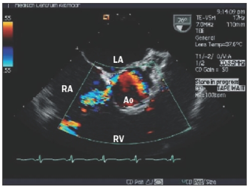
Transoesophageal still frame demonstrating turbulent flow from the aortic root into the right atrium (LA=left atrium, RA=right atrium, Ao=aortic root, RV=right ventricle).
Figure 2.

Transoesophageal still frame demonstrating the ruptured sinus Valsalva aneurysm of the non-coronary cusp of the aortic valve (LA=left atrium, RA=right atrium, Ao=aortic root, RV=right ventricle).
Discussion
Aneurysm of the sinus of Valsalva is a rare congenital anomaly that usually involves the right coronary sinus (76.8%), the non-coronary sinus (20.2%) or the left coronary sinus (3.0%).1 The male-to-female ratio is 3.5:1 and symptoms usually develop in the adult population.2 Sometimes symptoms are caused by pressure on structures such as the conduction system or coronary arteries, but most patients remain asymptomatic until rupture occurs. Rupture usually occurs into the right atrium or ventricle, but rupture to the left ventricle, pericardium, superior vena cava or pulmonary artery have also been described.3 In this section a remarkable ‘image’ is presented and a short comment is given.
We invite you to send in images (in triplicate) with a short comment (one page at the most) to Bohn
Stafleu van Loghum, PO Box 246, 3990 GA Houten, E-mail: l.jagers@bsl.nl.
‘Moving images’ are also welcomed and (after acceptance) will be published as a Web Site Feature and shown on our website: www.cardiologie.nl
This section is edited by M.J.M. Cramer and J.J. Bax.
References
- 1.Chu SH, Hung CR, How SS, Chang H, Wang SS, Tsai CH, et al. Ruptured aneurysms of the sinus of Valsalva in Oriental patients. J Thorac Cardiovasc Surg 1990;99:288-98. [PubMed] [Google Scholar]
- 2.Choudhary SK, Bhan A, Sharma R, et al. Sinus of Valsalva aneurysm: 20 years’ experience. J Card Surg 1997;12:300-8. [DOI] [PubMed] [Google Scholar]
- 3.Kucukoglu S, Ural E, Mutlu H, Ural D, Sonmez B, Uner S. Ruptured aneurysm of the Sinus of Valsalva into the left ventricle: a case report and review of the literature. J Am Soc Echocardiogr 1997;10:862-5. [DOI] [PubMed] [Google Scholar]