

Abstract
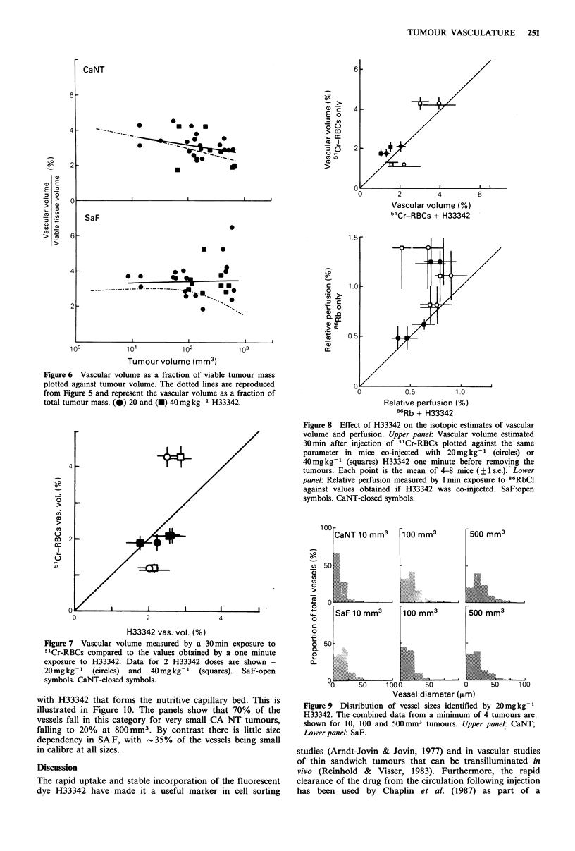
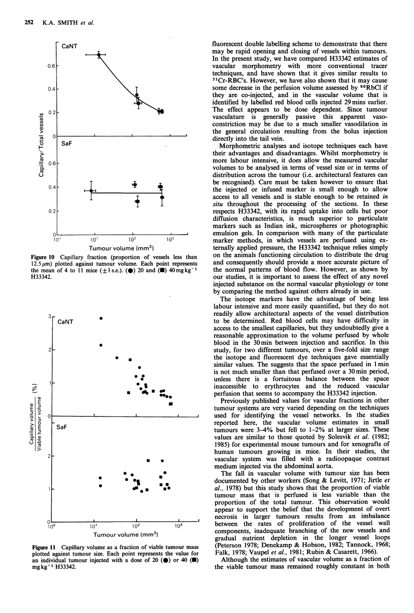
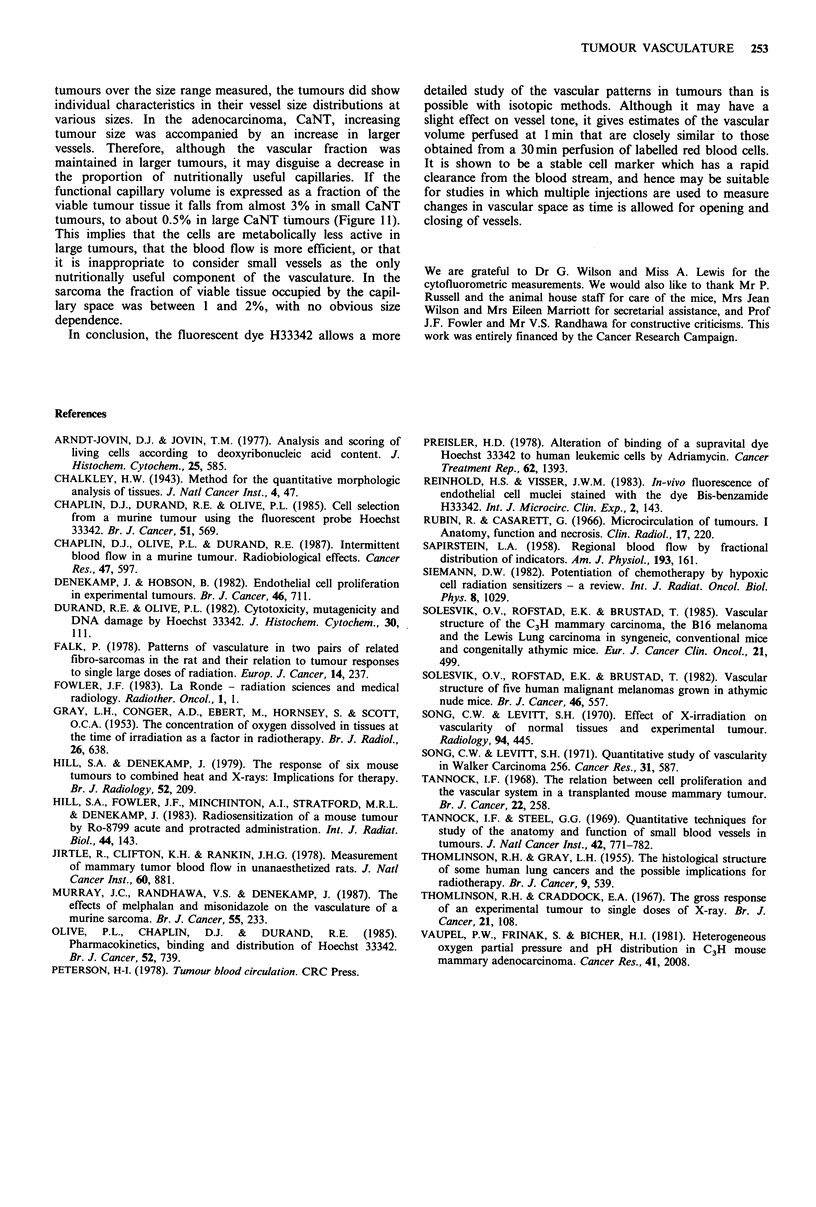
The DNA-binding fluorescent dye Hoechst 33342 (H33342) has been used in a series of investigations of the vascular parameters of two murine tumours. This dye has been shown, to have a short half-life in the circulation (T1/2 less than 2 min), but is stably bound for at least 2 h after it enters cells. It can be used in morphometric studies on frozen sections to determine the effective vascular volume, the capillary fraction and the size distribution of blood vessels in each tumour. These latter two parameters cannot be deduced from the less labour intensive techniques using radioactive isotopes. The effective vascular volume perfused in 1 min by H33342 was compared with the volume perfused in 30 min with 51Cr labelled erythrocytes. Similar volumes were estimated with the two techniques in a murine carcinoma and in a sarcoma. Both techniques showed that the vascular volume decreased in larger tumours. The H33342 analysis of vessel size showed the decrease in capillary vessels in the carcinomas was even greater, falling from 70% in small tumours to 20% in larger tumours. The deteriorating vascular network in larger tumours is associated with an increasing fraction of necrotic tissue. Experiments in which the isotopes and dye were co-injected suggest that at 40 mgkg-1 the dye may rapidly lead to a partial shutdown of the tumour vascular bed. This is less marked with 20 mg kg-1. In spite of this effect there is in general a close correlation between the volumes perfused by labelled red blood cells and the fluorescent dye.
Full text
PDF
Images in this article
Selected References
These references are in PubMed. This may not be the complete list of references from this article.
- Arndt-Jovin D. J., Jovin T. M. Analysis and sorting of living cells according to deoxyribonucleic acid content. J Histochem Cytochem. 1977 Jul;25(7):585–589. doi: 10.1177/25.7.70450. [DOI] [PubMed] [Google Scholar]
- Chaplin D. J., Durand R. E., Olive P. L. Cell selection from a murine tumour using the fluorescent probe Hoechst 33342. Br J Cancer. 1985 Apr;51(4):569–572. doi: 10.1038/bjc.1985.79. [DOI] [PMC free article] [PubMed] [Google Scholar]
- Chaplin D. J., Olive P. L., Durand R. E. Intermittent blood flow in a murine tumor: radiobiological effects. Cancer Res. 1987 Jan 15;47(2):597–601. [PubMed] [Google Scholar]
- Denekamp J., Hobson B. Endothelial-cell proliferation in experimental tumours. Br J Cancer. 1982 Nov;46(5):711–720. doi: 10.1038/bjc.1982.263. [DOI] [PMC free article] [PubMed] [Google Scholar]
- Durand R. E., Olive P. L. Cytotoxicity, Mutagenicity and DNA damage by Hoechst 33342. J Histochem Cytochem. 1982 Feb;30(2):111–116. doi: 10.1177/30.2.7061816. [DOI] [PubMed] [Google Scholar]
- Falk P. Patterns of vasculature in two pairs of related fibrosarcomas in the rat and their relation to tumour responses to single large doses of radiation. Eur J Cancer. 1978 Mar;14(3):237–250. doi: 10.1016/0014-2964(78)90187-1. [DOI] [PubMed] [Google Scholar]
- Fowler J. F. The second Klaas Breur memorial lecture. La Ronde--radiation sciences and medical radiology. Radiother Oncol. 1983 Aug;1(1):1–22. doi: 10.1016/s0167-8140(83)80003-6. [DOI] [PubMed] [Google Scholar]
- GRAY L. H., CONGER A. D., EBERT M., HORNSEY S., SCOTT O. C. The concentration of oxygen dissolved in tissues at the time of irradiation as a factor in radiotherapy. Br J Radiol. 1953 Dec;26(312):638–648. doi: 10.1259/0007-1285-26-312-638. [DOI] [PubMed] [Google Scholar]
- Hill S. A., Denekamp J. The response of six mouse tumours to combined heat and X rays: implications for therapy. Br J Radiol. 1979 Mar;52(615):209–218. doi: 10.1259/0007-1285-52-615-209. [DOI] [PubMed] [Google Scholar]
- Hill S. A., Fowler J. F., Minchinton A. I., Stratford M. R., Denekamp J. Radiosensitization of a mouse tumour by Ro 03-8799: acute and protracted administration. Int J Radiat Biol Relat Stud Phys Chem Med. 1983 Aug;44(2):143–150. doi: 10.1080/09553008314550941. [DOI] [PubMed] [Google Scholar]
- Jirtle R., Clifton K. H., Rankin J. H. Measurement of mammary tumor blood flow in unanesthetized rats. J Natl Cancer Inst. 1978 Apr;60(4):881–886. doi: 10.1093/jnci/60.4.881. [DOI] [PubMed] [Google Scholar]
- Murray J. C., Randhawa V., Denekamp J. The effects of melphalan and misonidazole on the vasculature of a murine sarcoma. Br J Cancer. 1987 Mar;55(3):233–238. doi: 10.1038/bjc.1987.45. [DOI] [PMC free article] [PubMed] [Google Scholar]
- Olive P. L., Chaplin D. J., Durand R. E. Pharmacokinetics, binding and distribution of Hoechst 33342 in spheroids and murine tumours. Br J Cancer. 1985 Nov;52(5):739–746. doi: 10.1038/bjc.1985.252. [DOI] [PMC free article] [PubMed] [Google Scholar]
- Preisler H. D. Alteration of binding of the supravital dye Hoechst 33342 to human leukemic cells by adriamycin. Cancer Treat Rep. 1978 Sep;62(9):1393–1396. [PubMed] [Google Scholar]
- Reinhold H. S., Visser J. W. In vivo fluorescence of endothelial cell nuclei stained with the dye Bis-benzamide H 33342. Int J Microcirc Clin Exp. 1983;2(2):143–146. [PubMed] [Google Scholar]
- Rubin P., Casarett G. Microcirculation of tumors. I. Anatomy, function, and necrosis. Clin Radiol. 1966 Jul;17(3):220–229. doi: 10.1016/s0009-9260(66)80027-2. [DOI] [PubMed] [Google Scholar]
- SAPIRSTEIN L. A. Regional blood flow by fractional distribution of indicators. Am J Physiol. 1958 Apr;193(1):161–168. doi: 10.1152/ajplegacy.1958.193.1.161. [DOI] [PubMed] [Google Scholar]
- Siemann D. W. Potentiation of chemotherapy by hypoxic cell radiation sensitizers--a review. Int J Radiat Oncol Biol Phys. 1982 Jun;8(6):1029–1034. doi: 10.1016/0360-3016(82)90172-9. [DOI] [PubMed] [Google Scholar]
- Solesvik O. V., Rofstad E. K., Brustad T. Vascular structure of five human malignant melanomas grown in athymic nude mice. Br J Cancer. 1982 Oct;46(4):557–567. doi: 10.1038/bjc.1982.240. [DOI] [PMC free article] [PubMed] [Google Scholar]
- Solesvik O. V., Rofstad E. K., Brustad T. Vascular structure of the C3H mammary carcinoma, the B16 melanoma and the Lewis lung carcinoma in syngeneic, conventional mice and congenitally athymic mice. Eur J Cancer Clin Oncol. 1985 Apr;21(4):499–505. doi: 10.1016/0277-5379(85)90044-6. [DOI] [PubMed] [Google Scholar]
- Song C. W., Levitt S. H. Effect of x irradiation on vascularity of normal tissues and experimental tumor. Radiology. 1970 Feb;94(2):445–447. doi: 10.1148/94.2.445. [DOI] [PubMed] [Google Scholar]
- Song C. W., Levitt S. H. Quantitative study of vascularity in Walker carcinoma 256. Cancer Res. 1971 May;31(5):587–589. [PubMed] [Google Scholar]
- THOMLINSON R. H., GRAY L. H. The histological structure of some human lung cancers and the possible implications for radiotherapy. Br J Cancer. 1955 Dec;9(4):539–549. doi: 10.1038/bjc.1955.55. [DOI] [PMC free article] [PubMed] [Google Scholar]
- Tannock I. F., Steel G. G. Quantitative techniques for study of the anatomy and function of small blood vessels in tumors. J Natl Cancer Inst. 1969 May;42(5):771–782. [PubMed] [Google Scholar]
- Tannock I. F. The relation between cell proliferation and the vascular system in a transplanted mouse mammary tumour. Br J Cancer. 1968 Jun;22(2):258–273. doi: 10.1038/bjc.1968.34. [DOI] [PMC free article] [PubMed] [Google Scholar]
- Thomlinson R. H., Craddock E. A. The gross response of an experimental tumour to single doses of x-rays. Br J Cancer. 1967 Mar;21(1):108–123. doi: 10.1038/bjc.1967.10. [DOI] [PMC free article] [PubMed] [Google Scholar]
- Vaupel P. W., Frinak S., Bicher H. I. Heterogeneous oxygen partial pressure and pH distribution in C3H mouse mammary adenocarcinoma. Cancer Res. 1981 May;41(5):2008–2013. [PubMed] [Google Scholar]