

Summary
In slightly over a period of twenty years, our comprehension of the cellular and molecular mechanisms that govern the Wnt signaling pathway continue to unfold. The Wnt proteins were initially implicated in viral carcinogenesis experiments associated with mammary tumors, but since this period investigations focusing on the Wnt pathways and their transmembrane receptors termed Frizzled have been advanced to demonstrate the critical nature of Wnt for the development of a variety of cell populations as well as the potential of the Wnt pathway to avert apoptotic injury. In particular, Wnt signaling plays a significant role in both the cardiovascular and nervous systems during embryonic cell patterning, proliferation, differentiation, and orientation. Furthermore, modulation of Wnt signaling under specific cellular influences can either promote or prevent the early and late stages of apoptotic cellular injury in neurons, endothelial cells, vascular smooth muscle cells, and cardiomyocytes. A number of downstream signal transduction pathways can mediate the biological response of the Wnt proteins that include Dishevelled, β-catenin, intracellular calcium, protein kinase C, Akt, and glycogen synthase kinase-3β. Interestingly, these cellular cascades of the Wnt-Frizzled pathways can participate in several neurodegenerative, vascular, and cardiac disorders and may be closely integrated with the function of trophic factors. Identification of the critical elements that modulate the Wnt-Frizzled signaling pathway should continue to unlock the potential of Wnt pathway for the development of new therapeutic options against neurodegenerative and vascular diseases.
Keywords: Akt, Alzheimer’s, β-catenin, Cardiomyocytes, Dishevelled, Endothelial cells, Erythropoietin, Frizzled, GSK-3β, Myocardial infarction, Neurons, Retinal disease, Stem cells, VEG
Introduction
Wnt proteins, derived from Drosophilia Wingless (Wg) and the mouse Int-1 genes, represent a large family of secreted cysteine-rich glycosylated proteins. This novel family of proteins are intimately involved in cellular signaling pathways that play a role in a variety of processes that involve embryonic cell patterning, proliferation, differentiation, orientation, adhesion, survival, and apoptosis (Nusse and Varmus, 1982; Melkonyan et al., 1997; Wodarz and Nusse, 1998; Smalley and Dale, 1999; Patapoutian and Reichardt, 2000; Chong and Maiese, 2004; Nelson and Nusse, 2004). Until recently, nineteen of the twenty-four Wnt genes that express Wnt proteins have been identified in the human. In addition, more than eighty target genes of Wnt signaling pathways also have been demonstrated in human, mouse, Drosophilia, Xenopus, and Zebrafish. This representation encompasses several cellular populations, such as neurons, cardiomyocytes, endothelial cells, cancer cells, and pre-adipocytes.
Wnt binds to Frizzled transmembrane receptors on the cell surface to activate downstream signaling events. These involve at least two intracellular signaling pathways with two that are considered of particular importance. One pathway controls target gene transcription through β-catenin, generally referred to as the canonical pathway that involves Wnt1, Wnt3a, and Wnt8 and functions through β-catenin-dependent pathways. Another pathway pertains to intracellular calcium (Ca2+) release which is termed the non-canonical or Wnt/Ca2+ pathway consisting primarily of Wnt-4, Wnt-5a, and Wnt-11 that functions through non-β-catenin-dependent pathways, such as the planar cell polarity (PCP) pathway (Nusse, 1999; Salinas, 1999; Patapoutian and Reichardt, 2000; Tada and Smith, 2000) and the Wnt-Ca2+-dependent pathways (Slusarski et al., 1997; Nusse, 1999; Salinas, 1999; Kuhl et al., 2000; Patapoutian and Reichardt, 2000; Katoh, 2002). Recent work has illustrated that eleven members of the Frizzled transmembrane receptors have been identified in the human and mouse genomes that encode for seven-pass transmembrane domain-containing serpentine receptor proteins (Nelson and Nusse, 2004). If one examines the Wnt-Frizzled signaling transduction pathway, it becomes evident that these pathways play critical roles during embryonic, non-vertebrate and vertebrate development as well as tumorigenesis (Dale, 1998; Kawakami et al., 2001; Lustig and Behrens, 2003).
Multiple studies have shown the importance of Wnt-Frizzled transduction pathway in controlling the pattern of the body axis as well as the development and maturation of the central nervous system (Augustine et al., 1993; Ikeya et al., 1997; Salinas, 1999; Wheeler and Hoppler, 1999; Melton et al., 2004), cardiovascular system (Park et al., 1996; Wheeler and Hoppler, 1999; van Gijn et al., 2001), and the limbs (Kengaku et al., 1997). During embryological development, alternations of the Wnt-Frizzled pathway can lead to abnormal morphogenesis in animal models (Stark et al., 1994; Ikeya et al., 1997; Liu et al., 1999) and congenital defects in humans (Jordan et al., 2001; Rodova et al., 2002; Niemann et al., 2004). In mature tissues, the Wnt-Frizzled pathway is involved in the self-renewal of stem cells and may be responsible for the maintenance of many normal tissues (Ross et al., 2000; Reya et al., 2003; Willert et al., 2003; He et al., 2004). Studies have revealed that dysfunction of the Wnt-Frizzled pathway can lead to neurodegenerative disorders, such as Alzheimer’s disease (Anderton, 1999; Grilli et al., 2003; Maiese and Chong, 2004; Chong et al., 2005d) and heart failure (Schumann et al., 2000; Haq et al., 2001; van Gijn et al., 2002). This paper will focus upon the Wnt protein and its receptor, the Wnt-Frizzled signaling pathway and components, and the significant role of the Wnt-Frizzled signaling pathway during cellular survival and injury in a broad array of cells that involve neuronal, endothelial, and cardiac cell populations.
The structure and function of Wnt proteins and their receptors
Though they can show varying degrees of sequence identity, the molecular structural characteristics that all Wnt proteins share involve the 39–46kDa lipid-modified secreted glycoproteins containing 350-400 amino acids, with a highly conserved pattern of 23-24 cysteine residues and several asparagines-linked glycosylation sites (Nusse and Varmus, 1992; Miller, 2002). Yet, some of Wnt proteins have an additional domain, such as the Drosphila Wg that contains an 85-amino acid domain near the center of protein (Nusse and Varmus, 1992).
Several members of Wnt proteins have been identified to control proliferation, differentiation, and death of various cells. The cell populations can include stem cells as well as the development of various tissues that in the nervous and cardiovascular systems (Table 1). Early studies have demonstrated that ecotopic expression of specific Wnt genes in Xenopus embryos can result in distinct phenotypes. In the C57MG mouse, transient expression of Wnt1, Wnt2 and Wnt3a in mammary epithelial cells can cause morphological transformation while the other Wnt proteins have little effect on cell morphology (Wong et al., 1994). In addition, in Xenopus embryos, the injection of Wnt1, Wnt3a and Wnt8 into the ventral blastomeres of four-cell embryos can lead to duplication of the body axis, but the overexpression of Wnt4, Wnt5a and Wnt11 genes can interfere with morphogenetic movement without inducing axis duplication (Smith and Harland, 1991; Sokol et al., 1991; Christian et al., 1992; Moon et al., 1993; Wolda et al., 1993).
Table 1.
Neuronal and cardiac expression of the Wnt and the Wnt receptor with biological function.
| ORGAN | CELLULAR EXPRESSION
|
BIOLOGICAL RESPONSE | |
|---|---|---|---|
| Wnt | Wnt Receptor | ||
| Brain | Neurons | Neurons | Cell regulation and cytoprotection |
| Astrocytes | Astrocytes | Cell regulation and cytoprotection | |
| Progenitor stem cells | Progenitor stem cells | Cellular development and maturation | |
| Vascular System | Endothelial cells | Endothelial cells | Angiogenesis (cell proliferation, differentiation, migration) |
| Progenitor vascular stem cells | Progenitor vascular stem cells | Angiogenesis and cardiomyogenesis | |
| Vascular smooth muscle cells | Vascular smooth muscle cells | Angiogenesis, vascular remodeling, and cytoprotection | |
| Heart | Progenitor cardiac stem cells | Progenitor cardiac stem cells | Cardiomyogenesis and cardiac conduction cell development |
| Endocardial cells | Endocardial cells | Endocardial cushion formation | |
| Cardiomyocytes | Cardiomyocytes | Cardiac remodeling and cytoprotection | |
As a result, Wnt proteins are generally divided into two functional classes based on their ability to induce a secondary body axis in Xenopus embryos and to activate certain signaling cascades that consist of the Wnt1 class and the Wnt5a class. The members of the Wnt1 class are inducers of a secondary body axis in Xenopus and include Wnt1, Wnt2, Wnt3, Wnt3a, Wnt8 and Wnt8a. Wnt proteins of this class facilitate activation of the Frizzled transmembrane receptor and the co-receptor lipoprotein related protein 5 and 6 (LRP-5/6). Ultimately, this leads to the activation of the typical canonical Wnt/β-catenin pathway. The Wnt5a class cannot induce secondary axis formation in Xenopus and includes the Wnt proteins of Wnt4, Wnt5a, Wnt5b, Wnt6, Wnt7a and Wnt11. These Wnt proteins bind the Frizzled transmembrane receptor to activate heterotrimeric G proteins and increase intracellular calcium levels. Alternatively, they can induce Rho-dependent changes in the actin cytoskeleton. Several recent studies also have shown that the different subsets of Wnt proteins can contribute to distinct physiological changes through triggering various intracellular pathways (Heisenberg et al., 2000; Tada and Smith, 2000; Winklbauer et al., 2001; Hsieh, 2004).
The main receptors of the Wnt proteins consist of at least 10 family members termed the Frizzled proteins after the first member, Drosophila tissue polarity gene I (Vinson et al., 1989; Adler et al., 1990). All members of the Frizzled protein family share the following characteristics: a N-terminal signal peptide, an extracellular domain that contains a 120-amino acids, a cysteine-rich domain followed by a hydrophilic linker region that shows little sequence similarity among family members, a highly conserved seven-transmembrane domain separated by short extracellular and cytoplasmic loops, and a cytoplasmic domain of variable size and little sequence homology among family members (Vinson et al., 1989; Adler et al., 1990; Wang et al., 1996; Wodarz and Nusse, 1998; Hsieh, 2004). Some Wnt proteins, such as Wnt8, can directly bind with the full-length Frizzled receptor protein. A single Wnt protein also can bind to a combination of Frizzled receptor proteins, including homologous members from a different species (Bhanot et al., 1996; Hsieh et al., 1999; Hsieh, 2004). The Wnt proteins bind to the activity sites of Frizzled receptor proteins that are relevant to either the canonical and non- canonical Wnt-Frizzled signaling pathways leading to specific biological functions. Interestingly, the Frizzled cysteine-rich domain also exists in several other proteins that include the soluble secreted Frizzled-related proteins (Finch et al., 1997; Leyns et al., 1997; Mayr et al., 1997; Melkonyan et al., 1997; Rattner et al., 1997; Wang et al., 1997a,b; Ellies et al., 2000; Jones et al., 2000), some receptors of tyrosine kinases (Jennings et al., 1993; Wilson et al., 1993; Glass et al., 1996), carboxypeptidase Z (Song and Fricker, 1997), the membrane-bound serine protease Corin (Yan et al., 1999), and an isoform of collagen (Rehn and Pihlajaniemi, 1995). These proteins appear to function as important regulators during Wnt-Frizzled signaling. For example, the soluble secreted Frizzled-related proteins have been found to function as antagonists of the Wnt-Frizzled signaling pathway when ectopically expressed in Xenopus embryos (Finch et al., 1997; Leyns et al., 1997; Wang et al., 1997a,b; Xu et al., 1998; Ellies et al., 2000; Jones et al., 2000). In addition, the carboxypeptidase Z has been identified to modulate Wnt 4a activity through its cysteine-rich domain (Hsieh, 2004).
In addition to the Frizzled protein receptors, other obligate co-receptors also are necessary for canonical Wnt-Frizzled signaling pathway. An additional single-pass transmembrane protein named as LRP-5/6 from the low-density-lipoprotein receptor family is required for this process (Pinson et al., 2000; Tamai et al., 2000; Wehrli et al., 2000). In the canonical Wnt-Frizzled signaling pathway, Wnt binds to both the Frizzled transmembrane receptor and the co-receptor LRP-5/6 (Wehrli et al., 2000) resulting in the inhibition of the downstream component glycogen synthase kinase-3β (GSK-3β) (Ikeda et al., 1998; Papkoff and Aikawa, 1998). More recent studies also have suggested that the Wnt signaling can be transmitted through the binding of extracellular domain of LRP-5/6 to Axin, a key component in the GSK-3β complex, indicating that the LRP-5/6 receptor is an important part of the Wnt-Frizzled signaling pathway (Mao et al., 2001; Tolwinski et al., 2003).
Another newly identified co-receptor for the canonical and non- canonical Wnt-Frizzled signaling pathway is Ryk that belongs to one of divergent members of the receptor tyrosine kinase family. Ryk not only can form a complex with Frizzled proteins such as the co-receptor LRP-5/6 resulting in activation of the canonical Wnt-Frizzled signaling pathway, but also can regulate the non-canonical Wnt-Frizzled signaling pathway through Frizzled-independent pathways (Cheyette, 2004; Bejsovec, 2005). At this time, a single Ryk gene in mammals, one Ryk gene in the nematode C. elegans (Inoue et al., 2004; Deshpande et al., 2005), and three Ryk genes in Drosophila (Yoshikawa et al., 2003) have been identified in an atypical receptor tyrosine kinase family. The molecular structure of all Ryk genes is characterized by an extracellular domain with homology to Wnt inhibitory factor-1 (WIF1), a single transmembrane-spanning sequence to which Wnt proteins bind (Schneider et al., 1999; Patthy, 2000). In addition, a conserved intracellular PDZ-binding motif exists which links Ryk to downstream molecules of Wnt-Frizzled signaling pathway, such as Dishevelled (Cheyette, 2004; Lu et al., 2004; Bejsovec, 2005).
Wnt proteins can bind to the extracellular domain of the Ryk receptor through the intracellular PDZ-binding domain in the Ryk receptor to result in regulating cell proliferation, differentiation, migration, polarity, survival, and death through either the canonical or the non- canonical Wnt-Frizzled signaling pathway. As an example, mammalian Ryk can function as a co-receptor with Frizzled to bind to Wnt1 and Wnt3a through its WIF domain and interact with Dishevelled via its PDZ domain resulting in the stimulation of neurite outgrowth (Lu et al., 2004). Furthermore, in C. elegans, Wnt proteins can employ Frizzled encoded by lin-17 and Ryk encoded by lin-18 to regulate cell fate during C. elegans vulval development (Inoue et al., 2004; Deshpande et al., 2005). In the Drosophila embryonic nervous system, Wnt5 specifically binds to the Derailed receptor that is a member of the Ryk receptor subfamily to mediate axonal guidance (Yoshikawa et al., 2003).
The Wnt-Frizzled Signaling Pathway
The Wnt-Frizzled signaling pathway includes the canonical Wnt signaling pathway and the non-canonical Wnt signaling pathway, the latter consisting of the Wnt/PCP pathway and the Wnt/Ca2+ pathway. Upon binding to either the Frizzled receptor or a receptor complex consisting of Frizzled and LRP5/6, Wnt protein can activate one of three different signaling cascades. These cascades include the canonical Wnt signaling pathway (Rattner et al., 1997; Nusse, 1999), the Wnt/PCP pathway (Rattner et al., 1997; Nusse, 1999; Veeman et al., 2003), or the Wnt/Ca2+ pathway (Slusarski et al., 1997; Nusse, 1999; Salinas, 1999; Kuhl et al., 2000; Patapoutian and Reichardt, 2000; Katoh, 2002). Each of pathways, although distinct, appears to be transduced initially through Dishevelled, a cytoplasmic multifunctional phosphoprotein (Axelrod et al., 1998; Boutros et al., 1998; Boutros and Mlodzik, 1999). Yet, the ultimate response to Wnt-Frizzled interaction will most probably depend upon cellular context at that time (Ilyas, 2005). In mammals, the Dishevelled protein family members contains Dishevelled-1, Dishevelled-2, Dishevelled-3 in all organs. These family members have three highly conserved domains that include an N-terminal DIX domain named for Dishevelled and Axin, a central PDZ domain termed for Postsynaptic density-95, Discs-large and Zonula occludens-1, and a C-terminal DEP that is named for Dishevelled, Egl-10 and Pleckstrin (Wharton, 2003; Habas and Dawid, 2005). At the level of Dishevelled, the Wnt signaling pathway can be separated along one of three different cascades that are dependent upon the three highly conserved domains of Dishevelled. As a result, Dishevelled is a key transducer of the Wnt signal that acts at the plasma membrane or in the cytoplasm in all three Wnt-Frizzled signaling pathways. However, new work has suggested that Dishevelled also acts within the nucleus and nuclear location of Dishevelled is essential for its function in the Wnt-Frizzled signaling pathway (Itoh et al., 2005; Weitzman, 2005).
The canonical Wnt signaling pathway
The canonical Wnt signaling pathway is considered to be a typical Wnt-Frizzled signaling pathway. It is also referred to as the Wnt/β-catenin-dependent signaling pathway or the Wnt/β-catenin pathway since it can regulate β-catenin protein levels to control the activation of Wnt-responsive target genes. The majority of Wnt proteins activate gene transcription through this signaling pathway controlled by β-catenin. In general, all Wnt signaling pathways are initiated by interaction of Wnt proteins with Frizzled receptors, but in this pathway, the Wnt signaling pathway will only be activated if the binding of the Wnt protein to the Frizzled transmembrane receptor takes place in the presence of the co-receptor LRP-5/6 (Pinson et al., 2000; Wehrli et al., 2000; Mao et al., 2001) resulting in the formation of a Wnt-Frizzled-LRP5/6 trimolecular complex. Once Wnt protein binds to the Frizzled transmembrane receptor and the co-receptor LRP-5/6, this is followed by recruitment of Dishevelled. Dishevelled is phosphorylated by casein kinase Iε to form a complex with Frat1 and inhibit GSK-3β activity (Ikeda et al., 1998; Papkoff and Aikawa, 1998; Lee et al., 1999; Kishida et al., 2001; Lee et al., 2001). In addition, the formation of the Wnt-Frizzled-LRP5/6 complex also promotes the LRP5/6-mediated degradation of Axin (Mao et al., 2001).
As a result, the combined inhibition of GSK-3β activity with the degradation of Axin blocks the formation of the protein complex consisting of GSK-3β, Axin, and adenomatous polyposis coli (APC) tumor suppressor protein. Yet, during the absence of Wnt signaling, β-catenin is associated with the protein complex of GSK-3β, Axin and APC tumor suppressor protein. β-catenin is phosphorylated by activation of GSK-3β leading to its ubiquitination and subsequent degradation by proteosomes (Aberle et al., 1997; Hart et al., 1999; Latres et al., 1999; Winston et al., 1999; Patapoutian and Reichardt, 2000). As a result, β-catenin cannot translocate into the nucleus and physically bind to DNA in order to activate the transcription of its target genes. In the absence of β-catenin in the nucleus, the T cell factor (Tcf) and lymphocyte enhancer factor (Lef) (Tcf/Lef) family members are associated with transcriptional inhibitors, such as Groucho (Cavallo et al., 1998; Roose et al., 1998).
However, without the formation of the protein complex of GSK-3β, Axin and APC tumor suppressor protein, phosphorylation of β-catenin with its subsequent degradation does not occur and the accumulation of free β-catenin results for translocation to the nucleus (Cavallo et al., 1998; Ikeda et al., 1998; Roose et al., 1998; Akiyama, 2000). Once positioned in the nucleus, the free β-catenin acts as a transcription factor and activates Tcf and Lef by forming nuclear complexes with members of the Tcf/Lef transcription factor family (Ishitani et al., 2003). This leads to the transcription and expression of a variety of Wnt-responsive target genes such as c-Myc (He et al., 1998), cyclin D1 (Nusse, 1999; Shtutman et al., 1999; Tetsu and McCormick, 1999), and Axin 2 (Jho et al., 2002; Lustig et al., 2002). In addition, the complexes of Tcf/Lef and β-catenin may cooperate with factors activated by other signaling pathways to alter cellular remodeling processes. Therefore, the Wnt/β-catenin pathway affects a broad array of cellular functions and regulates cell fate, proliferation, differentiation, adhesion, and survival through increased β-catenin levels with gene modulation via Tcf/Lef transcription factors (Behrens et al., 1996).
The canonical Wnt signaling pathway also is activated by several other cellular mechanisms. The shifting of proteins from the cadherin-bound pool to the cytoplasmic pool can increase the amount of available free β-catenin for the activation of target genes. Several receptor tyrosine kinases can phosphorylate tyrosine residues of the β-catenin and cadherin-catenin complex to allow β-catenin to become dissociated from the complex and increase the amount of β-catenin in the cytoplasm for subsequent translocation to the nucleus (Ilyas, 2005). Furthermore, surface receptors, such as epidermal growth factor receptor, c-RON and cErbB2, can then stimulate the canonical Wnt signaling pathway (Bonvini et al., 2001; Danilkovitch-Miagkova et al., 2001; Graham and Asthagiri, 2004). In addition, the insulin-like growth factor causes tyrosine phosphorylation and β-catenin stabilization (Playford et al., 2000). Integrin-linked kinase also can activate the canonical Wnt signaling pathway through the inhibition of GSK-3β and cAMP-responsive element-binding protein-dependent pathway (D’Amico et al., 2000).
The Non-Canonical Wnt signaling pathway
The non-canonical Wnt signaling pathway, also termed the atypical Wnt-Frizzled signaling pathway, has two intracellular signaling cascades that consist of the Wnt/Ca2+ pathway and the Wnt/PCP pathway. In the Wnt/Ca2+ pathway, Wnt protein binds to Frizzled transmembrane receptors on the cell surface resulting in several cellular processes that involve stimulation of heterotrimeric G proteins, increased intracellular Ca2+ release, decreased cyclic guanosine monophosphate (cGMP) levels, and activation of the two kinases Ca2+-calmodulin-dependent protein kinase II (CamKII) or calcineurin (CaCN) and protein kinase C (PKC). These processes can stimulate nuclear factor (NF)-AT and other transcription factors (Veeman et al., 2003; Wang and Malbon, 2003; Kuhl, 2004). Thus, the Wnt/Ca2+ pathway is most likely a G-protein dependent signaling pathway (Kuhl et al., 2000; Kuhl, 2004; Wang and Malbon, 2004). In the Wnt/PCP pathway, Wnt proteins bind to Frizzled transmembrane receptors on the cell surface followed by activating Rho/Rac small GTPase (Habas et al., 2003) and Jun N-terminal kinase (JNK) (Moriguchi et al., 1999) to assist in the subsequent regulation of cytoskeletal organization and gene expression (Moulin and Widmann, 2004).
It is important to note that several of the downstream proteins identified in the Wnt-Frizzled signaling pathway can independently function with proteins from other cellular systems. For example, Dishevelled is able to directly regulate JNK activity (Li et al., 1999) in addition to GSK-3β (Boutros et al., 1998). Free β-catenin also can form a complex with α-catenin and members of cadherins family to function as a structural adaptor protein linking cadherins to the actin cytoskeleton in cell-cell adhesion processes (Jamora and Fuchs, 2002; Nelson and Nusse, 2004). Association with cadherins can effectively sequester β-catenin from the cytoplasmic pool that is responsive to Wnt-Frizzled signaling (Fagotto et al., 1996; Sadot et al., 1998; Simcha et al., 1998). Therefore, the modulation of cadherin expression and function can indirectly regulate the Wnt-Frizzled signaling pathway through β-catenin. Because both cadherins and Wnt proteins have been demonstrated to regulate aspects of synapse formation, the interaction between these proteins also may play a critical role in the development of nervous system (Hall et al., 2000; Patapoutian and Reichardt, 2000; Tanaka et al., 2000).
Wnt signaling and cellular development in neuronal and vascular systems
The Wnt-Frizzled signaling pathway plays an important role in the biology of the development of the nervous system. In particular, the Wnt-Frizzled signaling pathway is involved in the development of the neural plate with neuronal progenitor cells and with the subsequent anterior-posterior extension of the neural tube. Ultimately, the Wnt-Frizzled signaling pathway leads to the development of the brain, spinal cord, and the extension of numerous sub-populations of sensory and motor neurons.
Several studies have demonstrated that the Wnt-Frizzled signaling pathway can regulate a number of patterning events during development of nervous system. In Xenopus embryos, the Wnt-Frizzled signaling pathway has been shown to activate neuronal development through inhibition of the expression of bone morphogenetic protein (BMP) 4 (Baker et al., 1999). Yet, the inhibition of BMP alone is insufficient to account for neural crest induction in Xenopus. Other BMPs also play a role during the development of nervous system. BMP 5 is involved in dorsal specification, which may play an important role in dorsal-ventral patterning of the developing brain which is ultimately under control of the Wnt-Frizzled signaling pathway (Golden et al., 1999; Ellies et al., 2000). Other work has revealed that BMP and a Wnt-BMP signaling loop controls cell proliferation, migration, and axonal guidance of neurons in the developing nervous system (Yeo and Gautier, 2004; Chizhikov and Millen, 2005). It also appears that blockade of Wnt8 function, as shown by overexpression of a dominant negative Wnt8, can inhibit the expression of neural crest markers, suggesting that the Wnt-Frizzled signaling pathway also is necessary for neural crest induction (LaBonne and Bronner-Fraser, 1998; Lewis et al., 2004). Other studies that employ gain-and loss-of-function for Wnt signaling have demonstrated that the Wnt-Frizzled signaling pathway plays a critical role either in neural crest induction or in the specification of the neural crest competence territory (Bastidas et al., 2004).
The canonical Wnt -Frizzled signaling pathway appears to be required for anterior neural patterning in studies with Xenopus embryos (Kiecker and Niehrs, 2001). XIdax, an inhibitor of the canonical Wnt -Frizzled signaling pathway, can reduce the expression of anterior neural markers, indicating that the canonical Wnt -Frizzled signaling pathway is crucial for the anterior neural development in Xenopus (Michiue et al., 2004). Interestingly, the co-expression of Wnt1 and Wnt3a may be necessary for the development of the dorsal neural tube since loss of these two Wnt proteins results in fewer dorsal lateral neural precursors, suggesting that the Wnt-Frizzled signaling pathway plays a vital role in regulating dorsal neural patterning (Ikeya et al., 1997; Muroyama et al., 2002; Chizhikov and Millen, 2005).
In fact, several cellular proteins in the Wnt -Frizzled signaling pathway that have been shown to be involved in the dorsal-ventral patterning of the neural tube also directly regulate patterning of the telencephalon (Grove and Tole, 1999) and also can contribute to forebrain patterning in the developing brain (Braun et al., 2003; Abu-Khalil et al., 2004). For example, Wnt 3a, Wnt 5a, and Wnt 2b contribute to the development of the cortical hem which forms the boundary between the hippocampus and choroids plexus in the embryonic cerebral cortex (Grove et al., 1998; Lee et al., 2000). Wnt genes, genes encoding Frizzled Wnt receptors, or secreted Frizzled-related proteins and Tcf/Lef-1 transcription factors, also are expressed in postnatal mouse cerebral cortex lasting into young adulthood, further indicating that the Wnt/β-catenin signaling pathway represents a major cortical input during embryonic brain development (Shimogori et al., 2004).
The Wnt -Frizzled signaling pathway also functions as a regulator of specific precursor populations in the developing brain. Ectopic expression of Wnt1 can strongly induce overproliferation of precursor cells in the caudal midbrain during midgestation. Yet, in the mature organism, Wnt 1 exhibits a cell size promoting effect specifically on neurons, suggesting modulation of the development process during different periods of gestation by the same Wnt protein (Panhuysen et al., 2004). Additional work also demonstrates that autoregulation of canonical Wnt/β-catenin signaling pathway can control midbrain development through the expression of transcription factor Tcf-4 isoforms that require Wnt2b, but also control Wnt2b (Kunz et al., 2004). It also is clear that Lef1/Tcf proteins regulate the generation of dentate gyrus granule cells and the development of the hippocampus (Galceran et al., 2000). Mouse embryos homozygous for a Lef1-lacZ fusion gene, which encodes a protein that not only is deficient in DNA binding, but also interferes with β-catenin-mediated transcriptional activation by other Lef1/Tcf proteins, are absent of the hippocampal structure. Recent studies further demonstrate roles for Dishevelled, Rac, and JNK signaling pathways during neuronal development. Wnt7β and Dishevelled can activate Rac and JNK signaling pathways to promote dendritic branching growth in cultured hippocampal neurons, since application of dominant-negative Rac, administration of dominant-negative JNK, or inhibition of JNK activity can inhibit Dishevelled-mediated dendritic growth (Rosso et al., 2005). These multiple studies illustrate that the Wnt -Frizzled signaling pathway can employ β-catenin and Lef1/Tcf transcription factors to control the proliferation and differentiation of almost all cells of nervous system. The Wnt -Frizzled signaling pathway regulates the development of the nervous system through multiple cell populations that can involve neural stem cells (Bronner-Fraser, 2004; Hirabayashi et al., 2004; Lee et al., 2004; Muroyama et al., 2004), cortical and hippocampal neurons (Patapoutian and Reichardt, 2000; Machon et al., 2003), dopaminergic neurons (Castelo-Branco et al., 2003), and sensory neurons (Bronner-Fraser, 2004; Lee et al., 2004).
In consideration of the extensive involvement of the Wnt system during development of the brain, it should come as no surprise that Wnt proteins also have a significant role in the vascular system. In work that has examined β-catenin expression in the avian mesonephros, a transitory embryonic kidney that is used in the study of vascular development and degeneration, degenerating mesonephros and glomerular capillary tufts had significantly depressed β-catenin expression. This was in contrast to viable cells with prominent β-catenin expression, suggesting that β-catenin expression was linked to remodeling of the vascular system (Nacher et al., 2005). Other studies show that targeted disruption of LRP-5, a Wnt co-receptor, results in persistent embryonic eye vascularization, further supporting a role for Wnt during postnatal vascular regression (Katoh, 2002). In regards to cardiac development, it has been shown that conditional targeting of APC, a protein that can down-regulate intracellular levels of β-catenin, in the neural crest yields apoptosis of cardiac neural crest cells, resulting in cardiac anomalies at birth (Hasegawa et al., 2002). This work suggests that Wnt signaling and β-catenin is required for proper development of cardiac tissue. Several studies also provide support for the ability of the Wnt system through GSK-3β to regulate cardiac development and hypertophy (Hardt and Sadoshima, 2002). Additional studies demonstrate that Wnt 11 can promote cardiomyogenic differentiation of human circulating endothelial progenitor cells through activating the non-canonical PKC-dependent signaling pathway (Koyanagi et al., 2005).
Control of apoptotic pathways by Wnt signaling
Apoptosis, also termed programmed cell death, is considered to be an active component of cell death that contributes to neuronal, myocardial and vascular destruction. Apoptosis, along with the proliferation and differentiation of cells, has long been recognized to play an important role in processes such as tissue homeostasis and morphogenesis during normal vertebrate development. During the development of the nervous system, the neurotrophic apoptosis that occurs within undifferentiated and differentiated cells has served as a paradigm to illustrate the importance of apoptosis for the normal development of nervous system (Yeo and Gautier, 2004). Dysfunctions in the regulation or execution of apoptosis are implicated in a wide range of developmental abnormalities and diseases (Maiese and Chong, 2004; Mattson, 2004). Apoptosis also serves as a central pathway that can lead to a cell’s demise in a variety of tissues and has recently been identified in organisms as diverse as plants (Hatsugai et al., 2004).
Apoptosis consists of membrane phosphatidylserine (PS) exposure and DNA fragmentation (Maiese et al., 2004) (Fig. 1). Apoptotic injury is believed to contribute significantly to a variety of disease states that especially involve the nervous system such as cerebral ischemic disease, Alzheimer’s disease, and trauma (Chong et al., 2004b; Doonan and Cotter, 2004; Ferretti, 2004; Koyama and Ikegaya, 2004; Li et al., 2004a). Outside of the nervous system, such as during cardiovascular injury, apoptosis also may be a significant precipitant of cell death (Maiese, 2001; Maiese and Chong, 2003), since ischemic injury has been shown to result in apoptosis in cardiomyocytes (Cai et al., 2003).
Fig. 1.
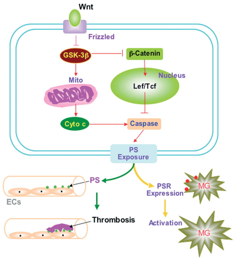
Wnt maintains cellular integrity in endothelial cells and potentially fosters a novel capacity to block thrombosis and microglial activation. Wnt functions through activation of its receptor Frizzled resulting in the inhibition of glycogen synthase kinase-3β (GSK-3β), which induces mitochondrial (Mito) membrane depolarization followed by cytochrome c (Cyto c) release and prevents the activity of β-catenin. Without Wnt, GSK-3β can activate caspases and subsequently leads membrane phosphatidylserine (PS) exposure, resulting in the induction of thrombosis in vascular system and PS receptor (PSR) expression on microglia with microglial activation.
As an early event in the dynamics of cellular apoptosis, PS exposure may be required for embryogenesis (Bose et al., 2004). Yet, in mature tissues, membrane PS externalization can become a signal for the phagocytosis of cells (Hong et al., 2004; Chong et al., 2005a) (Fig. 1). In the nervous system, cells expressing externalized PS may be removed by microglia (Lin and Maiese, 2001; Li et al., 2004b). An additional role for membrane PS externalization in the vascular cell system is the activation of coagulation cascades (Chong et al., 2002b, 2004a). The externalization of membrane PS residues in endothelial cells (ECs) can promote the formation of a procoagulant surface (Bombeli et al., 1997) (Fig. 1).
In contrast to the early externalization of membrane PS residues, the cleavage of genomic DNA into fragments is considered to be a delayed event that occurs late during apoptosis (Dombroski et al., 2000; Maiese and Vincent, 2000; Jessel et al., 2002; Kang et al., 2003b). Several enzymes responsible for DNA degradation have been differentiated based on their ionic sensitivities to zinc (Torriglia et al., 1997) and magnesium (Sun and Cohen, 1994). Calcium, a critical independent component that can determine cell survival (Weber, 2004), also may determine endonuclease activity through calcium/magnesium - dependent endonucleases such as DNase I (Madaio et al., 1996). Other enzymes that may degrade DNA include the acidic, cation independent endonuclease (DNase II) (Torriglia et al., 1995), cyclophilins (Montague et al., 1997), and the 97 kDa magnesium-dependent endonuclease (Pandey et al., 1997). In the nervous system, three separate endonuclease activities are present that include a constitutive acidic cation-independent endonuclease, a constitutive calcium/magnesium-dependent endonuclease, and an inducible magnesium dependent endonuclease (Vincent and Maiese, 1999b). The physiologic characteristics of the magnesium dependent endonuclease, such as a pH range of 7.4–8.0, a dependence on magnesium, and a molecular weight of 95–108 kDa, are consistent with a recently described constitutive 97 kDa endonuclease in non-neuronal tissues.
Oxidative stress can lead to apoptosis in neurons, ECs, cardiomyocytes, and smooth muscle cells through multiple cellular pathways (Chong et al., 2005c). Oxidative stress, such as nitric oxide (NO) exposure results in nuclei condensation and DNA fragmentation (Vincent and Maiese, 1999b; Goldshmit et al., 2001; Chong et al., 2003b; Pugazhenthi et al., 2003). In neurons, NO exposure produces apoptotic death in hippocampal and dopaminergic neurons (Vincent and Maiese, 1999a; Witting et al., 2000; Chong et al., 2003a; Sharma and Ebadi, 2003). Injury during NO exposure also can become synergistic with hydrogen peroxide to render neurons more sensitive to oxidative injury (de la Monte et al., 2003; Wang et al., 2003). Externalization of membrane PS residues also occurs in neurons during anoxia (Chong et al., 2002b), NO exposure (Chong et al., 2003d), or during the administration of agents that induce the production of reactive oxygen species, such as 6-hydroxydopamine (Salinas et al., 2003).
The Wnt-Frizzled signaling pathway controls apoptosis in a variety of cell populations during both development and injury (Fig. 2). During development, Wnt signaling can either facilitate or prevent apoptosis depending upon the environmental stimuli present. For example, Wnt proteins can regulate apoptosis within rhombomeres 3 and 5 in the developing hindbrain and in limb buds during vertebrate limb development to control development of the hindbrain and the limb (Ellies et al., 2000; Grotewold and Ruther, 2002a,b).
Fig. 2.
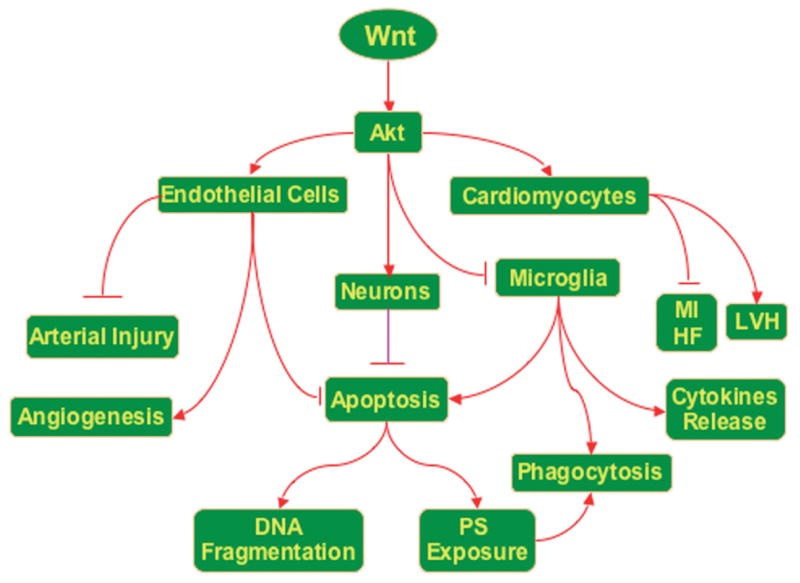
The Wnt-Frizzled signaling pathway employs Akt to regulate an array of vital cellular functions that involve cellular protection, angiogenesis, cardiovascular remodeling, DNA repair, and the maintenance of membrane asymmetry in neuronal and vascular systems. The Wnt-Frizzled signaling pathway employs Akt to impact upon the survival of neurons, endothelial cells, and cardiomyocytes to prevent cellular injury and promote angiogenesis. Through the activation of Akt, microglial activity is modulated to block harmful cytokine release and prevent the phagocytosis of cells “tagged” by cellular phosphatidylserine (PS) membrane exposure. The Wnt-Frizzled signaling pathway function in cardiovascular remodeling that involves regulation of LVH (left ventricular hypertrophy) and reduction in MI (myocardial infarction) and HF (heart failure).
A number of studies in a variety of organisms have demonstrated that the Wnt-Frizzled signaling pathway can regulate apoptosis through a variety of mechanisms that include the Wnt-BMP signaling loop (Golden et al., 1999; Ellies et al., 2000), secreted Frizzled-related protein-2 (SFRP2) expression (Ellies et al., 2000; Jones et al., 2000), Wnt-β-catenin signaling (Galceran et al., 2000; Brault et al., 2001; Ahmed et al., 2002; Hari et al., 2002), c-Jun N-Terminal kinase signaling (Grotewold and Ruther, 2002b; Lisovsky et al., 2002; Yeo and Gautier, 2004), GSK-3β-NF-kB signaling (Bournat et al., 2000; Kozlovsky et al., 2002) and gene expression such as human Dickkopf-1 (hDkk-1) (Shou et al., 2002), nemo (Mirkovic et al., 2002), sox 10 (Honore et al., 2003) and tau (Jackson et al., 2002).
As one of the best characterized members of the Wnt family, Wnt1 signaling has been associated with the control of apoptosis during injury in some cell systems. Wnt1 prevents apoptosis through β-catenin/Tcf transcription mediated pathways (Chen et al., 2001; Rhee et al., 2002). Overexpression of exogenous Wnt1 results in the protection of cells against c-myc induced apoptosis through induction of β-catenin, cyclooxygenase-2, and Wnt1 induced secreted protein (WISP-1) (You et al., 2002). Wnt1 signaling also can inhibit apoptosis through prevention of cytochrome c release from mitochondria and the subsequent inhibition of caspase 9 activation (Chen et al., 2001). The APC gene also appears to represent another mechanism that regulates apoptosis. The APC gene functions to cleave β-catenin leading to the down-regulation of transactivation of Tcf/Lef (Munemitsu et al., 1995). Without Tcf/Lef activity, APC is then permitted to increase the activities of caspase 3, caspase 7, and caspase 9 and lead to the cleavage of poly (ADP-ribose) polymerase (PARP) to enhance the vulnerability of cells to apoptosis (Chen et al., 2003).
SFRP2 is one of the family members of secreted Frizzled-related proteins, also known as secreted apoptosis-related protein 1 (SARP1), that functions as an antagonist of the Wnt-Frizzled signaling pathway to reduce apoptosis during development (Melkonyan et al., 1997; Rattner et al., 1997). For example, there exists a negative relationship between the expression of SFRP2 and the occurrence of apoptosis in rhombomeres 3 and 5. The overexpression of SFRP2 in the rhombencephalic neural crest can prevent the apoptosis of premigratory neural crest cells from rhombomeres 3 and 5 by inhibiting the expression of Wnt1 and BMP 4. In contrast, depleting SFRP2 function or overexpressing Wnt1 in rhombomeres results in apoptosis (Ellies et al., 2000). As an example of how the cellular environment can influence the modulation of apoptosis by the Wnt system, SFRP2 can be associated with increased apoptotic injury during degenerative disease processes such as retinitis pigmentosa that is characterized by progressive death of the photoreceptors due to apoptosis. SFRP2 has been shown to have significantly increased expression in areas of retinal degeneration, suggesting that during neurodegenerative disease blockade of Wnt signaling can lead to cell injury (Jones et al., 2000). In a similar vein, human Dkk-1 and Nemo also are Wnt antagonists similar to the SFRPs, but they can contribute to promote apoptosis by inhibiting the Wnt-Frizzled signaling pathway (Mirkovic et al., 2002; Shou et al., 2002). The results of exogenous application of Dkk-1 have identified that the apoptosis in the limb bud is induced by Dkk-1 through the prevention of the Wnt -Frizzled signaling pathway (Glinka et al., 1998; Grotewold and Ruther, 2002b).
The Wnt-β-catenin signaling pathway is a typical Wnt-Frizzled signaling pathway that regulates multiple cellular and development processes including control of cell proliferation and differentiation, cell polarity, and specification of cell fate, such as apoptosis. Yet, different from the Wnt-BMP signaling loop, the Wnt-β-catenin signaling pathway can prevent apoptosis through the regulation of β-catenin and Tcf/Lef. In β-catenin mutant embryos, the removal of β-catenin can lead to apoptotic loss of the hindbrain, the melanocyte lineage, neural crest cells, sensory neurons and dorsal root ganglia (Brault et al., 2001; Hari et al., 2002). Over-expression of exogenous Wnt-1 results in the protection of cells against c-Myc induced apoptosis through induction of β-catenin, cyclooxygenase-2, and Wnt-1 induced secreted protein (WISP-1) (You et al., 2002).
Additional work has revealed that the Wnt signaling pathway can decrease apoptosis and increase survival of neurons or neuronal cell lines by activating NF-kB (Bournat et al., 2000), inhibiting GSK-3β (Kozlovsky et al., 2002), or blocking the release of cytochrome c. In the absence of Wnt activity, GSK-3β phosphorylates β-catenin at serine or threonine residues of the N-terminal region to predispose degradation of β-catenin through ubiquination (Fig. 3). GSK-3β dependent phosphorylation of β-catenin can be promoted through phosphorylation of Axin (Yamamoto et al., 1999). In studies with chemotherapeutic agents, Wnt1 signaling also can inhibit apoptosis through prevention of cytochrome c release from mitochondria and the subsequent inhibition of caspase 9 activation (Chen et al., 2001).
Fig. 3.
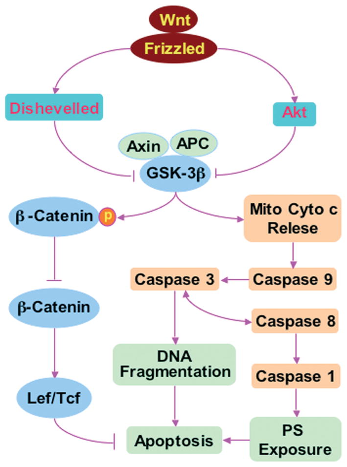
The Wnt-Frizzled signaling pathway regulates PS exposure and apoptosis. The Wnt protein binds to its Frizzled receptors resulting in the activation of Dishevelled followed by the inhibition of GSK-3β (glycogen synthase kinase), Axin and APC (adenomatous polyposis coli) tumor suppressor protein complex. The suppressed GSK-3β, Axin and APC complex prevents phosphorylation (p) of β-catenin and leads to the accumulation of β-catenin. β-catenin enters into cellular nucleus and contributes to the formation of Lef/Tcf (lymphocyte enhancer factor/T cell factor) and β-catenin complex that may cooperate with factors activated by other signaling pathways resulting in cellular proliferation, differentiation, survival and apoptosis through the induction of target nuclear gene transcription. In addition, the binding of Wnt to Frizzled receptors also can directly activate Akt that prevents the activity of GSK-3β and blocks Mito (mitochondrial) Cyto (cytochrome) c release. The inhibition of Cyto c release from Mito leads to the reduction of caspase activity followed by the decrease of DNA fragmentation and phosphatidylserine (PS) exposure resulting in the prevention of apoptosis.
The Wnt pathway also may rely upon protein kinase B (Akt) to promote cellular differentiation and survival (Chong et al., 2005b) (Fig. 3). Since Wnt can inactivate GSK-3β and block the phosphorylation of β-catenin (Ikeda et al., 1998; Papkoff and Aikawa, 1998), this leads to the activation of β-catenin followed by transcription of its target genes for cellular protection. Akt may be necessary in pathways that involve Wnt1, since Akt inhibits the activity of GSK-3β through phosphorylation of this protein to promote cell survival (Crowder and Freeman, 2000). Furthermore, neuronal cell differentiation that is dependent upon Wnt signaling appears to become stalled without Akt phosphorylation and the subsequent inactivation of GSK-3β (Fukumoto et al., 2001). In addition, Wnt has been demonstrated through WISP-1 to activate the anti-apoptotic signaling pathway of Akt following genomic DNA damage (Su et al., 2002) and to block cell injury during serum withdrawal through increased Akt phosphorylation and activity (Longo et al., 2002).
Wnt and neurodegenerative disease
In line with the “anti-apoptotic” effects of Wnt signaling, absence or dysfunction in Wnt signaling can lead to neuronal injury. Wnt1 expression has been demonstrated in the brains of individuals affected by neuropsychiatric disorders (Miyaoka et al., 1999). Furthermore, retinal degeneration during retinitis pigmentosa with the progressive loss of photoreceptors has been associated with increased secretion of Frizzled-related protein-2, a Wnt inhibitory protein, suggesting that loss of Wnt signaling may contribute to retinal neurodegeneration (Jones et al., 2000). Additional work demonstrates that a mutation in the membrane-type Frizzled-related protein gene may be involved in retinal photoreceptor degeneration (Kameya et al., 2002).
During Alzheimer’s disease, neurotoxicity of Aβ in hippocampal neurons has been linked to increased levels of GSK-3β and loss of β-catenin. Decreased production of Ab can occur during the enhancement of PKC activity (Savage et al., 1998) which may be controlled by the Wnt pathway (Garrido et al., 2002). The proteolytic processing of amyloid precursor protein (APP) during Alzheimer’s disease also has been closely linked to the Wnt pathway through presenilin 1 (PS1) and Dishevelled. PS1 is required for the processing of APP and has been shown to down-regulate Wnt signaling and interact with β-catenin to promote its turnover (Soriano et al., 2001). Dishevelled also can regulate the α-secretase cleavage of APP through PKC/mitogen-activated protein kinase dependent pathways, increasing soluble production of APP (sAPP) (Mudher et al., 2001). Overexpression of mouse Dishevelled-1 and -2 inhibits GSK-3β mediated phosphorylation of tau protein and may thus prevent formation of neurofibrillary tangles during Alzheimer’s disease (Wagner et al., 1997). Thus, Dishevelled may increase neuronal protection during neurodegenerative disorders through sAPP production and reduction in tau phosphorylation.
Wnt, Vascular regeneration, and vascular injury
Complimentary to the potential protective role the Wnt system may provide in neuronal populations is the ability of the Wnt pathway to modulate angiogenesis (Table 2), a process that consists of new capillary formation from pre-existing vessels into an avascular area (Chong et al., 2002a). This process involves vascular basal lamina formation, migration of ECs, and alignment of migrating cells for tubular formation (Chong et al., 2002c; Li et al., 2004b; Sakamaki, 2004). There are at least two types of angiogenesis. Sprouting angiogenesis is characterized by the proliferation and migration of ECs into vascular sites (Risau, 1997). In contrast, non-sprouting angiogenesis or intussusceptive microvascular growth occurs by splitting the existing vasculature into transluminal pillars or transendothelial bridges (Burri and Djonov, 2002). Angiogenesis takes place in various physiological and pathophysiological conditions. It is physiologically active during embryogenesis (Risau, 1997). In the adult, it occurs during more limited periods such as during menstruation and during some pathological conditions such as wound healing, chronic inflammation, and tumor growth.
Table 2.
Wnt-Frizzled signaling pathway in the cardiovascular system.
| CARDIOVASCULAR EVENTS | Wnt SIGNALING COMPONENTS | OUTCOME | REFERENCES |
|---|---|---|---|
| Angiogenesis | Wnt1 expression increased | HUVEC proliferation inhibited; HUVEC morphology altered | Cheng et al., 2003 |
| Wnt1 and β-catenin expression increased | EC and VSMC proliferation increased | Wright et al., 1999 | |
| FrzA expression increased | Vessel density increased | Barandon et al., 2003; Dufourcq et al., 2002 | |
| GSK-3β inhibited and β-catenin increased | New vessel formation increased | Holnthoner et al., 2002 | |
| Fzd 4 increased; CAMKII and PKC activated increased | Retinal angiogenesis increased | Robitaille et al., 2002 | |
| Dishevelled expression and β-catenin | Neovascularization increased | Blankesteijn et al., 2000 | |
| CYR 61 expression increased | VSMC adhesion and chemotaxis increased; angiogenesis increased | Brigstock et al., 2002 | |
| Arterial Injury | β-catenin accumulation increased | VSMC survival increased and apoptosis decreased | Wang et al., 2002 |
| FrzA expression increased | Proliferating vascular ECs decreased | Duplaa et al., 1999 | |
| Myocardial Infarction | FrzA expression increased | Infarct size, leukocyte infiltration, and apoptosis decreased; capillary density increased, and cardiac function improved | Barandon et al., 2003 |
| GSK-3β expression inhibited | Cardiomyocyte apoptosis decreased | Bergmann et al., 2004 | |
| Cardiac Hypertrophy | GSK-3β expression inhibited | Cardiomyocycte hypertrophy increased | Haq et al., 2000 |
| Cardiac Injury | SFRP 3/4 increased and β-catenin decreased | Pro-apoptotic Fas increased; anti-apoptotic Bcl-xL decreased; cardiomyocyte apoptosis increased | Schumann et al., 2000 |
| FrzA expression increased | Cardiac apoptosis decreased; cardiac function improved | Barandon et al., 2003 |
CAMKII, calcium/calmodulin-dependent protein kinase II; CYR 61, cysteine-rich 61; EC, endothelial cell; Fzd, Frizzled; GSK-3β, glycogen synthase kinase-3β; HUVEC, human umbilical vein endothelial cell; PKC, protein kinase C; SFRP, secreted Frizzled related protein; VSMC, vascular smooth muscle cell.
Several Wnt ligands, such as Wnt2, Wnt5a, Wnt7a, and Wnt10b, are expressed endogenously in ECs and vascular smooth muscle cells (VSMCs). More importantly, Wnt receptors that involve Fzd1, Fzd2, Fzd3, and Fzd5, as well as cysteine-rich 61 that contains Wnt-induced secreted proteins-1, 2 and 3 are also expressed in these cell populations for Wnt to exert a direct biological effect (Brigstock, 2002; Goodwin and D’Amore, 2002). It has been demonstrated that mice deficient in Wnt2 and Fzd5 display vascular abnormalities that include defective placental vasculature as well as embryonic lethal mutations (Ishikawa et al., 2001; Goodwin and D’Amore, 2002), suggesting that the Wnt pathway is essential for vessel development.
In several vascular cell populations, Wnt signaling plays a significant part in the modulation of new vessel formation. Overexpression of Wnt1 can lead to the proliferation of cultured primary ECs, increase the free pool of β-catenin, and activate transcription through Tcf/Lef, suggesting that the Wnt-Frizzled signaling pathway is closely involved in the proliferation of ECs (Wright et al., 1999). Yet, Wnt1 signaling also can have a regulatory role, such as during the proliferation of umbilical vein ECs, to block further growth through mechanisms that involve cell-cell contact (Cheng et al., 2003). Studies with mice homozygous for the deletion of the Wnt receptor ligand Fzd5 that can synergize with Wnt2, Wnt5a, and Wnt10b lead to embryos that died in utero approximately 10 days post coitum as a result of defects in yolk sac angiogenesis, supporting a critical role for Wnt during embryo vascular development (Ishikawa et al., 2001). In contrast, mice heterozygotes were found to be viable, fertile and appear normal. In disease that involve failure of peripheral retinal vascularization, mutations in Fzd4, a gene encoding the Wnt receptor Frizzled-4, are believed to account for the vascular failure. Studies have shown that injection of only wildtype Fzd4, but not mutated Fzd4, into Xenopus embryos can activate CamKII and PKC, components of the Wnt/Ca2+ signaling pathway, to control retinal angiogenesis (Robitaille et al., 2002), supporting that Wnt-Frizzled signaling pathway broadly controls vascular development and function in a number of organ systems. In addition, FrzA, as a member of secreted Frizzled-related protein, can increase migration and tube formation of ECs to result in enlarged, longer, and mature of vessels, further supporting the necessity of the Wnt-Frizzled signaling pathway during development of vasculature (Dufourcq et al., 2002).
In addition to vascular development, the Wnt pathway participates in the remodeling of vascular structure and the regulation of apoptosis during vascular injury (Table 2). Using a rat aorta balloon injury model, the Frizzled receptor (rFrz) genes, rFrz1 and rFrz 2, have been shown to be transiently down-regulated as early as one hour following balloon. Yet, Frzb-1, a secreted protein that acts as an antagonist of Wnt signaling, can be increased and appears to coincide with the arrest of aortic smooth muscle cell proliferation (Mao et al., 2000). Similarly, the secreted protein FrzA can be elevated in ECs during traumatic manipulation and subsequently block the proliferation of ECs (Duplaa et al., 1999). In regards to apoptotic vascular injury, it has been shown that within eight hours following EC shear stress, expression of β-catenin is significantly increased at the cell-cell junctions of ECs (Noria et al., 1999). In addition, transfection of a degradation-resistant β-catenin into rat VSMCs blocked apoptosis during vascular balloon injury and was associated with cell cycle progression by activating cyclin D1. Furthermore, use of a dominant negative Tcf-4 transgene lacking the β-catenin binding domain, Tcf4(N31), abolished cytoprotection (Wang et al., 2002). The Wnt- Frizzled signaling pathway also is involved with venous injury. In a crush injury vein model, Wnt5a can have increased expression that may be beneficial coupled to the repression of inhibitor proteins of the Wnt system (Price et al., 2004).
Wnt and cardiac infarction
In regards to ischemic heart disease, new vessel formation through angiogenesis is considered to be critical for effective reparative processes (Table 2). The neovascularization following myocardial infarction contributes to the restoration of blood supply to the infarct area. In transgenic mice following myocardial infarction, overexpression of FrzA can result in the reduction of infarct size, prevention of cardiac rupture, and improvement in cardiac function following the increase of capillary density and the decrease of apoptotic cardiomyocytes (Barandon et al., 2003). Additional work also has demonstrated that β-catenin is translocated from the plasma membrane to the cytoplasm of ECs following the expression of Dishevelled during the phase of the neovascularization after myocardial infarction (Blankesteijn et al., 2000). These data suggest that the Wnt system and its components that involve β-catenin can prevent cardiomyocyte injury and preserve potential myocardial function.
Following myocardial infarction, the remodeling of the injured heart tissue involves cellular proliferation, migration, and neovascularization. The proliferation and migration of fibroblast-like cells into the infarcted area can contribute to the deposition of extracellular matrix to promote granulation tissue extension. Subsequently, myofibroblasts emerge that contribute to the preservation of cardiac function by preventing the dilatation of the infarcted area (Gabbiani, 1998). The neovascularization following myocardial infarction serves to restore blood supply to the infarcted area to preserve cardiac function.
During the complicated process of wound healing in the heart, abnormalities of some components of the Wnt-Frizzled signaling pathway, such as Wnt (Barandon et al., 2003), Frizzled (van Gijn et al., 2001), Dishevelled-1 (Chen et al., 2004), GSK-3β (El Jamali et al., 2004), β-catenin (Bergmann et al., 2004), and SFRP (Barandon et al., 2003), have been observed. For example, during myoblast proliferation and migration following myocardial infarction, elevated expression of Frizzled genes including Frizzled 1, 2, 5, 6, 7, 8, and 10 have been identified during heart remodeling. The overexpression of some Frizzled genes may be associated with reduction in infarct size and the prevention of cardiac rupture to improve cardiac function (Barandon et al., 2003). It is important to note that different members of the Wnt family may contribute to distinct events during cardiac injury. Both Wnt8a and Wnt10b are upregulated following myocardial infarction while Wnt7b is down-regulated to undetectable levels during this period (Barandon et al., 2003).
Furthermore, the expression of another component, Dishevelled-1, of the Wnt-Frizzled signaling pathway also has been found in the myofibroblasts during cardiac infarction. Absence of Dishevelled-1 can not only induce the formation of myocardial infarction in mice, but also enhance the development of infarct rupture in the first week after myocardial infarction (Blankesteijn et al., 1999). In addition, the expression of Dishevelled-1 mRNA and cytoplasmic Dishevelled-1 protein are significantly enhanced within days in the myofibroblasts, vascular ECs, and smooth muscle cells of newly formed and pre-existing blood vessels in the region of a myocardial infarction. As a result, Dishevelled-1 may play a critical role in the wound healing process following myocardial infarction through the regulation of proliferation and migration of myofibroblasts and vascular ECs (Chen et al., 2004).
In addition to Frizzled, Dishevelled-1, and several Wnt genes, other members of the Wnt signaling cascade also appear to have relevant roles during myocardial infarction. Expression of SFRP3 and SFRP4 in failing human ventricular myocardium is increased when compared to donor hearts. This increased expression of SFRP is associated with the expression of the pro-apoptotic Fas proteins, but inversely linked to the expression of the anti-apoptotic protein Bcl-xL (Schumann et al., 2000). Other studies illustrate that infarct rupture following myocardial infarction is correlated with decreased levels of β-catenin in cardiomyocytes (Blankesteijn et al., 1999). The lack of β-catenin in the adherence junctions of cardiomyocytes may lead to impaired structural integrity of the heart, since β-catenin may play a vital role in a structural adaptor protein linking cadherins to the actin cytoskeleton in cell- cell adhesion (Ligon et al., 2001). Yet, the role of β-catenin during cardiac infarction is not entirely clear, since cytosolic β-catenin exists in vascular ECs and smooth muscle cells that reside in the area of myocardial infarction (Barandon et al., 2003).
As an additional parameter that can lead to cardiac failure, pathological cardiac hypertrophy may be dependent upon modulation of the Wnt pathway. In volume-overloaded rabbit hearts, the cardiac hypertrophy is accompanied by suppressed mRNA expression of β-catenin (Itoh et al., 2002). The suppressed levels of β-catenin may be a result of increased GSK-3β activity. Ectopic expression of GSK-3β can lead to embryonic heart development (Marvin et al., 2001; Schneider and Mercola, 2001), suggesting that GSK-3β my function as a negative regulator of cardiac hypertrophy. Additional work has demonstrated that inhibition of GSK-3β by Akt through phosphorylation of a serine residue at position 9 can play an important role in the development of cardiac hypertrophy (Haq et al., 2000; Hardt and Sadoshima, 2002).
Wnt and trophic factor regulation
Interestingly, the angiogenic effects of some growth factors may be closely linked to the Wnt- Frizzled signaling pathway. Vascular endothelial growth factor (VEGF) can initiate increased EC growth and stimulate new vessel formation by promoting tyrosine phosphorylation of β-catenin in ECs (Cohen et al., 1999; Roura et al., 1999). In addition, fibroblast growth factor 2 (FGF2) can inhibit GSK-3 activity, augment nuclear levels of β-catenin, and enhance Tcf/Lef-dependent transcription of a cyclin D1-luciferase construct, suggesting that the angiogenic properties of FGF2 are tightly regulated by β-catenin activation in the Wnt-Frizzled signaling pathway (Dono et al., 2002; Holnthoner et al., 2002).
Other cytokines and trophic factors also may have some dependence on the Wnt signaling pathway (Maiese et al., 2004, 2005). One potential candidate is erythropoietin (EPO). EPO is currently approved by the Food and Drug Administration for the treatment of anemia that can be the result of a variety of conditions. Yet, in a relatively short time period, the premise that EPO is required only for erythropoiesis has been advanced by studies demonstrating the existence of EPO and its receptor in other organs and tissues outside of the liver and the kidney, such as the heart and brain. As a result, EPO has been identified as a possible candidate in the formulation of therapeutic strategies for both cardiac and nervous system diseases.
EPO appears to be efficacious during cardiac ischemia and reperfusion injury (Li et al., 2004a; Maiese et al., 2005). EPO administration either prior to or during myocardial ischemia/reperfusion can decrease infarct size, resulting in enhanced cardiac function and improved left ventricular contractility (Parsa et al., 2004). EPO also can promote microvascular growth in the heart, suggesting that functional cardiac recovery with EPO may ensue also from the generation of new blood vessels (Ribatti et al., 1999). Other experimental studies have illustrated a potential direct protection of myocardial cells during EPO administration that can increase cardiac cell proliferation in neonatal rats, reduce myocardiocyte apoptosis during ischemia-reperfusion injury, and improve left ventricular function (Moon et al., 2003; Parsa et al., 2003).
In the nervous system, both cell culture and animal model work have demonstrated neuronal and vascular protection with EPO (Chong et al., 2002a,c; Genc et al., 2004). Systemic administration of EPO before or immediately after a retinal insult can protect retinal ganglion cells from apoptosis (Grimm et al., 2002) and can improve functional outcome and reduce lipid peroxidation during spinal cord injury (Kaptanoglu et al., 2004). EPO also can block microglial cell activation and proliferation to prevent phagocytosis of injured cells through pathways that involve cellular membrane PS exposure (Chong et al., 2004a) and the regulation of caspases (Chong et al., 2003b,c). EPO can directly address cellular inflammation by inhibiting several pro-inflammatory cytokines, such as IL-6, tumor necrosis factor-α(TNF-α), and monocyte chemoattractant protein 1 (Chong et al., 2002c; Genc et al., 2004).
A number of trophic factors and cytokines, such as EPO, may depend upon a member of the Wnt signaling pathway. For example, new work illustrates that EPO can maintain the subcellular localization of β-catenin in the nucleus of microglia during oxidative stress that involves oxygen-glucose deprivation, suggesting that EPO protects β-catenin from degradation (Fig. 4). In addition, other components of the Wnt pathway that are utilized by EPO, such as Akt, can offer cellular protection (Maiese et al., 2003). EPO can phosphorylate Akt and is dependent upon the activation of PI 3-K and Janus Kinase 2 (Jak2) (Witthuhn et al., 1993; Chong et al., 2002b). One of the principal pathways through which EPO prevents cellular apoptosis is through the activation of Akt (Maiese et al., 2004, 2005) During anoxia or free radical exposure, expression of the active form of Akt (phospho-Akt) is increased (Kang et al., 2003a,b). EPO can significantly enhance the activity of Akt during oxidative stress and prevent inflammatory activation of microglia (Chong et al., 2003a-c). This up-regulation of Akt activity during injury paradigms appears to be vital for EPO protection, since prevention of Akt phosphorylation blocks cellular protection by EPO (Chong et al., 2003a-c). Through the regulation of the PI 3-K/Akt dependent pathway, EPO can prevent cellular apoptosis following N-methyl-D-aspartate toxicity (Dzietko et al., 2004), hypoxia (Chong et al., 2002b), and oxidative stress (Chong et al., 2003a-c).
Fig. 4.
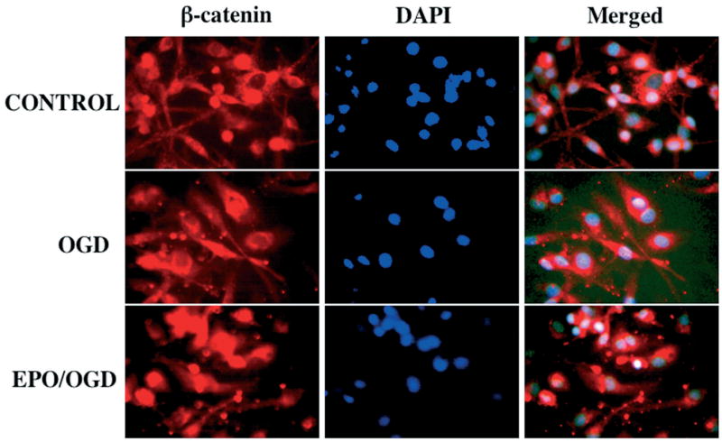
Eythropoietin (EPO) employs the Wnt pathway to modulate β-catenin. The pretreatment of EPO at 1 hour before oxygen-glucose deprivation (OGD) regulates the β-catenin location in microglia following OGD. Immunohistochemical double staining for β-catenin was obtained in microglia twelve hours following an six hour period of OGD by using a primary rabbit anti-β-catenin antibody with Texas-red avidin. OGD in microglia was performed by replacing media with glucose-free HBSS containing 116 mM NaCl, 5.4 mM KCl, 0.8 mM MgSO4, 1 mM NaH2PO4, 0.9 mM CaCl2, and 10 mg/L phenol red (pH 7.4) and cultures were maintained in an anoxic environment (95% N2 and 5% CO2) at 37 C for 6 hours. Cell nuclei were stained with DAPI (4′-6-diamidino-2-phenylinodole). Representative images illustrate the accumulation of β-catenin in the microglial cytoplasm during OGD and the translocation of β-catenin from the cytoplasm to nuclei following the application of EPO, but the expression of β-catenin exists in both cytoplasm and nuclei without cellular translocation in control (untreated) microglia.
Conclusion
In recent years, our understanding of the Wnt-Frizzled signaling pathway has advanced at a tremendous pace. The Wnt-Frizzled signaling pathway that consists of the pathways of Wnt/β-catenin, Wnt/Ca2+, and Wnt/planar polarity have significant roles during embryonic development, neurodegenerative disease, and cardiovascular disease. In particular, the Wnt-Frizzled signaling pathway is involved with the development of the neural plate and with the subsequent anterior-posterior extension of the neural tube. Ultimately, the Wnt-Frizzled signaling pathway leads to the development of the brain, spinal cord, and the extension of numerous sub-populations of sensory and motor neurons. If one examines the cardiovascular system, members of the Wnt family are intimately involved in vascular remolding, cardiac development, and cardiac hypertophy. The Wnt-Frizzled signaling pathway also controls both early and late apoptotic injury paradigms in a variety of cell populations during the development of an organism as well as during acute and chronic injury. Diseases of the nervous system, such as Alzheimer’s disease or retinal degeneration, and disorders of the vascular system, such as cardiac infarction and EC injury, are closely regulated by the Wnt pathway. Furthermore, both angiogenic factors and other trophic factors rely upon members of the Wnt family. Through further identification and targeting of the critical elements that shape and control the Wnt-Frizzled signaling pathway, a greater understanding of the biological potential of Wnt-Frizzled signaling pathway can emerge for the development of new therapeutic options against neurodegenerative and vascular diseases.
Acknowledgments
This research was supported by the following grants (KM): American Heart Association (National), Bugher Foundation Award, Janssen Neuroscience Award, Johnson and Johnson Focused Investigator Award, LEARN Foundation Award, MI Life Sciences Challenge Award, and NIH NIEHS (P30 ES06639).
References
- Aberle H, Bauer A, Stappert J, Kispert A, Kemler R. beta-catenin is a target for the ubiquitin-proteasome pathway. EMBO J. 1997;16:3797–3804. doi: 10.1093/emboj/16.13.3797. [DOI] [PMC free article] [PubMed] [Google Scholar]
- Abu-Khalil A, Fu L, Grove EA, Zecevic N, Geschwind DH. Wnt genes define distinct boundaries in the developing human brain: implications for human forebrain patterning. J Comp Neurol. 2004;474:276–288. doi: 10.1002/cne.20112. [DOI] [PubMed] [Google Scholar]
- Adler PN, Vinson C, Park WJ, Conover S, Klein L. Molecular structure of frizzled, a Drosophila tissue polarity gene. Genetics. 1990;126:401–416. doi: 10.1093/genetics/126.2.401. [DOI] [PMC free article] [PubMed] [Google Scholar]
- Ahmed Y, Nouri A, Wieschaus E. Drosophila Apc1 and Apc2 regulate Wingless transduction throughout development. Development. 2002;129:1751–1762. doi: 10.1242/dev.129.7.1751. [DOI] [PubMed] [Google Scholar]
- Akiyama T. Wnt/beta-catenin signaling. Cytokine growth factor Rev. 2000;11:273–282. doi: 10.1016/s1359-6101(00)00011-3. [DOI] [PubMed] [Google Scholar]
- Anderton BH. Alzheimer’s disease: clues from flies and worms. Curr Biol. 1999;9:R106–109. doi: 10.1016/s0960-9822(99)80062-1. [DOI] [PubMed] [Google Scholar]
- Augustine K, Liu ET, Sadler TW. Antisense attenuation of Wnt-1 and Wnt-3a expression in whole embryo culture reveals roles for these genes in craniofacial, spinal cord, and cardiac morphogenesis. Dev Genet. 1993;14:500–520. doi: 10.1002/dvg.1020140611. [DOI] [PubMed] [Google Scholar]
- Axelrod JD, Miller JR, Shulman JM, Moon RT, Perrimon N. Differential recruitment of Dishevelled provides signaling specificity in the planar cell polarity and Wingless signaling pathways. Genes Dev. 1998;12:2610–2622. doi: 10.1101/gad.12.16.2610. [DOI] [PMC free article] [PubMed] [Google Scholar]
- Baker JC, Beddington RS, Harland RM. Wnt signaling in Xenopus embryos inhibits bmp4 expression and activates neural development. Genes Dev. 1999;13:3149–3159. doi: 10.1101/gad.13.23.3149. [DOI] [PMC free article] [PubMed] [Google Scholar]
- Barandon L, Couffinhal T, Ezan J, Dufourcq P, Costet P, Alzieu P, Leroux L, Moreau C, Dare D, Duplaa C. Reduction of infarct size and prevention of cardiac rupture in transgenic mice overexpressing FrzA. Circulation. 2003;108:2282–2289. doi: 10.1161/01.CIR.0000093186.22847.4C. [DOI] [PubMed] [Google Scholar]
- Bastidas F, De Calisto J, Mayor R. Identification of neural crest competence territory: role of Wnt signaling. Dev Dyn. 2004;229:109–117. doi: 10.1002/dvdy.10486. [DOI] [PubMed] [Google Scholar]
- Behrens J, von Kries JP, Kuhl M, Bruhn L, Wedlich D, Grosschedl R, Birchmeier W. Functional interaction of beta-catenin with the transcription factor LEF-1. Nature. 1996;382:638–642. doi: 10.1038/382638a0. [DOI] [PubMed] [Google Scholar]
- Bejsovec A. Wnt pathway activation: new relations and locations. Cell. 2005;120:11–14. doi: 10.1016/j.cell.2004.12.021. [DOI] [PubMed] [Google Scholar]
- Bergmann MW, Rechner C, Freund C, Baurand A, El Jamali A, Dietz R. Statins inhibit reoxygenation-induced cardiomyocyte apoptosis: role for glycogen synthase kinase 3beta and transcription factor beta-catenin. J Mol Cell Cardiol. 2004;37:681–690. doi: 10.1016/j.yjmcc.2004.05.025. [DOI] [PubMed] [Google Scholar]
- Bhanot P, Brink M, Samos CH, Hsieh JC, Wang Y, Macke JP, Andrew D, Nathans J, Nusse R. A new member of the frizzled family from Drosophila functions as a Wingless receptor. Nature. 1996;382:225–230. doi: 10.1038/382225a0. [DOI] [PubMed] [Google Scholar]
- Blankesteijn WM, Gijn MEV, Daemen MJAP, Wynshaw-Boris, Smits JFM, Pratt RE. Alterations in the cadherin-catenin complex of dishevelled-1 knockout mice lead to infarct rupture after myocardial infarction. Circulation. 1999;100(suppl):156. [Google Scholar]
- Blankesteijn WM, van Gijn ME, Essers-Janssen YP, Daemen MJ, Smits JF. Beta-catenin, an inducer of uncontrolled cell proliferation and migration in malignancies, is localized in the cytoplasm of vascular endothelium during neovascularization after myocardial infarction. Am J Pathol. 2000;157:877–883. doi: 10.1016/s0002-9440(10)64601-9. [DOI] [PMC free article] [PubMed] [Google Scholar]
- Bombeli T, Karsan A, Tait JF, Harlan JM. Apoptotic vascular endothelial cells become procoagulant. Blood. 1997;89:2429–2442. [PubMed] [Google Scholar]
- Bonvini P, An WG, Rosolen A, Nguyen P, Trepel J, Garcia de Herreros A, Dunach M, Neckers LM. Geldanamycin abrogates ErbB2 association with proteasome-resistant beta-catenin in melanoma cells, increases beta-catenin-E-cadherin association, and decreases beta-catenin-sensitive transcription. Cancer Res. 2001;61:1671–1677. [PubMed] [Google Scholar]
- Bose J, Gruber AD, Helming L, Schiebe S, Wegener I, Hafner M, Beales M, Kontgen F, Lengeling A. The phosphatidylserine receptor has essential functions during embryogenesis but not in apoptotic cell removal. J Biol. 2004;3:15. doi: 10.1186/jbiol10. [DOI] [PMC free article] [PubMed] [Google Scholar]
- Bournat JC, Brown AM, Soler AP. Wnt-1 dependent activation of the survival factor NF-kappaB in PC12 cells. J Neurosci Res. 2000;61:21–32. doi: 10.1002/1097-4547(20000701)61:1<21::AID-JNR3>3.0.CO;2-7. [DOI] [PubMed] [Google Scholar]
- Boutros M, Mlodzik M. Dishevelled: at the crossroads of divergent intracellular signaling pathways. Mech Dev. 1999;83:27–37. doi: 10.1016/s0925-4773(99)00046-5. [DOI] [PubMed] [Google Scholar]
- Boutros M, Paricio N, Strutt DI, Mlodzik M. Dishevelled activates JNK and discriminates between JNK pathways in planar polarity and wingless signaling. Cell. 1998;94:109–118. doi: 10.1016/s0092-8674(00)81226-x. [DOI] [PubMed] [Google Scholar]
- Brault V, Moore R, Kutsch S, Ishibashi M, Rowitch DH, McMahon AP, Sommer L, Boussadia O, Kemler R. Inactivation of the beta-catenin gene by Wnt1-Cre-mediated deletion results in dramatic brain malformation and failure of craniofacial development. Development. 2001;128:1253–1264. doi: 10.1242/dev.128.8.1253. [DOI] [PubMed] [Google Scholar]
- Braun MM, Etheridge A, Bernard A, Robertson CP, Roelink H. Wnt signaling is required at distinct stages of development for the induction of the posterior forebrain. Development. 2003;130:5579–5587. doi: 10.1242/dev.00685. [DOI] [PubMed] [Google Scholar]
- Brigstock DR. Regulation of angiogenesis and endothelial cell function by connective tissue growth factor (CTGF) and cysteine-rich 61 (CYR61) Angiogenesis. 2002;5:153–165. doi: 10.1023/a:1023823803510. [DOI] [PubMed] [Google Scholar]
- Bronner-Fraser M. Development. Making sense of the sensory lineage Science. 2004;303:966–968. doi: 10.1126/science.1094732. [DOI] [PubMed] [Google Scholar]
- Burri PH, Djonov V. Intussusceptive angiogenesis--the alternative to capillary sprouting. Mol Aspects Med. 2002;23:S1–27. doi: 10.1016/s0098-2997(02)00096-1. [DOI] [PubMed] [Google Scholar]
- Cai Z, Manalo DJ, Wei G, Rodriguez ER, Fox-Talbot K, Lu H, Zweier JL, Semenza GL. Hearts from rodents exposed to intermittent hypoxia or erythropoietin are protected against ischemia-reperfusion injury. Circulation. 2003;108:79–85. doi: 10.1161/01.CIR.0000078635.89229.8A. [DOI] [PubMed] [Google Scholar]
- Castelo-Branco G, Wagner J, Rodriguez FJ, Kele J, Sousa K, Rawal N, Pasolli HA, Fuchs E, Kitajewski J, Arenas E. Differential regulation of midbrain dopaminergic neuron development by Wnt-1, Wnt-3a, and Wnt-5a. Proc Natl Acad Sci USA. 2003;100:12747–12752. doi: 10.1073/pnas.1534900100. [DOI] [PMC free article] [PubMed] [Google Scholar]
- Cavallo RA, Cox RT, Moline MM, Roose J, Polevoy GA, Clevers H, Peifer M, Bejsovec A. Drosophila Tcf and Groucho interact to repress Wingless signalling activity. Nature. 1998;395:604–608. doi: 10.1038/26982. [DOI] [PubMed] [Google Scholar]
- Chen L, Wu Q, Guo F, Xia B, Zuo J. Expression of Dishevelled-1 in wound healing after acute myocardial infarction: possible involvement in myofibroblast proliferation and migration. J Cell Mol Med. 2004;8:257–264. doi: 10.1111/j.1582-4934.2004.tb00281.x. [DOI] [PMC free article] [PubMed] [Google Scholar]
- Chen S, Guttridge DC, You Z, Zhang Z, Fribley A, Mayo MW, Kitajewski J, Wang CY. Wnt-1 signaling inhibits apoptosis by activating beta-catenin/T cell factor-mediated transcription. J Cell Biol. 2001;152:87–96. doi: 10.1083/jcb.152.1.87. [DOI] [PMC free article] [PubMed] [Google Scholar]
- Chen T, Yang I, Irby R, Shain KH, Wang HG, Quackenbush J, Coppola D, Cheng JQ, Yeatman TJ. Regulation of caspase expression and apoptosis by adenomatous polyposis coli. Cancer Res. 2003;63:4368–4374. [PubMed] [Google Scholar]
- Cheng CW, Smith SK, Charnock-Jones DS. Wnt-1 signaling inhibits human umbilical vein endothelial cell proliferation and alters cell morphology. Exp Cell Res. 2003;291:415–425. doi: 10.1016/j.yexcr.2003.07.006. [DOI] [PubMed] [Google Scholar]
- Cheyette BN. Ryk: another heretical Wnt receptor defies the canon. Sci STKE 2004. 2004:pe54. doi: 10.1126/stke.2632004pe54. [DOI] [PubMed] [Google Scholar]
- Chizhikov VV, Millen KJ. Roof plate-dependent patterning of the vertebrate dorsal central nervous system. Dev Biol. 2005;277:287–295. doi: 10.1016/j.ydbio.2004.10.011. [DOI] [PubMed] [Google Scholar]
- Chong ZZ, Maiese K. Targeting WNT, protein kinase B, and mitochondrial membrane integrity to foster cellular survival in the nervous system. Histol Histopathol. 2004;19:495–504. doi: 10.14670/hh-19.495. [DOI] [PMC free article] [PubMed] [Google Scholar]
- Chong ZZ, Kang JQ, Maiese K. Angiogenesis and plasticity: role of erythropoietin in vascular systems. J Hematother Stem Cell Res. 2002a;11:863–871. doi: 10.1089/152581602321080529. [DOI] [PubMed] [Google Scholar]
- Chong ZZ, Kang JQ, Maiese K. Erythropoietin is a novel vascular protectant through activation of Akt1 and mitochondrial modulation of cysteine proteases. Circulation. 2002b;106:2973–2979. doi: 10.1161/01.cir.0000039103.58920.1f. [DOI] [PubMed] [Google Scholar]
- Chong ZZ, Kang JQ, Maiese K. Hematopoietic factor erythropoietin fosters neuroprotection through novel signal transduction cascades. J Cereb Blood Flow Metab. 2002c;22:503–514. doi: 10.1097/00004647-200205000-00001. [DOI] [PubMed] [Google Scholar]
- Chong ZZ, Kang JQ, Maiese K. Apaf-1, Bcl-xL, Cytochrome c, and Caspase-9 Form the Critical Elements for Cerebral Vascular Protection by Erythropoietin. J Cereb Blood Flow Metab. 2003a;23:320–330. doi: 10.1097/01.WCB.0000050061.57184.AE. [DOI] [PubMed] [Google Scholar]
- Chong ZZ, Kang JQ, Maiese K. Erythropoietin fosters both intrinsic and extrinsic neuronal protection through modulation of microglia, Akt1, Bad, and caspase-mediated pathways. Br J Pharmacol. 2003b;138:1107–1118. doi: 10.1038/sj.bjp.0705161. [DOI] [PMC free article] [PubMed] [Google Scholar]
- Chong ZZ, Kang JQ, Maiese K. Akt1 drives endothelial cell membrane asymmetry and microglial activation through Bcl-x(L) and caspase 1, 3, and 9. Exp Cell Res. 2004a;296:196–207. doi: 10.1016/j.yexcr.2004.01.021. [DOI] [PubMed] [Google Scholar]
- Chong ZZ, Kang JQ, Maiese K. Essential cellular regulatory elements of oxidative stress in early and late phases of apoptosis in the central nervous system. Antioxid Redox Signal. 2004b;6:277–287. doi: 10.1089/152308604322899341. [DOI] [PubMed] [Google Scholar]
- Chong ZZ, Lin SH, Kang JQ, Maiese K. Erythropoietin prevents early and late neuronal demise through modulation of Akt1 and induction of caspase 1, 3, and 8. J Neurosci Res. 2003c;71:659–669. doi: 10.1002/jnr.10528. [DOI] [PubMed] [Google Scholar]
- Chong ZZ, Lin SH, Kang JQ, Maiese K. The tyrosine phosphatase SHP2 modulates MAP kinase p38 and caspase 1 and 3 to foster neuronal survival. Cell Mol Neurobiol. 2003d;23:561–578. doi: 10.1023/A:1025158314016. [DOI] [PMC free article] [PubMed] [Google Scholar]
- Chong ZZ, Kang J-Q, Li F, Maiese K. mGluRI targets microglial activation and selectively prevents neuronal cell engulfment through Akt and caspase dependent pathways. Curr Neurovasc Res. 2005a;2:197–211. doi: 10.2174/1567202054368317. [DOI] [PMC free article] [PubMed] [Google Scholar]
- Chong ZZ, Li F, Maiese K. Activating Akt and the brain’s resources to drive cellular survival and prevent inflammatory injury. Histol Histopathol. 2005b;20:299–315. doi: 10.14670/hh-20.299. [DOI] [PMC free article] [PubMed] [Google Scholar]
- Chong ZZ, Li F, Maiese K. Stress in the brain: novel cellular mechanisms of injury linked to Alzheimer’s disease. Brain Res Brain Res Rev. 2005c;49:1–21. doi: 10.1016/j.brainresrev.2004.11.005. [DOI] [PMC free article] [PubMed] [Google Scholar]
- Chong ZZ, Li FQ, Maiese K. Employing new cellular therapeutic targets for Alzheimer’s disease: A change for the better? Curr Neurovasc Res. 2005d;2:55–72. doi: 10.2174/1567202052773508. [DOI] [PMC free article] [PubMed] [Google Scholar]
- Christian JL, Olson DJ, Moon RT. Xwnt-8 modifies the character of mesoderm induced by bFGF in isolated Xenopus ectoderm. EMBO J. 1992;11:33–41. doi: 10.1002/j.1460-2075.1992.tb05024.x. [DOI] [PMC free article] [PubMed] [Google Scholar]
- Cohen AW, Carbajal JM, Schaeffer RC., Jr VEGF stimulates tyrosine phosphorylation of beta-catenin and small-pore endothelial barrier dysfunction. Am J Physiol. 1999;277:H2038–2049. doi: 10.1152/ajpheart.1999.277.5.H2038. [DOI] [PubMed] [Google Scholar]
- Crowder RJ, Freeman RS. Glycogen synthase kinase-3 beta activity is critical for neuronal death caused by inhibiting phosphatidylinositol 3-kinase or Akt but not for death caused by nerve growth factor withdrawal. J Biol Chem. 2000;275:34266–34271. doi: 10.1074/jbc.M006160200. [DOI] [PubMed] [Google Scholar]
- D’Amico M, Hulit J, Amanatullah DF, Zafonte BT, Albanese C, Bouzahzah B, Fu M, Augenlicht LH, Donehower LA, Takemaru K, Moon RT, Davis R, Lisanti MP, Shtutman M, Zhurinsky J, Ben-Ze’ev A, Troussard AA, Dedhar S, Pestell RG. The integrin-linked kinase regulates the cyclin D1 gene through glycogen synthase kinase 3beta and cAMP-responsive element-binding protein-dependent pathways. J Biol Chem. 2000;275:32649–32657. doi: 10.1074/jbc.M000643200. [DOI] [PubMed] [Google Scholar]
- Dale TC. Signal transduction by the Wnt family of ligands. Biochem J. 1998;329(Pt 2):209–223. doi: 10.1042/bj3290209. [DOI] [PMC free article] [PubMed] [Google Scholar]
- Danilkovitch-Miagkova A, Miagkov A, Skeel A, Nakaigawa N, Zbar B, Leonard EJ. Oncogenic mutants of RON and MET receptor tyrosine kinases cause activation of the beta-catenin pathway. Mol Cell Biol. 2001;21:5857–5868. doi: 10.1128/MCB.21.17.5857-5868.2001. [DOI] [PMC free article] [PubMed] [Google Scholar]
- de la Monte SM, Chiche J, von dem Bussche A, Sanyal S, Lahousse SA, Janssens SP, Bloch KD. Nitric oxide synthase-3 overexpression causes apoptosis and impairs neuronal mitochondrial function: relevance to Alzheimer’s-type neurodegeneration. Lab Invest. 2003;83:287–298. doi: 10.1097/01.lab.0000056995.07053.c0. [DOI] [PubMed] [Google Scholar]
- Deshpande R, Inoue T, Priess JR, Hill RJ. lin-17/Frizzled and lin-18 regulate POP-1/TCF-1 localization and cell type specification during C. elegans vulval development. Dev Biol. 2005;278:118–129. doi: 10.1016/j.ydbio.2004.10.020. [DOI] [PubMed] [Google Scholar]
- Dombroski D, Balasubramanian K, Schroit AJ. Phosphatidylserine expression on cell surfaces promotes antibody-dependent aggregation and thrombosis in beta2-glycoprotein I-immune mice. J Autoimmun. 2000;14:221–229. doi: 10.1006/jaut.2000.0365. [DOI] [PubMed] [Google Scholar]
- Dono R, Faulhaber J, Galli A, Zuniga A, Volk T, Texido G, Zeller R, Ehmke H. FGF2 signaling is required for the development of neuronal circuits regulating blood pressure. Circ Res. 2002;90:E5–E10. [PubMed] [Google Scholar]
- Doonan F, Cotter TG. Apoptosis: A potential therapeutic target for retinal degenerations. Curr Neurovasc Res. 2004;1:41–53. doi: 10.2174/1567202043480215. [DOI] [PubMed] [Google Scholar]
- Dufourcq P, Couffinhal T, Ezan J, Barandon L, Moreau C, Daret D, Duplaa C. FrzA, a secreted frizzled related protein, induced angiogenic response. Circulation. 2002;106:3097–3103. doi: 10.1161/01.cir.0000039342.85015.5c. [DOI] [PubMed] [Google Scholar]
- Duplaa C, Jaspard B, Moreau C, D’Amore PA. Identification and cloning of a secreted protein related to the cysteine-rich domain of frizzled. Evidence for a role in endothelial cell growth control. Circ Res. 1999;84:1433–1445. doi: 10.1161/01.res.84.12.1433. [DOI] [PubMed] [Google Scholar]
- Dzietko M, Felderhoff-Mueser U, Sifringer M, Krutz B, Bittigau P, Thor F, Heumann R, Buhrer C, Ikonomidou C, Hansen HH. Erythropoietin protects the developing brain against N-methyl-D-aspartate receptor antagonist neurotoxicity. Neurobiol Dis. 2004;15:177–187. doi: 10.1016/j.nbd.2003.10.006. [DOI] [PubMed] [Google Scholar]
- El Jamali A, Freund C, Rechner C, Scheidereit C, Dietz R, Bergmann MW. Reoxygenation after severe hypoxia induces cardiomyocyte hypertrophy in vitro: activation of CREB downstream of GSK3beta. FASEB J. 2004;18:1096–1098. doi: 10.1096/fj.03-1054fje. [DOI] [PubMed] [Google Scholar]
- Ellies DL, Church V, Francis-West P, Lumsden A. The WNT antagonist cSFRP2 modulates programmed cell death in the developing hindbrain. Development. 2000;127:5285–5295. doi: 10.1242/dev.127.24.5285. [DOI] [PubMed] [Google Scholar]
- Fagotto F, Funayama N, Gluck U, Gumbiner BM. Binding to cadherins antagonizes the signaling activity of beta-catenin during axis formation in Xenopus. J Cell Biol. 1996;132:1105–1114. doi: 10.1083/jcb.132.6.1105. [DOI] [PMC free article] [PubMed] [Google Scholar]
- Ferretti P. Neural stem cell plasticity: Recruitment of endogenous populations for regeneration. Curr Neurovasc Res. 2004;1:215–229. doi: 10.2174/1567202043362397. [DOI] [PubMed] [Google Scholar]
- Finch PW, He X, Kelley MJ, Uren A, Schaudies RP, Popescu NC, Rudikoff S, Aaronson SA, Varmus HE, Rubin JS. Purification and molecular cloning of a secreted, Frizzled-related antagonist of Wnt action. Proc Natl Acad Sci USA. 1997;94:6770–6775. doi: 10.1073/pnas.94.13.6770. [DOI] [PMC free article] [PubMed] [Google Scholar]
- Fukumoto S, Hsieh CM, Maemura K, Layne MD, Yet SF, Lee KH, Matsui T, Rosenzweig A, Taylor WG, Rubin JS, Perrella MA, Lee ME. Akt participation in the Wnt signaling pathway through Dishevelled. J Biol Chem. 2001;276:17479–17483. doi: 10.1074/jbc.C000880200. [DOI] [PubMed] [Google Scholar]
- Gabbiani G. Evolution and clinical implications of the myofibroblast concept. Cardiovasc Res. 1998;38:545–548. doi: 10.1016/s0008-6363(98)00065-0. [DOI] [PubMed] [Google Scholar]
- Galceran J, Miyashita-Lin EM, Devaney E, Rubenstein JL, Grosschedl R. Hippocampus development and generation of dentate gyrus granule cells is regulated by LEF1. Development. 2000;127:469–482. doi: 10.1242/dev.127.3.469. [DOI] [PubMed] [Google Scholar]
- Garrido JL, Godoy JA, Alvarez A, Bronfman M, Inestrosa NC. Protein kinase C inhibits amyloid beta peptide neurotoxicity by acting on members of the Wnt pathway. Faseb J. 2002;16:1982–1984. doi: 10.1096/fj.02-0327fje. [DOI] [PubMed] [Google Scholar]
- Genc S, Koroglu TF, Genc K. Erythropoietin as a novel neuroprotectant. Res Neur Neurosci. 2004;22:105–119. [PubMed] [Google Scholar]
- Glass DJ, Bowen DC, Stitt TN, Radziejewski C, Bruno J, Ryan TE, Gies DR, Shah S, Mattsson K, Burden SJ, DiStefano PS, Valenzuela DM, DeChiara TM, Yancopoulos GD. Agrin acts via a MuSK receptor complex. Cell. 1996;85:513–523. doi: 10.1016/s0092-8674(00)81252-0. [DOI] [PubMed] [Google Scholar]
- Glinka A, Wu W, Delius H, Monaghan AP, Blumenstock C, Niehrs C. Dickkopf-1 is a member of a new family of secreted proteins and functions in head induction. Nature. 1998;391:357–362. doi: 10.1038/34848. [DOI] [PubMed] [Google Scholar]
- Golden JA, Bracilovic A, McFadden KA, Beesley JS, Rubenstein JL, Grinspan JB. Ectopic bone morphogenetic proteins 5 and 4 in the chicken forebrain lead to cyclopia and holoprosencephaly. Proc Natl Acad Sci USA. 1999;96:2439–2444. doi: 10.1073/pnas.96.5.2439. [DOI] [PMC free article] [PubMed] [Google Scholar]
- Goldshmit Y, Erlich S, Pinkas-Kramarski R. Neuregulin rescues PC12-ErbB4 cells from cell death induced by H(2)O(2). Regulation of reactive oxygen species levels by phosphatidylinositol 3-kinase. J Biol Chem. 2001;276:46379–46385. doi: 10.1074/jbc.M105637200. [DOI] [PubMed] [Google Scholar]
- Goodwin AM, D’Amore PA. Wnt signaling in the vasculature. Angiogenesis. 2002;5:1–9. doi: 10.1023/a:1021563510866. [DOI] [PubMed] [Google Scholar]
- Graham NA, Asthagiri AR. Epidermal growth factor-mediated T-cell factor/lymphoid enhancer factor transcriptional activity is essential but not sufficient for cell cycle progression in nontransformed mammary epithelial cells. J Biol Chem. 2004;279:23517–23524. doi: 10.1074/jbc.M314055200. [DOI] [PubMed] [Google Scholar]
- Grilli M, Toninelli GF, Uberti D, Spano P, Memo M. Alzheimer’s disease linking neurodegeneration with neurodevelopment. Funct Neurol. 2003;18:145–148. [PubMed] [Google Scholar]
- Grimm C, Wenzel A, Groszer M, Mayser H, Seeliger M, Samardzija M, Bauer C, Gassmann M, Reme CE. HIF-1-induced erythropoietin in the hypoxic retina protects against light-induced retinal degeneration. Nat Med. 2002;8:718–724. doi: 10.1038/nm723. [DOI] [PubMed] [Google Scholar]
- Grotewold L, Ruther U. Bmp, Fgf and Wnt signalling in programmed cell death and chondrogenesis during vertebrate limb development: the role of Dickkopf-1. Int J Dev Biol. 2002a;46:943–947. [PubMed] [Google Scholar]
- Grotewold L, Ruther U. The Wnt antagonist Dickkopf-1 is regulated by Bmp signaling and c-Jun and modulates programmed cell death. EMBO J. 2002b;21:966–975. doi: 10.1093/emboj/21.5.966. [DOI] [PMC free article] [PubMed] [Google Scholar]
- Grove EA, Tole S. Patterning events and specification signals in the developing hippocampus. Cereb Cortex. 1999;9:551–561. doi: 10.1093/cercor/9.6.551. [DOI] [PubMed] [Google Scholar]
- Grove EA, Tole S, Limon J, Yip L, Ragsdale CW. The hem of the embryonic cerebral cortex is defined by the expression of multiple Wnt genes and is compromised in Gli3-deficient mice. Development. 1998;125:2315–2325. doi: 10.1242/dev.125.12.2315. [DOI] [PubMed] [Google Scholar]
- Habas R, Dawid IB. Dishevelled and Wnt signaling: is the nucleus the final frontier? J Biol. 2005;4:2. doi: 10.1186/jbiol22. [DOI] [PMC free article] [PubMed] [Google Scholar]
- Habas R, Dawid IB, He X. Coactivation of Rac and Rho by Wnt/Frizzled signaling is required for vertebrate gastrulation. Genes Dev. 2003;17:295–309. doi: 10.1101/gad.1022203. [DOI] [PMC free article] [PubMed] [Google Scholar]
- Hall AC, Lucas FR, Salinas PC. Axonal remodeling and synaptic differentiation in the cerebellum is regulated by WNT-7a signaling. Cell. 2000;100:525–535. doi: 10.1016/s0092-8674(00)80689-3. [DOI] [PubMed] [Google Scholar]
- Haq S, Choukroun G, Kang ZB, Ranu H, Matsui T, Rosenzweig A, Molkentin JD, Alessandrini A, Woodgett J, Hajjar R, Michael A, Force T. Glycogen synthase kinase-3beta is a negative regulator of cardiomyocyte hypertrophy. J Cell Biol. 2000;151:117–130. doi: 10.1083/jcb.151.1.117. [DOI] [PMC free article] [PubMed] [Google Scholar]
- Haq S, Choukroun G, Lim H, Tymitz KM, del Monte F, Gwathmey J, Grazette L, Michael A, Hajjar R, Force T, Molkentin JD. Differential activation of signal transduction pathways in human hearts with hypertrophy versus advanced heart failure. Circulation. 2001;103:670–677. doi: 10.1161/01.cir.103.5.670. [DOI] [PubMed] [Google Scholar]
- Hardt SE, Sadoshima J. Glycogen synthase kinase-3beta: a novel regulator of cardiac hypertrophy and development. Circ Res. 2002;90:1055–1063. doi: 10.1161/01.res.0000018952.70505.f1. [DOI] [PubMed] [Google Scholar]
- Hari L, Brault V, Kleber M, Lee HY, Ille F, Leimeroth R, Paratore C, Suter U, Kemler R, Sommer L. Lineage-specific requirements of beta-catenin in neural crest development. J Cell Biol. 2002;159:867–880. doi: 10.1083/jcb.200209039. [DOI] [PMC free article] [PubMed] [Google Scholar]
- Hart M, Concordet JP, Lassot I, Albert I, del los Santos R, Durand H, Perret C, Rubinfeld B, Margottin F, Benarous R, Polakis P. The F-box protein beta-TrCP associates with phosphorylated beta-catenin and regulates its activity in the cell. Curr Biol. 1999;9:207–210. doi: 10.1016/s0960-9822(99)80091-8. [DOI] [PubMed] [Google Scholar]
- Hasegawa S, Sato T, Akazawa H, Okada H, Maeno A, Ito M, Sugitani Y, Shibata H, Miyazaki Ji J, Katsuki M, Yamauchi Y, Yamamura Ki K, Katamine S, Noda T. Apoptosis in neural crest cells by functional loss of APC tumor suppressor gene. Proc Natl Acad Sci USA. 2002;99:297–302. doi: 10.1073/pnas.012264999. [DOI] [PMC free article] [PubMed] [Google Scholar]
- Hatsugai N, Kuroyanagi M, Yamada K, Meshi T, Tsuda S, Kondo M, Nishimura M, Hara-Nishimura I. A plant vacuolar protease, VPE, mediates virus-induced hypersensitive cell death. Science. 2004;305:855–858. doi: 10.1126/science.1099859. [DOI] [PubMed] [Google Scholar]
- He TC, Sparks AB, Rago C, Hermeking H, Zawel L, da Costa LT, Morin PJ, Vogelstein B, Kinzler KW. Identification of c-MYC as a target of the APC pathway. Science. 1998;281:1509–1512. doi: 10.1126/science.281.5382.1509. [DOI] [PubMed] [Google Scholar]
- He XC, Zhang J, Tong WG, Tawfik O, Ross J, Scoville DH, Tian Q, Zeng X, He X, Wiedemann LM, Mishina Y, Li L. BMP signaling inhibits intestinal stem cell self-renewal through suppression of Wnt-beta-catenin signaling. Nat Genet. 2004;36:1117–1121. doi: 10.1038/ng1430. [DOI] [PubMed] [Google Scholar]
- Heisenberg CP, Tada M, Rauch GJ, Saude L, Concha ML, Geisler R, Stemple DL, Smith JC, Wilson SW. Silberblick/Wnt11 mediates convergent extension movements during zebrafish gastrulation. Nature. 2000;405:76–81. doi: 10.1038/35011068. [DOI] [PubMed] [Google Scholar]
- Hirabayashi Y, Itoh Y, Tabata H, Nakajima K, Akiyama T, Masuyama N, Gotoh Y. The Wnt/beta-catenin pathway directs neuronal differentiation of cortical neural precursor cells. Development. 2004;131:2791–2801. doi: 10.1242/dev.01165. [DOI] [PubMed] [Google Scholar]
- Holnthoner W, Pillinger M, Groger M, Wolff K, Ashton AW, Albanese C, Neumeister P, Pestell RG, Petzelbauer P. Fibroblast growth factor-2 induces Lef/Tcf-dependent transcription in human endothelial cells. J Biol Chem. 2002;277:45847–45853. doi: 10.1074/jbc.M209354200. [DOI] [PubMed] [Google Scholar]
- Hong JR, Lin GH, Lin CJ, Wang WP, Lee CC, Lin TL, Wu JL. Phosphatidylserine receptor is required for the engulfment of dead apoptotic cells and for normal embryonic development in zebrafish. Development. 2004;131:5417–5427. doi: 10.1242/dev.01409. [DOI] [PubMed] [Google Scholar]
- Honore SM, Aybar MJ, Mayor R. Sox10 is required for the early development of the prospective neural crest in Xenopus embryos. Dev Biol. 2003;260:79–96. doi: 10.1016/s0012-1606(03)00247-1. [DOI] [PubMed] [Google Scholar]
- Hsieh JC. Specificity of WNT-receptor interactions. Front Biosci. 2004;9:1333–1338. doi: 10.2741/1321. [DOI] [PubMed] [Google Scholar]
- Hsieh JC, Rattner A, Smallwood PM, Nathans J. Biochemical characterization of Wnt-frizzled interactions using a soluble, biologically active vertebrate Wnt protein. Proc Natl Acad Sci USA. 1999;96:3546–3551. doi: 10.1073/pnas.96.7.3546. [DOI] [PMC free article] [PubMed] [Google Scholar]
- Ikeda S, Kishida S, Yamamoto H, Murai H, Koyama S, Kikuchi A. Axin, a negative regulator of the Wnt signaling pathway, forms a complex with GSK-3beta and beta-catenin and promotes GSK-3beta-dependent phosphorylation of beta-catenin. EMBO J. 1998;17:1371–1384. doi: 10.1093/emboj/17.5.1371. [DOI] [PMC free article] [PubMed] [Google Scholar]
- Ikeya M, Lee SM, Johnson JE, McMahon AP, Takada S. Wnt signalling required for expansion of neural crest and CNS progenitors. Nature. 1997;389:966–970. doi: 10.1038/40146. [DOI] [PubMed] [Google Scholar]
- Ilyas M. Wnt signalling and the mechanistic basis of tumour development. J Pathol. 2005;205:130–144. doi: 10.1002/path.1692. [DOI] [PubMed] [Google Scholar]
- Inoue T, Oz HS, Wiland D, Gharib S, Deshpande R, Hill RJ, Katz WS, Sternberg PW. C. elegans LIN-18 is a Ryk ortholog and functions in parallel to LIN-17/Frizzled in Wnt signaling. Cell. 2004;118:795–806. doi: 10.1016/j.cell.2004.09.001. [DOI] [PubMed] [Google Scholar]
- Ishikawa T, Tamai Y, Zorn AM, Yoshida H, Seldin MF, Nishikawa S, Taketo MM. Mouse Wnt receptor gene Fzd5 is essential for yolk sac and placental angiogenesis. Development. 2001;128:25–33. doi: 10.1242/dev.128.1.25. [DOI] [PubMed] [Google Scholar]
- Ishitani T, Ninomiya-Tsuji J, Matsumoto K. Regulation of lymphoid enhancer factor 1/T-cell factor by mitogen-activated protein kinase-related Nemo-like kinase-dependent phosphorylation in Wnt/beta-catenin signaling. Mol Cell Biol. 2003;23:1379–1389. doi: 10.1128/MCB.23.4.1379-1389.2003. [DOI] [PMC free article] [PubMed] [Google Scholar]
- Itoh K, Brott BK, Bae GU, Ratcliffe MJ, Sokol SY. Nuclear localization is required for Dishevelled function in Wnt/beta-catenin signaling. J Biol. 2005;4:3. doi: 10.1186/jbiol20. [DOI] [PMC free article] [PubMed] [Google Scholar]
- Itoh M, Takeishi Y, Nakada S, Miyamoto T, Tsunoda Y, Takahashi H, Kubota I, Tomoike H. Long-term treatment with angiotensin II type 1 receptor antagonist, CV-11974, restores beta-catenin mRNA expression in volume-overloaded rabbit hearts. Heart Vessels. 2002;17:36–41. doi: 10.1007/s003800200040. [DOI] [PubMed] [Google Scholar]
- Jackson GR, Wiedau-Pazos M, Sang TK, Wagle N, Brown CA, Massachi S, Geschwind DH. Human wild-type tau interacts with wingless pathway components and produces neurofibrillary pathology in Drosophila. Neuron. 2002;34:509–519. doi: 10.1016/s0896-6273(02)00706-7. [DOI] [PubMed] [Google Scholar]
- Jamora C, Fuchs E. Intercellular adhesion, signalling and the cytoskeleton. Nat Cell Biol. 2002;4:E101–108. doi: 10.1038/ncb0402-e101. [DOI] [PubMed] [Google Scholar]
- Jennings CG, Dyer SM, Burden SJ. Muscle-specific trk-related receptor with a kringle domain defines a distinct class of receptor tyrosine kinases. Proc Natl Acad Sci USA. 1993;90:2895–2899. doi: 10.1073/pnas.90.7.2895. [DOI] [PMC free article] [PubMed] [Google Scholar]
- Jessel R, Haertel S, Socaciu C, Tykhonova S, Diehl HA. Kinetics of apoptotic markers in exogeneously induced apoptosis of EL4 cells. J Cell Mol Med. 2002;6:82–92. doi: 10.1111/j.1582-4934.2002.tb00313.x. [DOI] [PMC free article] [PubMed] [Google Scholar]
- Jho EH, Zhang T, Domon C, Joo CK, Freund JN, Costantini F. Wnt/beta-catenin/Tcf signaling induces the transcription of Axin2, a negative regulator of the signaling pathway. Mol Cell Biol. 2002;22:1172–1183. doi: 10.1128/MCB.22.4.1172-1183.2002. [DOI] [PMC free article] [PubMed] [Google Scholar]
- Jones SE, Jomary C, Grist J, Stewart HJ, Neal MJ. Modulated expression of secreted frizzled-related proteins in human retinal degeneration. Neuroreport. 2000;11:3963–3967. doi: 10.1097/00001756-200012180-00012. [DOI] [PubMed] [Google Scholar]
- Jordan BK, Mohammed M, Ching ST, Delot E, Chen XN, Dewing P, Swain A, Rao PN, Elejalde BR, Vilain E. Up-regulation of WNT-4 signaling and dosage-sensitive sex reversal in humans. Am J Hum Genet. 2001;68:1102–1109. doi: 10.1086/320125. [DOI] [PMC free article] [PubMed] [Google Scholar]
- Kameya S, Hawes NL, Chang B, Heckenlively JR, Naggert JK, Nishina PM. Mfrp, a gene encoding a frizzled related protein, is mutated in the mouse retinal degeneration 6. Hum Mol Genet. 2002;11:1879–1886. doi: 10.1093/hmg/11.16.1879. [DOI] [PubMed] [Google Scholar]
- Kang JQ, Chong ZZ, Maiese K. Akt1 protects against inflammatory microglial activation through maintenance of membrane asymmetry and modulation of cysteine protease activity. J Neurosci Res. 2003a;74:37–51. doi: 10.1002/jnr.10740. [DOI] [PubMed] [Google Scholar]
- Kang JQ, Chong ZZ, Maiese K. Critical role for Akt1 in the modulation of apoptotic phosphatidylserine exposure and microglial activation. Mol Pharmacol. 2003b;64:557–569. doi: 10.1124/mol.64.3.557. [DOI] [PubMed] [Google Scholar]
- Kaptanoglu E, Solaroglu I, Okutan O, Surucu HS, Akbiyik F, Beskonakli E. Erythropoietin exerts neuroprotection after acute spinal cord injury in rats: effect on lipid peroxidation and early ultrastructural findings. Neurosurg Rev. 2004;27:113–120. doi: 10.1007/s10143-003-0300-y. [DOI] [PubMed] [Google Scholar]
- Katoh M. Regulation of WNT signaling molecules by retinoic acid during neuronal differentiation in NT2 cells: threshold model of WNT action. Int J Mol Med. 2002;10:683–687. [PubMed] [Google Scholar]
- Kawakami Y, Capdevila J, Buscher D, Itoh T, Rodriguez Esteban C, Izpisua Belmonte JC. WNT signals control FGF-dependent limb initiation and AER induction in the chick embryo. Cell. 2001;104:891–900. doi: 10.1016/s0092-8674(01)00285-9. [DOI] [PubMed] [Google Scholar]
- Kengaku M, Twombly V, Tabin C. Expression of Wnt and Frizzled genes during chick limb bud development. Cold Spring Harb Symp Quant Biol. 1997;62:421–429. [PubMed] [Google Scholar]
- Kiecker C, Niehrs C. A morphogen gradient of Wnt/beta-catenin signalling regulates anteroposterior neural patterning in Xenopus. Development. 2001;128:4189–4201. doi: 10.1242/dev.128.21.4189. [DOI] [PubMed] [Google Scholar]
- Kishida M, Hino S, Michiue T, Yamamoto H, Kishida S, Fukui A, Asashima M, Kikuchi A. Synergistic activation of the Wnt signaling pathway by Dvl and casein kinase Iepsilon. J Biol Chem. 2001;276:33147–33155. doi: 10.1074/jbc.M103555200. [DOI] [PubMed] [Google Scholar]
- Koyama R, Ikegaya Y. Mossy fiber sprouting as a potential therapeutic target for epilepsy. Curr Neurovasc Res. 2004;1:3–10. doi: 10.2174/1567202043480242. [DOI] [PubMed] [Google Scholar]
- Koyanagi M, Haendeler J, Badorff C, Brandes RP, Hoffmann J, Pandur P, Zeiher AM, Kuhl M, Dimmeler S. Non-canonical Wnt signaling enhances differentiation of human circulating progenitor cells to cardiomyogenic cells. J Biol Chem. 2005;280:16838–16842. doi: 10.1074/jbc.M500323200. [DOI] [PubMed] [Google Scholar]
- Kozlovsky N, Belmaker RH, Agam G. GSK-3 and the neurodevelopmental hypothesis of schizophrenia. Eur Neuropsychopharmacol. 2002;12:13–25. doi: 10.1016/s0924-977x(01)00131-6. [DOI] [PubMed] [Google Scholar]
- Kuhl M. The WNT/calcium pathway: biochemical mediators, tools and future requirements. Front Biosci. 2004;9:967–974. doi: 10.2741/1307. [DOI] [PubMed] [Google Scholar]
- Kuhl M, Sheldahl LC, Park M, Miller JR, Moon RT. The Wnt/Ca2+ pathway: a new vertebrate Wnt signaling pathway takes shape. Trends Genet. 2000;16:279–283. doi: 10.1016/s0168-9525(00)02028-x. [DOI] [PubMed] [Google Scholar]
- Kunz M, Herrmann M, Wedlich D, Gradl D. Autoregulation of canonical Wnt signaling controls midbrain development. Dev Biol. 2004;273:390–401. doi: 10.1016/j.ydbio.2004.06.015. [DOI] [PubMed] [Google Scholar]
- LaBonne C, Bronner-Fraser M. Neural crest induction in Xenopus: evidence for a two-signal model. Development. 1998;125:2403–2414. doi: 10.1242/dev.125.13.2403. [DOI] [PubMed] [Google Scholar]
- Latres E, Chiaur DS, Pagano M. The human F box protein beta-Trcp associates with the Cul1/Skp1 complex and regulates the stability of beta-catenin. Oncogene. 1999;18:849–854. doi: 10.1038/sj.onc.1202653. [DOI] [PubMed] [Google Scholar]
- Lee E, Salic A, Kirschner MW. Physiological regulation of [beta]-catenin stability by Tcf3 and CK1epsilon. J Cell Biol. 2001;154:983–993. doi: 10.1083/jcb.200102074. [DOI] [PMC free article] [PubMed] [Google Scholar]
- Lee HY, Kleber M, Hari L, Brault V, Suter U, Taketo MM, Kemler R, Sommer L. Instructive role of Wnt/beta-catenin in sensory fate specification in neural crest stem cells. Science. 2004;303:1020–1023. doi: 10.1126/science.1091611. [DOI] [PubMed] [Google Scholar]
- Lee JS, Ishimoto A, Yanagawa S. Characterization of mouse dishevelled (Dvl) proteins in Wnt/Wingless signaling pathway. J Biol Chem. 1999;274:21464–21470. doi: 10.1074/jbc.274.30.21464. [DOI] [PubMed] [Google Scholar]
- Lee SM, Tole S, Grove E, McMahon AP. A local Wnt-3a signal is required for development of the mammalian hippocampus. Development. 2000;127:457–467. doi: 10.1242/dev.127.3.457. [DOI] [PubMed] [Google Scholar]
- Lewis JL, Bonner J, Modrell M, Ragland JW, Moon RT, Dorsky RI, Raible DW. Reiterated Wnt signaling during zebrafish neural crest development. Development. 2004;131:1299–1308. doi: 10.1242/dev.01007. [DOI] [PubMed] [Google Scholar]
- Leyns L, Bouwmeester T, Kim SH, Piccolo S, De Robertis EM. Frzb-1 is a secreted antagonist of Wnt signaling expressed in the Spemann organizer. Cell. 1997;88:747–756. doi: 10.1016/s0092-8674(00)81921-2. [DOI] [PMC free article] [PubMed] [Google Scholar]
- Li L, Yuan H, Xie W, Mao J, Caruso AM, McMahon A, Sussman DJ, Wu D. Dishevelled proteins lead to two signaling pathways. Regulation of LEF-1 and c-Jun N-terminal kinase in mammalian cells. J Biol Chem. 1999;274:129–134. doi: 10.1074/jbc.274.1.129. [DOI] [PubMed] [Google Scholar]
- Li F, Chong ZZ, Maiese K. Erythropoietin on a tightrope: balancing neuronal and vascular protection between Iitrinsic and extrinsic pathways. Neurosignals. 2004a;13:265–289. doi: 10.1159/000081963. [DOI] [PubMed] [Google Scholar]
- Li F, Chong ZZ, Maiese K. Navigating novel mechanisms of cellular plasticity with the NAD+ precursor and nutrient nicotinamide. Front Biosci. 2004b;9:2500–2520. doi: 10.2741/1412. [DOI] [PubMed] [Google Scholar]
- Ligon LA, Karki S, Tokito M, Holzbaur EL. Dynein binds to beta-catenin and may tether microtubules at adherens junctions. Nat Cell Biol. 2001;3:913–917. doi: 10.1038/ncb1001-913. [DOI] [PubMed] [Google Scholar]
- Lin SH, Maiese K. The metabotropic glutamate receptor system protects against ischemic free radical programmed cell death in rat brain endothelial cells. J Cereb Blood Flow Metab. 2001;21:262–275. doi: 10.1097/00004647-200103000-00010. [DOI] [PubMed] [Google Scholar]
- Lisovsky M, Itoh K, Sokol SY. Frizzled receptors activate a novel JNK-dependent pathway that may lead to apoptosis. Curr Biol. 2002;12:53–58. doi: 10.1016/s0960-9822(01)00628-5. [DOI] [PubMed] [Google Scholar]
- Liu P, Wakamiya M, Shea MJ, Albrecht U, Behringer RR, Bradley A. Requirement for Wnt3 in vertebrate axis formation. Nat Genet. 1999;22:361–365. doi: 10.1038/11932. [DOI] [PubMed] [Google Scholar]
- Longo KA, Kennell JA, Ochocinska MJ, Ross SE, Wright WS, MacDougald OA. Wnt signaling protects 3T3-L1 preadipocytes from apoptosis through induction of insulin-like growth factors. J Biol Chem. 2002;277:38239–38244. doi: 10.1074/jbc.M206402200. [DOI] [PubMed] [Google Scholar]
- Lu W, Yamamoto V, Ortega B, Baltimore D. Mammalian Ryk is a Wnt coreceptor required for stimulation of neurite outgrowth. Cell. 2004;119:97–108. doi: 10.1016/j.cell.2004.09.019. [DOI] [PubMed] [Google Scholar]
- Lustig B, Behrens J. The Wnt signaling pathway and its role in tumor development. J Cancer Res Clin Oncol. 2003;129:199–221. doi: 10.1007/s00432-003-0431-0. [DOI] [PMC free article] [PubMed] [Google Scholar]
- Lustig B, Jerchow B, Sachs M, Weiler S, Pietsch T, Karsten U, van de Wetering M, Clevers H, Schlag PM, Birchmeier W, Behrens J. Negative feedback loop of Wnt signaling through upregulation of conductin/axin2 in colorectal and liver tumors. Mol Cell Biol. 2002;22:1184–1193. doi: 10.1128/MCB.22.4.1184-1193.2002. [DOI] [PMC free article] [PubMed] [Google Scholar]
- Machon O, van den Bout CJ, Backman M, Kemler R, Krauss S. Role of beta-catenin in the developing cortical and hippocampal neuroepithelium. Neuroscience. 2003;122:129–143. doi: 10.1016/s0306-4522(03)00519-0. [DOI] [PubMed] [Google Scholar]
- Madaio MP, Fabbi M, Tiso M, Daga A, Puccetti A. Spontaneously produced anti-DNA/DNase I autoantibodies modulate nuclear apoptosis in living cells. Eur J Immunol. 1996;26:3035–3041. doi: 10.1002/eji.1830261232. [DOI] [PubMed] [Google Scholar]
- Maiese K. The dynamics of cellular injury: transformation into neuronal and vascular protection. Histol Histopathol. 2001;16:633–644. doi: 10.14670/HH-16.633. [DOI] [PubMed] [Google Scholar]
- Maiese K, Chong ZZ. Nicotinamide: necessary nutrient emerges as a novel cytoprotectant for the brain. Trends Pharmacol Sci. 2003;24:228–232. doi: 10.1016/S0165-6147(03)00078-6. [DOI] [PubMed] [Google Scholar]
- Maiese K, Chong ZZ. Insights into oxidative stress and potential novel therapeutic targets for Alzheimer disease. Restor Neurol Neurosci. 2004;22:87–104. [PubMed] [Google Scholar]
- Maiese K, Chong ZZ, Kang J. Transformation into treatment: Novel therapeutics that begin within the cell. In: Maiese K, editor. Neuronal and vascular plasticity: Elucidating basic cellular mechanisms for future therapeutic discovery. Kluwer Academic Publishers; Norwell, MA: 2003. pp. 1–26. [Google Scholar]
- Maiese K, Li F, Chong ZZ. Erythropoietin in the brain: can the promise to protect be fulfilled? Trends Pharmacol Sci. 2004;25:577–583. doi: 10.1016/j.tips.2004.09.006. [DOI] [PubMed] [Google Scholar]
- Maiese K, Li F, Chong ZZ. New avenues of exploration for erythropoietin. JAMA. 2005;293:90–95. doi: 10.1001/jama.293.1.90. [DOI] [PMC free article] [PubMed] [Google Scholar]
- Maiese K, Vincent AM. Membrane asymmetry and DNA degradation: functionally distinct determinants of neuronal programmed cell death. J Neurosci Res. 2000;59:568–580. doi: 10.1002/(SICI)1097-4547(20000215)59:4<568::AID-JNR13>3.0.CO;2-R. [DOI] [PubMed] [Google Scholar]
- Mao C, Malek OT, Pueyo ME, Steg PG, Soubrier F. Differential expression of rat frizzled-related frzb-1 and frizzled receptor fz1 and fz2 genes in the rat aorta after balloon injury. Arterioscler Thromb Vasc Biol. 2000;20:43–51. doi: 10.1161/01.atv.20.1.43. [DOI] [PubMed] [Google Scholar]
- Mao J, Wang J, Liu B, Pan W, Farr GH, 3rd, Flynn C, Yuan H, Takada S, Kimelman D, Li L, Wu D. Low-density lipoprotein receptor-related protein-5 binds to Axin and regulates the canonical Wnt signaling pathway. Mol Cell. 2001;7:801–809. doi: 10.1016/s1097-2765(01)00224-6. [DOI] [PubMed] [Google Scholar]
- Marvin MJ, Di Rocco G, Gardiner A, Bush SM, Lassar AB. Inhibition of Wnt activity induces heart formation from posterior mesoderm. Genes Dev. 2001;15:316–327. doi: 10.1101/gad.855501. [DOI] [PMC free article] [PubMed] [Google Scholar]
- Mattson MP. Pathways towards and away from Alzheimer’s disease. Nature. 2004;430:631–639. doi: 10.1038/nature02621. [DOI] [PMC free article] [PubMed] [Google Scholar]
- Mayr T, Deutsch U, Kuhl M, Drexler HC, Lottspeich F, Deutzmann R, Wedlich D, Risau W. Fritz: a secreted frizzled-related protein that inhibits Wnt activity. Mech Dev. 1997;63:109–125. doi: 10.1016/s0925-4773(97)00035-x. [DOI] [PubMed] [Google Scholar]
- Melkonyan HS, Chang WC, Shapiro JP, Mahadevappa M, Fitzpatrick PA, Kiefer MC, Tomei LD, Umansky SR. SARPs: a family of secreted apoptosis-related proteins. Proc Natl Acad Sci USA. 1997;94:13636–13641. doi: 10.1073/pnas.94.25.13636. [DOI] [PMC free article] [PubMed] [Google Scholar]
- Melton KR, Iulianella A, Trainor PA. Gene expression and regulation of hindbrain and spinal cord development. Front Biosci. 2004;9:117–138. doi: 10.2741/1202. [DOI] [PubMed] [Google Scholar]
- Michiue T, Fukui A, Yukita A, Sakurai K, Danno H, Kikuchi A, Asashima M. XIdax, an inhibitor of the canonical Wnt pathway, is required for anterior neural structure formation in Xenopus. Dev Dyn. 2004;230:79–90. doi: 10.1002/dvdy.20037. [DOI] [PubMed] [Google Scholar]
- Miller JR. The Wnts. Genome Biol. 2002;3:REVIEWS3001. doi: 10.1186/gb-2001-3-1-reviews3001. [DOI] [PMC free article] [PubMed] [Google Scholar]
- Mirkovic I, Charish K, Gorski SM, McKnight K, Verheyen EM. Drosophila nemo is an essential gene involved in the regulation of programmed cell death. Mech Dev. 2002;119:9–20. doi: 10.1016/s0925-4773(02)00289-7. [DOI] [PubMed] [Google Scholar]
- Miyaoka T, Seno H, Ishino H. Increased expression of Wnt-1 in schizophrenic brains. Schizophr Res. 1999;38:1–6. doi: 10.1016/s0920-9964(98)00179-0. [DOI] [PubMed] [Google Scholar]
- Montague JW, Hughes F, Jr, Cidlowski JA. Native recombinant cyclophilins A, B, and C degrade DNA independently of peptidylprolyl cis-trans-isomerase activity. Potential roles of cyclophilins in apoptosis. J Biol Chem. 1997;272:6677–6684. doi: 10.1074/jbc.272.10.6677. [DOI] [PubMed] [Google Scholar]
- Moon C, Krawczyk M, Ahn D, Ahmet I, Paik D, Lakatta EG, Talan MI. Erythropoietin reduces myocardial infarction and left ventricular functional decline after coronary artery ligation in rats. Proc Natl Acad Sci USA. 2003;100:11612–11617. doi: 10.1073/pnas.1930406100. [DOI] [PMC free article] [PubMed] [Google Scholar]
- Moon RT, Campbell RM, Christian JL, McGrew LL, Shih J, Fraser S. Xwnt-5A: a maternal Wnt that affects morphogenetic movements after overexpression in embryos of Xenopus laevis. Development. 1993;119:97–111. doi: 10.1242/dev.119.1.97. [DOI] [PubMed] [Google Scholar]
- Moriguchi T, Kawachi K, Kamakura S, Masuyama N, Yamanaka H, Matsumoto K, Kikuchi A, Nishida E. Distinct domains of mouse dishevelled are responsible for the c-Jun N-terminal kinase/stress-activated protein kinase activation and the axis formation in vertebrates. J Biol Chem. 1999;274:30957–30962. doi: 10.1074/jbc.274.43.30957. [DOI] [PubMed] [Google Scholar]
- Moulin N, Widmann C. Islet-Brain (IB)/JNK-interacting proteins (JIPs): Future targets for the treatment of neurodegenerative diseases? Curr Neurovasc Res. 2004;1:111–127. doi: 10.2174/1567202043480161. [DOI] [PubMed] [Google Scholar]
- Mudher A, Chapman S, Richardson J, Asuni A, Gibb G, Pollard C, Killick R, Iqbal T, Raymond L, Varndell I, Sheppard P, Makoff A, Gower E, Soden PE, Lewis P, Murphy M, Golde TE, Rupniak HT, Anderton BH, Lovestone S. Dishevelled regulates the metabolism of amyloid precursor protein via protein kinase C/mitogen-activated protein kinase and c-Jun terminal kinase. J Neurosci. 2001;21:4987–4995. doi: 10.1523/JNEUROSCI.21-14-04987.2001. [DOI] [PMC free article] [PubMed] [Google Scholar]
- Munemitsu S, Albert I, Souza B, Rubinfeld B, Polakis P. Regulation of intracellular beta-catenin levels by the adenomatous polyposis coli (APC) tumor-suppressor protein. Proc Natl Acad Sci USA. 1995;92:3046–3050. doi: 10.1073/pnas.92.7.3046. [DOI] [PMC free article] [PubMed] [Google Scholar]
- Muroyama Y, Fujihara M, Ikeya M, Kondoh H, Takada S. Wnt signaling plays an essential role in neuronal specification of the dorsal spinal cord. Genes Dev. 2002;16:548–553. doi: 10.1101/gad.937102. [DOI] [PMC free article] [PubMed] [Google Scholar]
- Muroyama Y, Kondoh H, Takada S. Wnt proteins promote neuronal differentiation in neural stem cell culture. Biochem Biophys Res Commun. 2004;313:915–921. doi: 10.1016/j.bbrc.2003.12.023. [DOI] [PubMed] [Google Scholar]
- Nacher V, Carretero A, Navarro M, Armengol C, Llombart C, Blasi J, Ruberte J. beta-Catenin expression during vascular development and degeneration of avian mesonephros. J Anat. 2005;206:165–174. doi: 10.1111/j.1469-7580.2005.00382.x. [DOI] [PMC free article] [PubMed] [Google Scholar]
- Nelson WJ, Nusse R. Convergence of Wnt, beta-catenin, and cadherin pathways. Science. 2004;303:1483–1487. doi: 10.1126/science.1094291. [DOI] [PMC free article] [PubMed] [Google Scholar]
- Niemann S, Zhao C, Pascu F, Stahl U, Aulepp U, Niswander L, Weber JL, Muller U. Homozygous WNT3 mutation causes tetra-amelia in a large consanguineous family. Am J Hum Genet. 2004;74:558–563. doi: 10.1086/382196. [DOI] [PMC free article] [PubMed] [Google Scholar]
- Noria S, Cowan DB, Gotlieb AI, Langille BL. Transient and steady-state effects of shear stress on endothelial cell adherens junctions. Circ Res. 1999;85:504–514. doi: 10.1161/01.res.85.6.504. [DOI] [PubMed] [Google Scholar]
- Nusse R. WNT targets. Repression and activation Trends Genet. 1999;15:1–3. doi: 10.1016/s0168-9525(98)01634-5. [DOI] [PubMed] [Google Scholar]
- Nusse R, Varmus HE. Many tumors induced by the mouse mammary tumor virus contain a provirus integrated in the same region of the host genome. Cell. 1982;31:99–109. doi: 10.1016/0092-8674(82)90409-3. [DOI] [PubMed] [Google Scholar]
- Nusse R, Varmus HE. Wnt genes. Cell. 1992;69:1073–1087. doi: 10.1016/0092-8674(92)90630-u. [DOI] [PubMed] [Google Scholar]
- Pandey S, Walker PR, Sikorska M. Identification of a novel 97 kDa endonuclease capable of internucleosomal DNA cleavage. Biochemistry. 1997;36:711–720. doi: 10.1021/bi962387h. [DOI] [PubMed] [Google Scholar]
- Panhuysen M, Vogt Weisenhorn DM, Blanquet V, Brodski C, Heinzmann U, Beisker W, Wurst W. Effects of Wnt1 signaling on proliferation in the developing mid-/hindbrain region. Mol Cell Neurosci. 2004;26:101–111. doi: 10.1016/j.mcn.2004.01.011. [DOI] [PubMed] [Google Scholar]
- Papkoff J, Aikawa M. WNT-1 and HGF regulate GSK3 beta activity and beta-catenin signaling in mammary epithelial cells. Biochem Biophys Res Commun. 1998;247:851–858. doi: 10.1006/bbrc.1998.8888. [DOI] [PubMed] [Google Scholar]
- Park M, Wu X, Golden K, Axelrod JD, Bodmer R. The wingless signaling pathway is directly involved in Drosophila heart development. Dev Biol. 1996;177:104–116. doi: 10.1006/dbio.1996.0149. [DOI] [PubMed] [Google Scholar]
- Parsa CJ, Matsumoto A, Kim J, Riel RU, Pascal LS, Walton GB, Thompson RB, Petrofski JA, Annex BH, Stamler JS, Koch WJ. A novel protective effect of erythropoietin in the infarcted heart. J Clin Invest. 2003;112:999–1007. doi: 10.1172/JCI18200. [DOI] [PMC free article] [PubMed] [Google Scholar]
- Parsa CJ, Kim J, Riel RU, Pascal LS, Thompson RB, Petrofski JA, Matsumoto A, Stamler JS, Koch WJ. Cardioprotective effects of erythropoietin in the reperfused ischemic heart: a potential role for cardiac fibroblasts. J Biol Chem. 2004;279:20655–20662. doi: 10.1074/jbc.M314099200. [DOI] [PubMed] [Google Scholar]
- Patapoutian A, Reichardt LF. Roles of Wnt proteins in neural development and maintenance. Curr Opin Neurobiol. 2000;10:392–399. doi: 10.1016/s0959-4388(00)00100-8. [DOI] [PMC free article] [PubMed] [Google Scholar]
- Patthy L. The WIF module. Trends Biochem Sci. 2000;25:12–13. doi: 10.1016/s0968-0004(99)01504-2. [DOI] [PubMed] [Google Scholar]
- Pinson KI, Brennan J, Monkley S, Avery BJ, Skarnes WC. An LDL-receptor-related protein mediates Wnt signalling in mice. Nature. 2000;407:535–538. doi: 10.1038/35035124. [DOI] [PubMed] [Google Scholar]
- Playford MP, Bicknell D, Bodmer WF, Macaulay VM. Insulin-like growth factor 1 regulates the location, stability, and transcriptional activity of beta-catenin. Proc Natl Acad Sci USA. 2000;97:12103–12108. doi: 10.1073/pnas.210394297. [DOI] [PMC free article] [PubMed] [Google Scholar]
- Price RM, Tulsyan N, Dermody JJ, Schwalb M, Soteropoulos P, Castronuovo JJ., Jr Gene expression after crush injury of human saphenous vein: using microarrays to define the transcriptional profile. J Am Coll Surg. 2004;199:411–418. doi: 10.1016/j.jamcollsurg.2004.04.023. [DOI] [PubMed] [Google Scholar]
- Pugazhenthi S, Nesterova A, Jambal P, Audesirk G, Kern M, Cabell L, Eves E, Rosner MR, Boxer LM, Reusch JE. Oxidative stress-mediated down-regulation of bcl-2 promoter in hippocampal neurons. J Neurochem. 2003;84:982–996. doi: 10.1046/j.1471-4159.2003.01606.x. [DOI] [PubMed] [Google Scholar]
- Rattner A, Hsieh JC, Smallwood PM, Gilbert DJ, Copeland NG, Jenkins NA, Nathans J. A family of secreted proteins contains homology to the cysteine-rich ligand-binding domain of frizzled receptors. Proc Natl Acad Sci USA. 1997;94:2859–2863. doi: 10.1073/pnas.94.7.2859. [DOI] [PMC free article] [PubMed] [Google Scholar]
- Rehn M, Pihlajaniemi T. Identification of three N-terminal ends of type XVIII collagen chains and tissue-specific differences in the expression of the corresponding transcripts. The longest form contains a novel motif homologous to rat and Drosophila frizzled proteins. J Biol Chem. 1995;270:4705–4711. doi: 10.1074/jbc.270.9.4705. [DOI] [PubMed] [Google Scholar]
- Reya T, Duncan AW, Ailles L, Domen J, Scherer DC, Willert K, Hintz L, Nusse R, Weissman IL. A role for Wnt signalling in self-renewal of haematopoietic stem cells. Nature. 2003;423:409–414. doi: 10.1038/nature01593. [DOI] [PubMed] [Google Scholar]
- Rhee CS, Sen M, Lu D, Wu C, Leoni L, Rubin J, Corr M, Carson DA. Wnt and frizzled receptors as potential targets for immunotherapy in head and neck squamous cell carcinomas. Oncogene. 2002;21:6598–6605. doi: 10.1038/sj.onc.1205920. [DOI] [PubMed] [Google Scholar]
- Ribatti D, Presta M, Vacca A, Ria R, Giuliani R, Dell’Era P, Nico B, Roncali L, Dammacco F. Human erythropoietin induces a pro-angiogenic phenotype in cultured endothelial cells and stimulates neovascularization in vivo. Blood. 1999;93:2627–2636. [PubMed] [Google Scholar]
- Risau W. Mechanisms of angiogenesis. Nature. 1997;386:671–674. doi: 10.1038/386671a0. [DOI] [PubMed] [Google Scholar]
- Robitaille J, MacDonald ML, Kaykas A, Sheldahl LC, Zeisler J, Dube MP, Zhang LH, Singaraja RR, Guernsey DL, Zheng B, Siebert LF, Hoskin-Mott A, Trese MT, Pimstone SN, Shastry BS, Moon RT, Hayden MR, Goldberg YP, Samuels ME. Mutant frizzled-4 disrupts retinal angiogenesis in familial exudative vitreoretinopathy. Nat Genet. 2002;32:326–330. doi: 10.1038/ng957. [DOI] [PubMed] [Google Scholar]
- Rodova M, Islam MR, Maser RL, Calvet JP. The polycystic kidney disease-1 promoter is a target of the beta-catenin/T-cell factor pathway. J Biol Chem. 2002;277:29577–29583. doi: 10.1074/jbc.M203570200. [DOI] [PubMed] [Google Scholar]
- Roose J, Molenaar M, Peterson J, Hurenkamp J, Brantjes H, Moerer P, van de Wetering M, Destree O, Clevers H. The Xenopus Wnt effector XTcf-3 interacts with Groucho-related transcriptional repressors. Nature. 1998;395:608–612. doi: 10.1038/26989. [DOI] [PubMed] [Google Scholar]
- Ross SE, Hemati N, Longo KA, Bennett CN, Lucas PC, Erickson RL, MacDougald OA. Inhibition of adipogenesis by Wnt signaling. Science. 2000;289:950–953. doi: 10.1126/science.289.5481.950. [DOI] [PubMed] [Google Scholar]
- Rosso SB, Sussman D, Wynshaw-Boris A, Salinas PC. Wnt signaling through Dishevelled, Rac and JNK regulates dendritic development. Nat Neurosci. 2005;8:34–42. doi: 10.1038/nn1374. [DOI] [PubMed] [Google Scholar]
- Roura S, Miravet S, Piedra J, Garcia de Herreros A, Dunach M. Regulation of E-cadherin/Catenin association by tyrosine phosphorylation. J Biol Chem. 1999;274:36734–36740. doi: 10.1074/jbc.274.51.36734. [DOI] [PubMed] [Google Scholar]
- Sadot E, Simcha I, Shtutman M, Ben-Ze’ev A, Geiger B. Inhibition of beta-catenin-mediated transactivation by cadherin derivatives. Proc Natl Acad Sci USA. 1998;95:15339–15344. doi: 10.1073/pnas.95.26.15339. [DOI] [PMC free article] [PubMed] [Google Scholar]
- Sakamaki K. Regulation of endothelial cell death and its role in angiogenesis and vascular regression. Curr Neurovasc Res. 2004;1:305–315. doi: 10.2174/1567202043362072. [DOI] [PubMed] [Google Scholar]
- Salinas M, Diaz R, Abraham NG, Ruiz de Galarreta CM, Cuadrado A. Nerve growth factor protects against 6-hydroxydopamine-induced oxidative stress by increasing expression of heme oxygenase-1 in a phosphatidylinositol 3-kinase-dependent manner. J Biol Chem. 2003;278:13898–13904. doi: 10.1074/jbc.M209164200. [DOI] [PubMed] [Google Scholar]
- Salinas PC. Wnt factors in axonal remodelling and synaptogenesis. Biochem Soc Symp. 1999;65:101–109. [PubMed] [Google Scholar]
- Savage MJ, Trusko SP, Howland DS, Pinsker LR, Mistretta S, Reaume AG, Greenberg BD, Siman R, Scott RW. Turnover of amyloid beta-protein in mouse brain and acute reduction of its level by phorbol ester. J Neurosci. 1998;18:1743–1752. doi: 10.1523/JNEUROSCI.18-05-01743.1998. [DOI] [PMC free article] [PubMed] [Google Scholar]
- Schneider S, Buchert M, Georgiev O, Catimel B, Halford M, Stacker SA, Baechi T, Moelling K, Hovens CM. Mutagenesis and selection of PDZ domains that bind new protein targets. Nat Biotechnol. 1999;17:170–175. doi: 10.1038/6172. [DOI] [PubMed] [Google Scholar]
- Schneider VA, Mercola M. Wnt antagonism initiates cardiogenesis in Xenopus laevis. Genes Dev. 2001;15:304–315. doi: 10.1101/gad.855601. [DOI] [PMC free article] [PubMed] [Google Scholar]
- Schumann H, Holtz J, Zerkowski HR, Hatzfeld M. Expression of secreted frizzled related proteins 3 and 4 in human ventricular myocardium correlates with apoptosis related gene expression. Cardiovasc Res. 2000;45:720–728. doi: 10.1016/s0008-6363(99)00376-4. [DOI] [PubMed] [Google Scholar]
- Sharma SK, Ebadi M. Metallothionein attenuates 3-morpholinosydnonimine (SIN-1)-induced oxidative stress in dopaminergic neurons. Antioxid Redox Signal. 2003;5:251–264. doi: 10.1089/152308603322110832. [DOI] [PubMed] [Google Scholar]
- Shimogori T, VanSant J, Paik E, Grove EA. Members of the Wnt, Fz, and Frp gene families expressed in postnatal mouse cerebral cortex. J Comp Neurol. 2004;473:496–510. doi: 10.1002/cne.20135. [DOI] [PubMed] [Google Scholar]
- Shou J, Ali-Osman F, Multani AS, Pathak S, Fedi P, Srivenugopal KS. Human Dkk-1, a gene encoding a Wnt antagonist, responds to DNA damage and its overexpression sensitizes brain tumor cells to apoptosis following alkylation damage of DNA. Oncogene. 2002;21:878–889. doi: 10.1038/sj.onc.1205138. [DOI] [PubMed] [Google Scholar]
- Shtutman M, Zhurinsky J, Simcha I, Albanese C, D’Amico M, Pestell R, Ben-Ze’ev A. The cyclin D1 gene is a target of the beta-catenin/LEF-1 pathway. Proc Natl Acad Sci USA. 1999;96:5522–5527. doi: 10.1073/pnas.96.10.5522. [DOI] [PMC free article] [PubMed] [Google Scholar]
- Simcha I, Shtutman M, Salomon D, Zhurinsky J, Sadot E, Geiger B, Ben-Ze’ev A. Differential nuclear translocation and transactivation potential of beta-catenin and plakoglobin. J Cell Biol. 1998;141:1433–1448. doi: 10.1083/jcb.141.6.1433. [DOI] [PMC free article] [PubMed] [Google Scholar]
- Slusarski DC, Corces VG, Moon RT. Interaction of Wnt and a Frizzled homologue triggers G-protein-linked phosphatidylinositol signalling. Nature. 1997;390:410–413. doi: 10.1038/37138. [DOI] [PubMed] [Google Scholar]
- Smalley MJ, Dale TC. Wnt signalling in mammalian development and cancer. Cancer Metastasis Rev. 1999;18:215–230. doi: 10.1023/a:1006369223282. [DOI] [PubMed] [Google Scholar]
- Smith WC, Harland RM. Injected Xwnt-8 RNA acts early in Xenopus embryos to promote formation of a vegetal dorsalizing center. Cell. 1991;67:753–765. doi: 10.1016/0092-8674(91)90070-f. [DOI] [PubMed] [Google Scholar]
- Sokol S, Christian JL, Moon RT, Melton DA. Injected Wnt RNA induces a complete body axis in Xenopus embryos. Cell. 1991;67:741–752. doi: 10.1016/0092-8674(91)90069-b. [DOI] [PubMed] [Google Scholar]
- Song L, Fricker LD. Cloning and expression of human carboxypeptidase Z, a novel metallocarboxypeptidase. J Biol Chem. 1997;272:10543–10550. doi: 10.1074/jbc.272.16.10543. [DOI] [PubMed] [Google Scholar]
- Soriano S, Kang DE, Fu M, Pestell R, Chevallier N, Zheng H, Koo EH. Presenilin 1 negatively regulates beta-catenin/T cell factor/lymphoid enhancer factor-1 signaling independently of beta-amyloid precursor protein and notch processing. J Cell Biol. 2001;152:785–794. doi: 10.1083/jcb.152.4.785. [DOI] [PMC free article] [PubMed] [Google Scholar]
- Stark K, Vainio S, Vassileva G, McMahon AP. Epithelial transformation of metanephric mesenchyme in the developing kidney regulated by Wnt-4. Nature. 1994;372:679–683. doi: 10.1038/372679a0. [DOI] [PubMed] [Google Scholar]
- Su F, Overholtzer M, Besser D, Levine AJ. WISP-1 attenuates p53-mediated apoptosis in response to DNA damage through activation of the Akt kinase. Genes Dev. 2002;16:46–57. doi: 10.1101/gad.942902. [DOI] [PMC free article] [PubMed] [Google Scholar]
- Sun XM, Cohen GM. Mg(2+)-dependent cleavage of DNA into kilobase pair fragments is responsible for the initial degradation of DNA in apoptosis. J Biol Chem. 1994;269:14857–14860. [PubMed] [Google Scholar]
- Tada M, Smith JC. Xwnt11 is a target of Xenopus Brachyury: regulation of gastrulation movements via Dishevelled, but not through the canonical Wnt pathway. Development. 2000;127:2227–2238. doi: 10.1242/dev.127.10.2227. [DOI] [PubMed] [Google Scholar]
- Tamai K, Semenov M, Kato Y, Spokony R, Liu C, Katsuyama Y, Hess F, Saint-Jeannet JP, He X. LDL-receptor-related proteins in Wnt signal transduction. Nature. 2000;407:530–535. doi: 10.1038/35035117. [DOI] [PubMed] [Google Scholar]
- Tanaka H, Shan W, Phillips GR, Arndt K, Bozdagi O, Shapiro L, Huntley GW, Benson DL, Colman DR. Molecular modification of N-cadherin in response to synaptic activity. Neuron. 2000;25:93–107. doi: 10.1016/s0896-6273(00)80874-0. [DOI] [PubMed] [Google Scholar]
- Tetsu O, McCormick F. Beta-catenin regulates expression of cyclin D1 in colon carcinoma cells. Nature. 1999;398:422–426. doi: 10.1038/18884. [DOI] [PubMed] [Google Scholar]
- Tolwinski NS, Wehrli M, Rives A, Erdeniz N, DiNardo S, Wieschaus E. Wg/Wnt signal can be transmitted through arrow/LRP5,6 and Axin independently of Zw3/Gsk3beta activity. Dev Cell. 2003;4:407–418. doi: 10.1016/s1534-5807(03)00063-7. [DOI] [PubMed] [Google Scholar]
- Torriglia A, Chaudun E, Chany-Fournier F, Jeanny JC, Courtois Y, Counis MF. Involvement of DNase II in nuclear degeneration during lens cell differentiation. J Biol Chem. 1995;270:28579–28585. doi: 10.1074/jbc.270.48.28579. [DOI] [PubMed] [Google Scholar]
- Torriglia A, Chaudun E, Courtois Y, Counis MF. On the use of Zn2+ to discriminate endonucleases activated during apoptosis. Biochimie. 1997;79:435–438. doi: 10.1016/s0300-9084(97)86153-6. [DOI] [PubMed] [Google Scholar]
- van Gijn ME, Blankesteijn WM, Smits JF, Hierck B, Gittenberger-de Groot AC. Frizzled 2 is transiently expressed in neural crest-containing areas during development of the heart and great arteries in the mouse. Anat Embryol (Berl) 2001;203:185–192. doi: 10.1007/s004290000152. [DOI] [PubMed] [Google Scholar]
- van Gijn ME, Daemen MJ, Smits JF, Blankesteijn WM. The wnt-frizzled cascade in cardiovascular disease. Cardiovasc Res. 2002;55:16–24. doi: 10.1016/s0008-6363(02)00221-3. [DOI] [PubMed] [Google Scholar]
- Veeman MT, Axelrod JD, Moon RT. A second canon. Functions and mechanisms of beta-catenin-independent Wnt signaling. Dev Cell. 2003;5:367–377. doi: 10.1016/s1534-5807(03)00266-1. [DOI] [PubMed] [Google Scholar]
- Vincent AM, Maiese K. Direct temporal analysis of apoptosis induction in living adherent neurons. J Histochem Cytochem. 1999a;47:661–672. doi: 10.1177/002215549904700508. [DOI] [PubMed] [Google Scholar]
- Vincent AM, Maiese K. Nitric oxide induction of neuronal endonuclease activity in programmed cell death. Exp Cell Res. 1999b;246:290–300. doi: 10.1006/excr.1998.4282. [DOI] [PubMed] [Google Scholar]
- Vinson CR, Conover S, Adler PN. A Drosophila tissue polarity locus encodes a protein containing seven potential transmembrane domains. Nature. 1989;338:263–264. doi: 10.1038/338263a0. [DOI] [PubMed] [Google Scholar]
- Wagner U, Brownlees J, Irving NG, Lucas FR, Salinas PC, Miller CC. Overexpression of the mouse dishevelled-1 protein inhibits GSK-3beta-mediated phosphorylation of tau in transfected mammalian cells. FEBS Lett. 1997;411:369–372. doi: 10.1016/s0014-5793(97)00733-3. [DOI] [PubMed] [Google Scholar]
- Wang HY, Malbon CC. Wnt signaling, Ca2+, and cyclic GMP: visualizing Frizzled functions. Science. 2003;300:1529–1530. doi: 10.1126/science.1085259. [DOI] [PubMed] [Google Scholar]
- Wang HY, Malbon CC. Wnt-frizzled signaling to G-protein-coupled effectors. Cell Mol Life Sci. 2004;61:69–75. doi: 10.1007/s00018-003-3165-x. [DOI] [PMC free article] [PubMed] [Google Scholar]
- Wang JY, Shum AY, Ho YJ. Oxidative neurotoxicity in rat cerebral cortex neurons: synergistic effects of H2O2 and NO on apoptosis involving activation of p38 mitogen-activated protein kinase and caspase-3. J Neurosci Res. 2003;72:508–519. doi: 10.1002/jnr.10597. [DOI] [PubMed] [Google Scholar]
- Wang S, Krinks M, Lin K, Luyten FP, Moos M., Jr Frzb, a secreted protein expressed in the Spemann organizer, binds and inhibits Wnt-8. Cell. 1997a;88:757–766. doi: 10.1016/s0092-8674(00)81922-4. [DOI] [PubMed] [Google Scholar]
- Wang S, Krinks M, Moos M., Jr Frzb-1, an antagonist of Wnt-1 and Wnt-8, does not block signaling by Wnts -3A, -5A, or -11. Biochem Biophys Res Commun. 1997b;236:502–504. doi: 10.1006/bbrc.1997.6995. [DOI] [PubMed] [Google Scholar]
- Wang X, Xiao Y, Mou Y, Zhao Y, Blankesteijn WM, Hall JL. A role for the beta-catenin/T-cell factor signaling cascade in vascular remodeling. Circ Res. 2002;90:340–347. doi: 10.1161/hh0302.104466. [DOI] [PubMed] [Google Scholar]
- Wang Y, Macke JP, Abella BS, Andreasson K, Worley P, Gilbert DJ, Copeland NG, Jenkins NA, Nathans J. A large family of putative transmembrane receptors homologous to the product of the Drosophila tissue polarity gene frizzled. J Biol Chem. 1996;271:4468–4476. doi: 10.1074/jbc.271.8.4468. [DOI] [PubMed] [Google Scholar]
- Weber JT. Calcium homeostasis following traumatic neuronal injury. Curr Neurovasc Res. 2004;1:151–171. doi: 10.2174/1567202043480134. [DOI] [PubMed] [Google Scholar]
- Wehrli M, Dougan ST, Caldwell K, O’Keefe L, Schwartz S, Vaizel-Ohayon D, Schejter E, Tomlinson A, DiNardo S. Arrow encodes an LDL-receptor-related protein essential for Wingless signalling. Nature. 2000;407:527–530. doi: 10.1038/35035110. [DOI] [PubMed] [Google Scholar]
- Weitzman JB. Dishevelled nuclear shuttling. J Biol. 2005;4:1. doi: 10.1186/jbiol21. [DOI] [PMC free article] [PubMed] [Google Scholar]
- Wharton KA., Jr Runnin’ with the Dvl: proteins that associate with Dsh/Dvl and their significance to Wnt signal transduction. Dev Biol. 2003;253:1–17. doi: 10.1006/dbio.2002.0869. [DOI] [PubMed] [Google Scholar]
- Wheeler GN, Hoppler S. Two novel Xenopus frizzled genes expressed in developing heart and brain. Mech Dev. 1999;86:203–207. doi: 10.1016/s0925-4773(99)00117-3. [DOI] [PubMed] [Google Scholar]
- Willert K, Brown JD, Danenberg E, Duncan AW, Weissman IL, Reya T, Yates JR, 3rd, Nusse R. Wnt proteins are lipid-modified and can act as stem cell growth factors. Nature. 2003;423:448–452. doi: 10.1038/nature01611. [DOI] [PubMed] [Google Scholar]
- Wilson C, Goberdhan DC, Steller H. Dror, a potential neurotrophic receptor gene, encodes a Drosophila homolog of the vertebrate Ror family of Trk-related receptor tyrosine kinases. Proc Natl Acad Sci USA. 1993;90:7109–7113. doi: 10.1073/pnas.90.15.7109. [DOI] [PMC free article] [PubMed] [Google Scholar]
- Winklbauer R, Medina A, Swain RK, Steinbeisser H. Frizzled-7 signalling controls tissue separation during Xenopus gastrulation. Nature. 2001;413:856–860. doi: 10.1038/35101621. [DOI] [PubMed] [Google Scholar]
- Winston JT, Strack P, Beer-Romero P, Chu CY, Elledge SJ, Harper JW. The SCFbeta-TRCP-ubiquitin ligase complex associates specifically with phosphorylated destruction motifs in IkappaBalpha and beta-catenin and stimulates IkappaBalpha ubiquitination in vitro. Genes Dev. 1999;13:270–283. doi: 10.1101/gad.13.3.270. [DOI] [PMC free article] [PubMed] [Google Scholar]
- Witthuhn BA, Quelle FW, Silvennoinen O, Yi T, Tang B, Miura O, Ihle JN. JAK2 associates with the erythropoietin receptor and is tyrosine phosphorylated and activated following stimulation with erythropoietin. Cell. 1993;74:227–236. doi: 10.1016/0092-8674(93)90414-l. [DOI] [PubMed] [Google Scholar]
- Witting A, Muller P, Herrmann A, Kettenmann H, Nolte C. Phagocytic clearance of apoptotic neurons by Microglia/Brain macrophages in vitro: involvement of lectin-, integrin-, and phosphatidylserine-mediated recognition. J Neurochem. 2000;75:1060–1070. doi: 10.1046/j.1471-4159.2000.0751060.x. [DOI] [PubMed] [Google Scholar]
- Wodarz A, Nusse R. Mechanisms of Wnt signaling in development. Annu Rev Cell Dev Biol. 1998;14:59–88. doi: 10.1146/annurev.cellbio.14.1.59. [DOI] [PubMed] [Google Scholar]
- Wolda SL, Moody CJ, Moon RT. Overlapping expression of Xwnt-3A and Xwnt-1 in neural tissue of Xenopus laevis embryos. Dev Biol. 1993;155:46–57. doi: 10.1006/dbio.1993.1005. [DOI] [PubMed] [Google Scholar]
- Wong GT, Gavin BJ, McMahon AP. Differential transformation of mammary epithelial cells by Wnt genes. Mol Cell Biol. 1994;14:6278–6286. doi: 10.1128/mcb.14.9.6278. [DOI] [PMC free article] [PubMed] [Google Scholar]
- Wright M, Aikawa M, Szeto W, Papkoff J. Identification of a Wnt-responsive signal transduction pathway in primary endothelial cells. Biochem Biophys Res Commun. 1999;263:384–388. doi: 10.1006/bbrc.1999.1344. [DOI] [PubMed] [Google Scholar]
- Xu Q, D’Amore PA, Sokol SY. Functional and biochemical interactions of Wnts with FrzA, a secreted Wnt antagonist. Development. 1998;125:4767–4776. doi: 10.1242/dev.125.23.4767. [DOI] [PubMed] [Google Scholar]
- Yamamoto H, Kishida S, Kishida M, Ikeda S, Takada S, Kikuchi A. Phosphorylation of axin, a Wnt signal negative regulator, by glycogen synthase kinase-3beta regulates its stability. J Biol Chem. 1999;274:10681–10684. doi: 10.1074/jbc.274.16.10681. [DOI] [PubMed] [Google Scholar]
- Yan W, Sheng N, Seto M, Morser J, Wu Q. Corin, a mosaic transmembrane serine protease encoded by a novel cDNA from human heart. J Biol Chem. 1999;274:14926–14935. doi: 10.1074/jbc.274.21.14926. [DOI] [PubMed] [Google Scholar]
- Yeo W, Gautier J. Early neural cell death: dying to become neurons. Dev Biol. 2004;274:233–244. doi: 10.1016/j.ydbio.2004.07.026. [DOI] [PubMed] [Google Scholar]
- Yoshikawa S, McKinnon RD, Kokel M, Thomas JB. Wnt-mediated axon guidance via the Drosophila Derailed receptor. Nature. 2003;422:583–588. doi: 10.1038/nature01522. [DOI] [PubMed] [Google Scholar]
- You Z, Saims D, Chen S, Zhang Z, Guttridge DC, Guan KL, MacDougald OA, Brown AM, Evan G, Kitajewski J, Wang CY. Wnt signaling promotes oncogenic transformation by inhibiting c-Myc-induced apoptosis. J Cell Biol. 2002;157:429–440. doi: 10.1083/jcb.200201110. [DOI] [PMC free article] [PubMed] [Google Scholar]