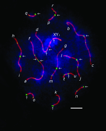
Figure 1.—
SC spread from a shrew spermatocyte at pachytene, stained with DAPI (blue) and immunolabeled with antibodies to SCP3 (red), MLH1 (green), and centromere proteins (green). Bar, 5 μm. Chromosome arms (indicated by letters next to their telomeres) were identified by DAPI banding. Centromeres (indicated by arrows) differ from MLH1 foci by their brighter and more diffuse staining. Note that the centromeres on the af bivalent and on the d arm of the sex trivalent are misaligned and therefore generate weaker signals than aligned centromeres.

