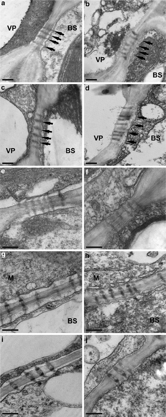
Fig. 3.
TEM images of cellular interfaces along the symplastic pathway of minor veins. a-d Bundle sheath–vascular parenchyma cells. Arrows indicate the location of the plasma membrane in the bundle sheath cell. a Wild-type plasmodesmata span the cell wall, lack occlusions, and connect with the plasma membrane. b sxd1-1 chlorotic leaf tips contain occlusions over the plasmodesmata on the bundle sheath cell side of the cell wall. c tdy1-R chlorotic sectors contain normal appearing plasmodesmata that lack occlusions. Note the plasmodesmata span the cell wall and connect with the plasma membrane. d tdy1-R; sxd1-1double mutant chlorotic tissues contain deposits over the plasmodesmata in the bundle sheath cell similar to sxd1-1 single mutants. Additional cellular interfaces along the symplastic pathway of wild type (e, g, i) and tdy1-R chlorotic tissues (f, h, j) are shown. In e–j the plasmodesmata spanned the cell wall connecting the cytoplasms of adjacent cells and lacked occlusions. e, f BS–BS cells, g, h BS–M cells, i, j M–M cells. BS bundle sheath, M mesophyll. Scale bars represent 200 nm

