Measurements of cartilage thickness in human ankles are important in evaluating the development of osteoarthritis, particularly following articular fractures. Unlike the knee, cartilage in the ankle is relatively thin (on the order of 1.5 mm), so it is difficult to image clearly using either MRI or CT techniques. Part of the problem is the lack of separation between the surfaces of the tibial and talar cartilage, making it essentially impossible to determine their respective thicknesses.
Ankle distraction has been achieved in the operating room by applying tension to the foot using a windlass attached to the operating table, while the patient’s upper body is restrained by straps. Trauma units use devices like the Hare Traction splint, which applies force to the foot while bracing against the posterior ischium, to temporarily distract fractures of the femur. Neither of these systems is suitable for use with MRI, and significant modifications would be required to use them during CT scans.
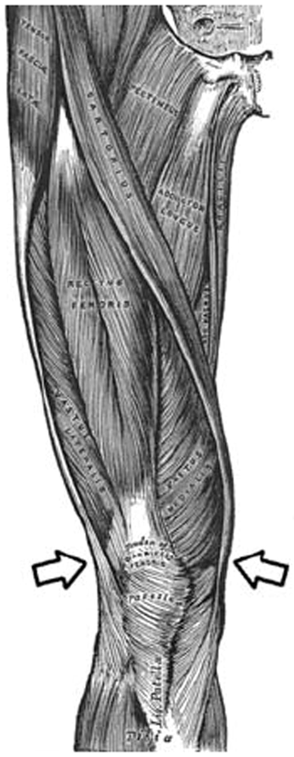
We have built a novel device that distracts the ankle (as well as the knee) using a specially modified brace above the knee to grip the distal surfaces of the thigh musculature where it narrows above the knee as anchor points (Figure 1) and a screw to apply tension to the foot strap. The distance between the tensioning mechanism and the brace is adjustable for patient size. The parts of the distractor adjacent to the ankle are radiolucent and the entire device is compatible with the MR and CT environments (Figure 2). In use, the device has been found to be minimally uncomfortable for the patient.
Fig 1.

Arrows indicate anchor points for the knee brace on the musculature of the distal thigh.
Fig 2.

The distractor in place on a subject leg
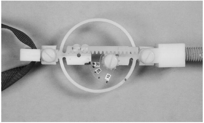
To facilitate application of consistent tension to the ankle, a simple proving ring-based gauge made entirely of plastic was added to the system (Figure 3). It is placed in the load train between the tensioning screw and the ankle strap. As the force increases, the circular ring distorts and the rack attached to one side of the hoop moves relative to the pinion on the other. This action turns the attached calibrated scale, and the force is read against a mark on the rim of the hoop.
Figure 3.

The tension gauge is connected between the foot strap (left) and the tensioning screw (right).
The gauge ensures that consistent loads are applied to the ankle and allows the technician to monitor any slippage that would cause the force to diminish. As a matter of patient safety, forces are not allowed to exceed 30 pounds (130N).
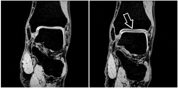
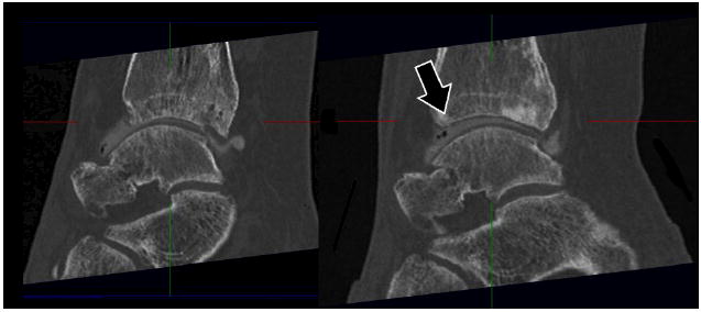
Using the device, we have greatly improved the quality of our MRI scans. Distraction of the ankle joint introduces enough separation between the cartilage surfaces for accurate segmentation of the digitized image for finite element modeling, as well as for direct visual analysis (Figure 4). CT scans also benefit from use of the device (Figure 5).
Figure 4.

Coronal plane MRI scans showing distraction of the ankle mortise. Anatomical (left), and distracted (right).
Figure 5.

Sagittal plane CT scans showing distraction between the tibia and talus. Anatomical (left), and distracted (right).
Acknowledgments
Research supported by NIH grant 5 P50 AR048939


