Abstract
Purpose
To review the parameters for quantitative assessment of the anterior segment and iridocorneal angle and to develop a comprehensive schematic for the evaluation of angle anatomy and pathophysiology by high-resolution imaging.
Methods
The published literature of the last 15 years was reviewed, analyzed, and organized into a construct for assessment of anterior segment processes.
Results
Modern anterior segment imaging techniques have allowed us to devise new quantitative parameters to improve the information obtained. Ultrasound biomicroscopy, slit-lamp optical coherence tomography, and anterior segment optical coherence tomography provide high-resolution images for analysis of physiologic and pathologic processes. These include iridocorneal angle analysis (eg, angle opening distance, angle recess area, trabecular-iris space area), anterior and posterior chamber depth and area, iris and ciliary body cross-sectional area and volume, quantitative anatomic relationships between structures, and videographic analysis of iris movement and accommodative changes under various conditions. Modern devices permit imaging of the entire anterior chamber, allowing calculation of anterior chamber and pupillary diameters and correlating these with measurement of anterior chamber dynamics in light vs dark conditions. We have tabulated all reported anterior segment measurement modalities and devised a construct for assessment of normal and abnormal conditions.
Conclusion
Quantitative measurement of static and dynamic anterior segment parameters, both normal and abnormal, provides a broad range of parameters for analysis of the numerous aspects of the pathophysiology of the anterior segment of the eye.
INTRODUCTION
Anterior segment imaging has significantly altered the diagnosis and evaluation of glaucoma. The information gained with new imaging modalities provides clinicians with both qualitative and quantitative information about anatomical relationships of the anterior segment.
High-frequency ultrasound biomicroscopy (UBM) is the most established anterior segment imaging device, providing objective, high-resolution images of angle structures. UBM allows for visualization of structures in the posterior chamber that are otherwise hidden from clinical observation and can augment gonioscopy in the qualitative and quantitative evaluation of pathologic changes leading to angle closure. Most commercially available instruments use a 50- to 80-MHz transducer with a lateral and axial physical resolution of approximately 50 μm and 25 μm, respectively.1 UBM can be used to demonstrate a wide variety of anterior segment pathology, including congenital glaucoma, and is also useful for presurgical and postsurgical evaluation. Newer UBM models on the market, with 100-MHz transducers, have been exclusively designed for real-time morphological assessment of the anterior chamber angle and for the evaluation of aqueous drainage passageways, including Schlemm’s canal (iScience Surgical, www.iscienceinterventional.com/US/iultrasound.htm, accessed March 2007).
Anterior segment optical coherence tomography (AS-OCT; Carl Zeiss Meditec, Dublin, California) and slit-lamp–adapted optical coherence tomography (SL-OCT; Heidelberg Engineering, Dossenheim, Germany) are recently developed methods that allow for objective and quantitative imaging of anterior segment structures and angle configuration.2 Advantages over UBM include noncontact methodology, with consequent reduction of patient discomfort and risk of corneal injury, and the ability to image the eye in the sitting position. AS-OCT, with an axial and transverse optical resolution of 18 μm and 60 μm, respectively, has better resolution than SL-OCT, which has an axial and transverse resolution of 9 μm and 15 μm, respectively. Both modalities rely on infrared light of 1310 nm wavelength to provide images of the anterior segment; however, AS-OCT and SL-OCT cannot image structures posterior to the pigment epithelium of the iris and ciliary body owing to absorption of light by this layer.
UBM software quantifies distance and area by counting the number of pixels along the measured line or inside the designated area and multiplying the pixel counts by the theoretical size of the pixel. Two factors, physical resolution and measurement precision, are important in image quantification. Resolution refers to how close together two objects can be placed and still appear distinct. As previously mentioned, most commercially available instruments have a lateral and axial physical resolution of approximately 50 μm and 25 μm, respectively. The measurement precision refers to the width and height of a single pixel on the screen identifiable by the operator using the screen cursor.2 The standard Humphrey and Paradigm UBM monitors (864 × 432 pixels) have lateral and axial measurement precision of approximately 6 and 12 μm, respectively.1 Using UBM software, measurement precision can be better than physical resolution by oversampling the signal.3
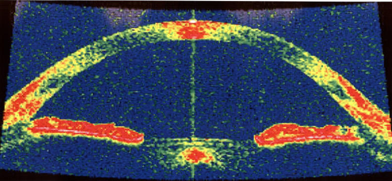
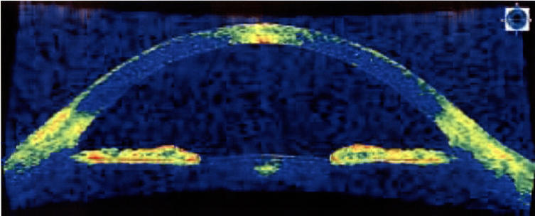
Limitations of both SL-OCT and AS-OCT include distortion from off-axis measurements, requiring a software correction for optical distortion and for changes in beam angulation due to passage of light through media with different refractive qualities. Sector scanning UBM devices, for example OTI and Sonomed models, also require correction for image distortion. In OCT, the scanning system is a fixed point on the mirror of the slit lamp. The center of the image is the only section scanned vertically, whereas all scans adjacent to the center are influenced by the so-called scanning angle. Using the fan correction, the software recalculates the data to eliminate the influence of the scanning angle with a resulting B-scan in the shape of a fan (Figure 1). Correction for the various refractive indexes of the cornea is also done with OCT software. The image must first undergo segmentation to identify the areas of different tissue. This step is essential for location of the exact position of the anterior and posterior cornea. Recalculating the image for both scanning angle and refractive error leads to a B-scan in a butterfly shape (Figure 2).
FIGURE 1.
Fan correction in optical coherence tomography software for elimination of influence of scanning angle. The resulting B-scan has the shape of a fan.
FIGURE 2.
Correction in optical coherence tomography software for refractive index and scanning angle yields a B-scan in the shape of a butterfly.
Several other imaging devices have been used to evaluate the anterior chamber. The Pentacam-Scheimpflug, a noncontact optical imaging system, has two camera components with a software program used to construct a 3-dimensional image. Important information about the cornea, anterior chamber, and lens may be obtained from these images.4 Several studies have shown good correlation of corneal thickness and anterior chamber depth measurements with “gold standard” devices.5–7 The Pentacam-Scheimpflug has also been used to estimate the anterior chamber angle8; however, anterior chamber angle was not found to be significantly correlated with anterior chamber depth or with other quantitative angle parameters. Also, direct visualization of the angle is not possible with this device, preventing morphological assessment of the anterior chamber in cases of plateau iris or lesions of the ciliary body.
Orbscan scanning–slit topography is an alternative noncontact optical system that has primarily been used to view the cornea, although it may also have applications in angle estimation.4 Similar to Pentacam-Scheimpflug, Orbscan cannot directly visualize the angle, preventing an assessment of the structural aspects of the anterior chamber. Orbscan has high reproducibility and significant correlation with other clinical parameters9 (ie, anterior chamber depth, axial length, corneal diameter). To the best of our knowledge, the quantitative angle parameters obtained with this device have not yet been correlated to those of more established modalities, for example, UBM. Although very promising, more information is needed regarding reproducibility of parameters and agreement with established devices, in order to routinely utilize Pentacam-Scheimpflug or Orbscan for quantitative assessment of the anterior chamber angle.
The purpose of this study was to examine the various existing parameters for quantification of the anterior segment in order to propose a new methodology for diagnosis of various ocular diseases. This discussion is limited to those parameters obtained with UBM and OCT, as these have been proven to be the most reproducible and accurate, and therefore the most relevant to clinical decision making and patient prognosis.
METHODS
The published literature on anterior segment imaging was reviewed, analyzed, and organized into a construct for assessment of anterior segment processes. UBM and OCT parameters were examined primarily, as these have proven to be the most reproducible based on prior studies. However, discussion is not limited to these two modalities. The procedures of UBM and OCT examination are briefly described.
The technique of performing UBM is similar to traditional immersion B-scan ultrasonography. The probe is suspended from an articulated arm to diminish motion artifacts. The Humphrey and Paradigm UBM models utilize a linear scan format that minimizes lateral distortion. Scanning is performed in the supine position. An eyecup, measuring 18, 20, or 22 mm in diameter, is inserted between the eyelids and holds methylcellulose or normal saline solution as the coupling agent. After insertion of the probe into the coupling medium, the real-time image is displayed on a video monitor and can be stored on videotape for later analysis. Inexperienced examiners with minimal practice can typically obtain good qualitative information; however, acquisition of highly reproducible distance measurements is strongly dependent on examiner technique and experience. The distance from the center of the anterior chamber, plane of section, and the orientation of the probe with respect to the perpendicular may affect the apparent structural configuration of the anterior segment.
There is some indication that use of the UBM eyecup may cause corneal indentation and a resultant widening of the iridocorneal angle. The most recent version of UBM, the Paradigm P60 model, does not require an eyecup due to modifications to the transducer head; however, the parameters obtained with this device have not yet been established as reproducible. The OTI and Sonomed units require a soft silicone eyecup, unlike older models made by Paradigm and Humphrey that utilize a stiffer eyecup. There have not been any studies with these new models regarding the effect of corneal indentation by the new transducer head.
Compared to UBM, SL-OCT and AS-OCT are both simpler to use, require much less operator skill, and provide noncontact scanning of the anterior segment. The benefits of noncontact scanning include absence of physical or mechanical distortion of the anterior segment as well as applications in postsurgical imaging of blebs and drains. Generally, the patient is seated in front of the machine while scans are taken of the anterior segment. As mentioned previously, these images are limited by their dependence on light waves, which are absorbed by the posterior pigment epithelium, thus preventing a reliable view of the ciliary body and more posterior structures. This prevents diagnosis of plateau iris and distinction of iris cysts from iris tumors.
RESULTS
ANTERIOR CHAMBER AND ANGLE PARAMETERS
Anterior segment OCT was used to image the entire anterior chamber, and angle dynamics were studied under both dark and light conditions using measurements of pupillary diameter and anterior chamber diameter (defined as scleral spur-to-spur distance). As expected, the pupillary diameter was significantly smaller in light (2.904 ± 0.84) than in dark (3.425 ± 1.34, P = .002), whereas the anterior chamber diameter did not change (P = .836). Angle opening distance 500 (AOD 500), for both superior and inferior angles, was greater in eyes with a larger anterior chamber depth (P < .05; r2 range, 0.10–0.28) (S.F. Sandler, unpublished data, 2007).
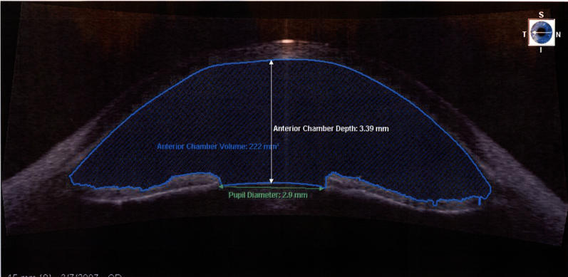
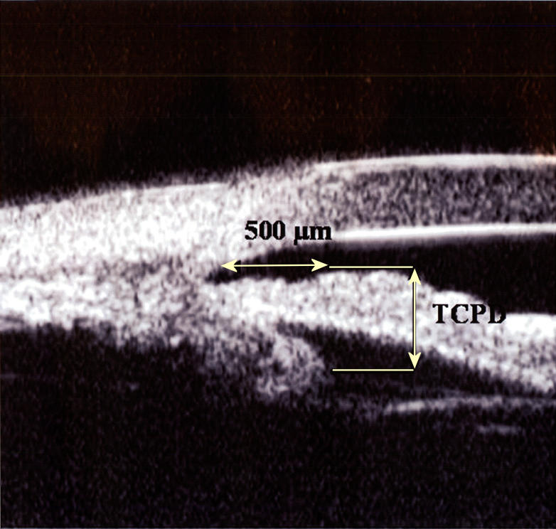
Quantification of angle parameters may provide important information about the underlying mechanism and the extent of apposition in cases of angle closure. Several parameters exist for description of the angle using both anterior segment OCT and UBM software. Pavlin and associates10 were the first to use UBM to quantify the anterior chamber and have suggested several parameters for objective measurement of the anterior segment. Anterior chamber depth is useful for evaluation of a number of conditions affecting the eye, including angle-closure glaucoma, and also for postoperative follow-up of cataract surgery with phakic intraocular lens implantation. With both UBM and OCT, anterior chamber depth is most often measured as the axial distance from the internal corneal surface to the lens surface, although measurements can be made at positions other than the axial position (Figure 3).1 Perhaps the most commonly used parameter is AOD, defined as the length of the line, perpendicular to the corneal endothelial surface, from a point on the endothelial surface 500 or 750 μm anterior to the scleral spur to the iris surface. Measurements are made in the iris plane, and AOD is affected by peripheral anterior synechiae. Both UBM and SL-OCT software are capable of calculating this parameter, used mainly to demonstrate apposition in angle-closure glaucoma11–13 and plateau iris.14 Pavlin and associates10 also proposed the trabecular-ciliary process distance (TCPD) for evaluation of angle closure; this is the length of a line drawn perpendicular to the iris, between a point 500 μm from the scleral spur to the ciliary body (Figure 4).
FIGURE 3.
Anterior chamber depth, volume, and pupillary diameter as calculated by SL-OCT software.
FIGURE 4.
Trabecular ciliary process distance. Ultrasound biomicroscopy image with measurement of trabecular-ciliary process distance (TCPD) (dotted closed arrow represents 500 μm from the scleral spur).
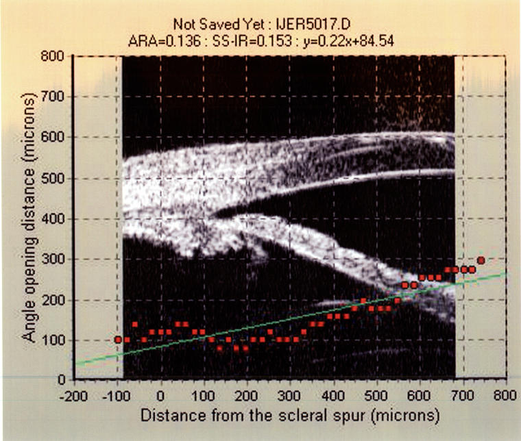
AOD treats the iris surface as a straight line, which may be problematic in cases of angle-closure glaucoma presenting with irregularities of the iris contour and curvature. The angle recess area (ARA) was devised by Ishikawa and colleagues15 in response to these limitations. The ARA is an area bordered by the anterior iris surface, corneal endothelium, and a line drawn perpendicular to the corneal endothelium from the iris surface to a point 750 μm anterior to the scleral spur (Figure 5).16 Ultrasound biomicroscopy software plots consecutive AODs from the base of the angle recess to 750 μm anterior to the scleral spur and performs a linear regression of the AODs. A formula is calculated with the basic form, y = ax + b. Two important figures in this formula, the acceleration (a) and the y-intercept (b), can be used to describe many possible types of angle configuration. The acceleration describes the rate at which the angle widens from the scleral spur, and the y-intercept describes the distance from the scleral spur to the iris. Negative values for a and b denote shallow depth at the central and peripheral parts of the angle, respectively.
FIGURE 5.
Angle recess area calculation using the ultrasound biomicroscopy pro 2000 software. After the observer selects the scleral spur, the program automatically detects the border, calculates the angle recession area at 750 μm anterior to the scleral spur, and performs linear regression analysis of consecutive angle opening distances, producing two figures; the acceleration and y-intercept. The equation of the linear regression is y = ax + b, where a is the acceleration and b is the y-intercept. The y-intercept is the estimated angle opening distance at the level of scleral spur, and the acceleration describes how rapidly the angle widens from the iris root.
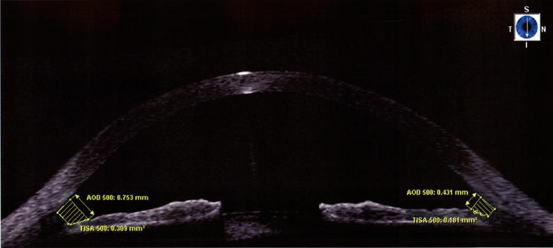
Radhakrishnan and colleagues2 proposed the trabecular-iris space area (TISA), which they state represents the actual filtering area more accurately compared to the ARA. The TISA excludes the nonfiltering region behind the scleral spur via its posterior border—a line drawn from the scleral spur to the opposing iris perpendicular to the plane of the inner scleral wall (Figure 6). TISA may be more sensitive in identifying narrow angles in eyes with deep angle recesses. The same researchers devised the trabecular-iris contact length (TICL) as a way to describe an anatomically closed angle. The TICL is defined as the linear distance of iris contact with corneoscleral surface beginning at the scleral spur and extending anteriorly and represents the measured length of contact between the iris and angle structures anterior to the scleral spur.
FIGURE 6.
Angle opening distance (AOD) and trabecular-iris space area (TISA) calculated using SL-OCT software. TISA differs from AOD by its posterior border, a line drawn from the scleral spur to the opposing iris perpendicular to the plane of the inner scleral wall.
Noncontact goniometry using anterior segment OCT has also been used by Wirbelauer and colleagues17 to calculate the anterior chamber angle. Using a measurement software program that facilitates the creation of an angle (OCTeval, version 1.1; 4Optics AG, Lübeck, Germany), the distance between the points of highest reflectivity at the tissue boundaries is measured. Using digital goniometry, with a classification of occludable angle as one with an anterior chamber angle of less than 22 degrees or gonioscopy grade ≤2, Wirbelauer and colleagues found a high correlation between anterior chamber angle measurements and gonioscopy (0.80, P = .001).
IRIS AND CILIARY BODY EVALUATION
Studying the changes in iris configuration is important for the characterization of certain types of open-angle glaucoma, particularly glaucoma caused by pigment dispersion syndrome. In this condition, friction between the posterior iris surface and the anterior zonular bundles causes mechanical disruption and release of pigment granules into the posterior chamber. Anterior segment imaging can provide important information regarding alterations in iris anatomy that may predispose to pigment dispersion syndrome.
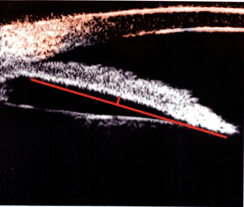
Several methods have been used to describe iris configuration. Using UBM, Sokol and colleagues18 have measured the scleral spur to iris root (SS-to-IR) distance to determine the effect of iris insertion on expression of pigment dispersion syndrome. Potash and colleagues19 defined a parameter, iris concavity, derived by creating a line from the most peripheral point to the most central point of the iris pigment epithelium, and extending a perpendicular line to the iris pigment epithelium at the point of greatest concavity or convexity (Figure 7). Iris thickness can be measured along the TCPD (ID1), as well as 2 mm from the iris root (ID2), and at its thickest point at the pupil (ID3).1 Cross-sectional area of the iris can be quantified by slit-lamp photography using the Scheimpflug principle; however, this method does not allow for area and volume analysis of the anterior segment due to limitations of image resolution. Ishikawa and colleagues20 developed UBM software for measuring iris volume and evaluated the changes in iris cross-sectional area and volume under dark and lighted conditions. Iris volume and cross-sectional area were measured using an automated software program that provides axial alignment using keratometry and corneal diameter information. They effectively demonstrated a decrease in iris volume after pharmacologic and physiologic dilation and a correlation of pupillary diameter with iris volume and iris cross-sectional area change after physiologic dilation.
FIGURE 7.
Iris concavity parameter. Iris concavity is determined by drawing a line connecting the most peripheral and most central points of the iris pigment epithelium and measuring the largest perpendicular distance from the line to the iris pigment epithelium.
Gohdo and colleagues21 have also measured ciliary body thickness (CBT) to assess its influence on eyes with narrow angles. Ciliary body thickness is measured from a meridional section of the temporal limbus taken at the valley in between ciliary processes, 1 mm and 2 mm posterior to the scleral spur.22
ACADEMIA
Leung and colleagues23 have proposed the ACADEMIA (anterior chamber angle detection with edge measurement and identification) algorithm for angle quantification. The steps of the algorithm include image preprocessing to facilitate subsequent detection of edge, detection of the edges lining the corneal endothelium and scleral-corneal and anterior iris surfaces at the iridocorneal angle, fitting of the edges at the scleral-corneal and iris boundaries by straight lines to a defined distance, and determination of the angle by the intersection of the two edges. They found a high correlation of AOD500 with median/average ACADEMIA angle (r = 0.90/0.91). ACADEMIA is less influenced by an irregular anterior iris contour than AOD and requires no subjective operator input. The algorithm may be limited in cases of appositional angle closure, however, in which the apex of the angle may be falsely identified by the software, and in images of poor quality with low signal-to-noise ratios.
REAL-TIME VIDEOGRAPHY
Modern anterior segment imaging devices allow for real-time video capture of the anterior segment. C. K. Leung used AS-OCT, with a scan speed of 2000 A-scans per second, and recording software capable of capturing one frame every 5 milliseconds (CamStudio 2.1, Rendersoft Software) to analyze the dynamic configurational changes of the anterior segment in light and dark (unpublished data, 2007). Video files provide both dynamic and static views of the iris and angle configuration changes.
DISCUSSION
The reliability of UBM measurements has been previously confirmed. Tello and colleagues24 have studied the reproducibility of Pavlin’s UBM parameters and found good intraobserver reproducibility for all parameters except AOD. Interobserver reproducibility was poor, and this finding was confirmed by Urbak and colleagues,25 who also reported poor interobserver reproducibility for all measurements except central corneal thickness. Most UBM software requires an element of observer input; for example, localization of the scleral spur by an operator is necessary for calculation of most angle parameters. Identification of this important landmark is also dependent on the presence of image quality criteria. It is possible that the combination of objective perception and variations in image quality can lead to low interobserver reproducibility, further highlighting the need for an automated software capable of detecting the scleral spur.
Both SL-OCT and AS-OCT are relatively new, and software for these devices is still being modified and improved. More studies are needed to evaluate the reliability and reproducibility of measurements taken using these modalities. In a comparison of SL-OCT and UBM, AOD as measured by UBM was found to be significantly less than AOD measured using SL-OCT in both the superior and inferior angles (P = .0001) (S. Dorairaj, unpublished data, 2007). In the superior angle, a highly significant difference was seen between AODs in light (P = .002) but not in the dark (P = .153). Also, there was good agreement between SL-OCT and UBM measurements of ITC, with kappa values ranging from 0.65 to 0.47. S. F. Sandler evaluated the intraobserver and interobserver reliability and reproducibility of SL-OCT for evaluation of anterior chamber depth and central corneal thickness (unpublished data, 2007). Although SL-OCT provided operator-independent, highly reproducible measures for both parameters, there was a discrepancy between the measurements as taken by SL-OCT, axial OCT biometry, and ultrasonic pachymetry, highlighting a potential need for calibration of measurement algorithms.
Müller and colleagues26 found low intraobserver and interobserver variability for AS-OCT measurements of anterior chamber angle and opening width, supporting its use for quantitative angle assessment. Nemeth and colleagues27 measured anterior chamber depth with both AS-OCT and a standard A-scan device using an immersion technique and assessed the repeatability, reproducibility, and correlations of the measurements. They reported significantly deeper anterior chamber measurements with AS-OCT than with immersion A-scan. They also found better repeatability of anterior chamber depth measurements than with immersion ultrasound, with equal reproducibility of both methods.
With respect to quantitative evaluation of anterior chamber angle, several factors may affect measurement, including lighting conditions, patient positioning, and variations in software algorithms. As previously described, quantitative parameters of UBM and SL-OCT vary with regard to the patient posture during examination. With UBM, the patient is in the supine position, which may cause posterior movement of the lens and other structures, thus showing fewer instances of apposition on UBM testing.28 However, UBM measurements have actually been found to be smaller in some comparative studies.2 It is possible that AOD, as measured in different sectors of the eye, will have varying measurements due to the varying effect of gravity on these different locations. In pigment dispersion syndrome, disruption of the posterior pigment epithelium of the iris is facilitated by a concave iris configuration, which can be reversed by inhibition of blinking, among other factors.29 It is thought that this iris concavity results from reverse pupillary block, and it has been hypothesized that blinking results in transfer of a bolus of aqueous humor into the anterior chamber.30 Prior studies have demonstrated that increased iris concavity, which can be quantified on UBM imaging, in pigment dispersion syndrome appears to be related to increased iridolenticular contact, creating an anatomic configuration that predisposes to reverse pupillary block. When blinking is prevented, accumulated aqueous humor in the posterior chamber alters iris position in pigment dispersion syndrome and increases iridozonular and iridociliary-process distances while minimizing iridolenticular contact. Normal blinking can create transient vector forces to promote aqueous humor flow from posterior to anterior chamber.31
AS-OCT was used to analyze 37 normal subjects with open angles and 18 subjects with narrow angles, and AOD 500 measurements taken in light were significantly higher than those measured in the dark for both the open-angle and narrow-angle groups (P = .001) (C. K. Leung, unpublished data, 2007). After adjusting for age, regression analysis identified a positive correlation between anterior chamber depth and AOD difference, and the majority of eyes (n = 47) showed a high correlation between AOD and pupil size. The differences of the angle width measured in the dark and in the light among individuals demonstrate the importance of standardized background light intensity during angle evaluation.
After examination of 17 normal eyes and 14 narrow-angle eyes using UBM, anterior segment OCT, and gonioscopy, Radhakrishnan and colleagues2 found that ARA and the TISA had similar discriminative abilities for detection of narrow angles. The areas under the receiver operating characteristic curve using UBM and SL-OCT for ARA 750 were 0.97 and 0.96, respectively, and for TISA 750 it was 0.96 for both devices. A limitation of TISA is that it uses a smaller sample of the AOD in the linear regression analysis. This may add variability to the description of the angle shape when this smaller area measurement is combined with acceleration and the y-intercept of the linear regression analysis. The TICL had a 100% and 79.3% specificity using SL-OCT and UBM, respectively, for ruling out angle closure. Previous UBM studies, however, have demonstrated variations in trabecular height among individuals with glaucoma, therefore making the use of a fixed distance from scleral spur to measure TICL somewhat inaccurate, as it may incorrectly include structures anterior to Schwalbe’s line.32
TCPD has been used to evaluate the eyes of patients with angle closure. Several studies have shown that a shorter TCPD is associated with anterior placement of the ciliary body.12,33 Based on this information, the TCPD may prove useful for evaluation of patients with plateau iris, who generally have a more anteriorly displaced ciliary body. Gohdo and colleagues21 report an association between eyes with narrow angles and the presence of a thinner ciliary body.
In cases of occludable angles, gonioscopy does not always guarantee a diagnosis. Provocative testing has been described for the detection of potential appositional angle closure in patients with asymptomatic narrow angles. Previous UBM and SL-OCT studies evaluating quantitative parameters by provocative testing from light to dark have concluded that there is increased probability of identifying occludable angles if the imaging tests are done in dark (S. Dorairaj, unpublished data, 2007).
The scleral ciliary process angle (SCPA), measured between the axis of the ciliary process and the line tangential to the scleral surface, has been suggested as a useful parameter for evaluation of anterior placement of the ciliary body, particularly in plateau iris.34 It is possible, however, that the incorrect placement of the reference point from which the tangential line is drawn may result in widely variant SCPA measurements. Until a repeatable and reliable method of reference point placement is established, the use of this parameter should be approached cautiously.
Quantitative measurements may also be useful for the diagnosis and classification of open-angle glaucomas. Potash and colleagues19 have used the iris concavity parameter to demonstrate a midperipheral iris concavity in 56% of eyes examined, supporting the hypothesis of reverse pupillary block in pigment dispersion syndrome. This information is not only useful for diagnostic purposes but provides the basis for subsequent treatment of iris concavity with miotic therapy and laser iridotomy. Kanadani and colleagues35 studied both eyes of patients with clinically asymmetric pigment dispersion syndrome using UBM and determined that the severely affected eye had a more concave iris configuration than the less affected eye (−0.28 vs 0.08 mm). These measurements confirm the predisposition of patients with a more posterior iris insertion to a phenotypic expression of pigment dispersion syndrome. Sokol and colleagues18 also measured the SS-to-IR distance to determine the effect of iris insertion on expression of pigment dispersion syndrome. They found that SS-to-IR distance was significantly greater in eyes with pigment dispersion syndrome (0.04 mm ± 0.04 mm) than in control eyes (0.28 mm ± 0.04 mm) (P = .01), as well as the overall distance from Schwalbe’s line to the iris root (0.98 ± 0.04 mm vs ± 0.04 mm). These findings support a more posterior insertion of the iris into the ciliary body in eyes with pigment dispersion syndrome than control eyes.
Quantitative measurement of static and dynamic anterior segment parameters, both normal and abnormal, provides a broad range of parameters for analysis of the numerous aspects of the pathophysiology of the anterior segment of the eye. Table 1 categorizes a comprehensive schematic of reproducible anterior segment parameters by UBM and AS-OCT/SL-OCT. SL-OCT and AS-OCT parameters are limited to the measurement of those structures anterior to the iris, and therefore are not particularly useful for the evaluation of the ciliary body and zonules.
TABLE 1.
QUANTITATIVE PARAMETERS OBTAINED WITH ULTRASOUND BIOMICROSCOPY (UBM), SLIT-LAMP–ADAPTED OPTICAL COHEREHNCE TOMOGRAPHY (SL-OCT) AND ANTERIOR SEGMENT OCT (AS-OCT)
| PARAMETER | UBM | SL-OCT/AS-OCT | RECOMMENDED CONDITIONS |
|---|---|---|---|
| Angle opening distance | ✓ | ✓ | RPB, PIC, PIS, PDS |
| Trabecular ciliary process distance | ✓ | PIC, PIS, PDS | |
| Trabecular iris space area | ✓ | ✓ | RPB |
| Trabecular iris contact length | ✓ | ✓ | RPB |
| Scleral spur to iris root distance | ✓ | × | RPB, PIC, PIS, PDS |
| Scleral ciliary process angle | ✓ | PIC, PIS, CT | |
| Angle recess area | ✓ | RPB, PIC, PIS, PDS | |
| Anterior chamber depth | ✓ | ✓ | RPB, PIC, PIS, PDS, MG |
| Pupillary diameter | × | ✓ | RPB, PIC, PIS, PDS, XFS |
| Anterior chamber diameter | × | ✓ | RPB, PIC, PIS, PDS |
| Iris thickness | ✓ | ✓ | RPB, PIC, PIS, PDS, IT, IC |
| Iris configuration | ✓ | × | PDS |
| Iris volume | ✓ | × | PDS, RPB, IC, IT |
| Iridociliary process distance | ✓ | PDS, PIC, PIS, IC, XFS | |
| Ciliary body thickness | ✓ | RPB, PIC, PIS, PDS, CT | |
| Iris lens contact distance | ✓ | RPB, PDS | |
| Iridozonular distance | ✓ | XFS |
CT, ciliary body tumors; IC, iris cyst; IT, iris tumors, MG, malignant glaucoma, PIC, plateau iris configuration, PIS, plateau iris syndrome; PDS, pigment dispersion syndrome; RPB, relative pupillary block, XFS, exfoliation syndrome.
✓ = Reproducible parameter.
× = Software capable of estimating the parameter but no proven reproducibility.
Quantitative parameters have greatly improved our understanding of the anatomical relationships of various anterior segment structures and have also given us useful tools for monitoring patient prognosis. Improvements in software, for example, the development of 3-dimensional UBM, may provide a better understanding of the spatial relationships of anterior segment.36 Modifications to the technology will also increase the image resolution, providing clinicians more precise measurements for clinical decision making. The quantitative information provided by modern anterior segment imaging devices, although useful by itself, should not overshadow the contribution of qualitative information gathered from the images acquired through these devices.
In the routine management of patients by the general ophthalmologist, anterior segment imaging devices can serve as an important screening tool for angle-closure glaucoma and may provide good images of structural defects of the anterior segment. The SL-OCT and AS-OCT are best suited for screening because they can image eyes quickly and in a noncontact manner. By routinely monitoring certain parameters, including anterior chamber depth, AOD, and anterior chamber diameter, physicians can observe any structural changes in anatomy and obtain important information for the diagnosis and treatment of a variety of anterior segment diseases, in particular, angle-closure glaucoma and pigment dispersion syndrome.
UBM is indispensable for imaging of pathology posterior to the iris, as anterior segment OCT is incapable of penetrating beyond this point. In cases of plateau iris, initially diagnosed with gonioscopy, immediate imaging with UBM is warranted for confirmation. Although anterior segment OCT may be able, in certain cases, to image cysts and tumors of the iris, UBM provides the most consistent and reliable method for viewing these structures. Likewise, anterior segment OCT is capable of demonstrating cases of angle closure by providing good views of the anterior chamber; however, for cases of angle closure originating posterior to the iris, for example, phacomorphic glaucoma or malignant glaucoma, UBM provides the best documentation of these findings. The combination of both these devices provides a comprehensive, qualititative picture of trabeculectomy blebs and glaucoma implants (Table 2).
TABLE 2.
QUANTITATIVE AND QUALITATIVE USES OF ULTRASOUND BIOMICROSCOPY AND SLIT-LAMP–ADAPTED OPTICAL COHEREHNCE TOMOGRAPHY (SL-OCT) AND ANTERIOR SEGMENT OCT (AS-OCT)
| ULTRASOUND BIOMICROSCOPY | AS-OCT/SL-OCT |
|---|---|
| Assessment of trabeculectomy blebs (eg, internal ostium) | Assessment of trabeculectomy blebs (eg, subconjunctival cysts) |
| Post-traumatic assessment of structures | Post-traumatic assessment of structures |
| Cyclodialysis cleft and supraciliary effusion | Cyclodialysis cleft and supraciliary effusion |
| Plateau iris configuration | Relative pupillary block |
| Malignant glaucoma | Pigment dispersion syndrome |
| Phacomorphic glaucoma | |
| Glaucoma implants in ciliary sulcus | |
| Iris and ciliary body cysts and tumors | |
| Uveitis-glaucoma-hyphema syndrome for assessment of haptics position |
In terms of patient positioning, contact, and comfort, anterior segment OCT is most useful in postsurgical patients, children, and those who are uncooperative with the UBM examination. Advantages of UBM include penetration beyond the posterior iris, established reliability of measurements, and good correlation of measurements with histological samples. Anterior segment OCT, however, provides better resolution of images, is noncontact, and can be done in the sitting position (Table 3).
TABLE 3.
ADVANTAGES AND DISADVANTAGES OF ULTRASOUND BIOMICROSCOPY AND SLIT-LAMP–ADAPTED OPTICAL COHEREHNCE TOMOGRAPHY (SL-OCT) AND ANTERIOR SEGMENT OCT (AS-OCT)
| MODALITY | ADVANTAGES | DISADVANTAGES |
|---|---|---|
| AS-OCT/SL-OCT | Noncontact
Sitting position Not dependent on operator skill Useful in children Better image resolution Postsurgical assessment |
Cannot image structures posterior to iris
Needs software correction to rectify optical distortion (dewarping) |
| Ultrasound biomicroscopy | Can image structures posterior to iris
Good correlation of measurements with histological samples |
Contact
Supine position Requires skilled operator Risk of corneal injury May not be used in immediate postsurgical period Needs general anesthesia for assessment in children Smaller eyecup may cause corneal indentation and widening of iridocorneal angle |
In summary, several tools are available for imaging the anterior segment. In clinical practice, the modality chosen is dependent on a number of factors, including clinical findings, differential diagnosis, patient comfort, and structures to be imaged. Taking into account these considerations, a number of quantitative parameters are available for use in diagnosis and treatment of many ocular diseases. With continued improvements to the technology, fewer limitations will exist for the routine use of these modalities in the clinical setting.
PEER DISCUSSION
DR MARK B. SHERWOOD
Many years ago, the development of clinical gonioscopy enabled the differentiation of closed from open-angle types of glaucoma. Scheie and Shaffer developed simple methods to quantify the angle width, and this angle quantification was significantly enhanced by Spaeth, whose grading system also records the shape and insertion point of the iris. We are now entering a new era in diagnosis of anterior segment pathology and quantifying of the anterior anatomy of the eye using ultrasound biomicroscopy (UBM) and optical coherence tomography (OCT). This paper focuses our attention on these rapidly developing technologies and provides an excellent, “state of the art” update comparison and literature review.
At present there are many different parts of the anterior chamber that can be measured; indeed Table 1 of this study lists 17 of them. Most current research has focused on reliability and reproducibility of these measurements, but future studies will need to determine how well these quantitative findings relate to clinical outcome. With the alphabet soup of parameters, a consensus of interested parties may be needed to decide which particular parameters are the most important to focus on for future studies. Significant differences in measurements are produced between sitting and supine positions, variations in lighting conditions and even the quadrant of the eye examined. In order to enhance comparisons between studies, these factors would also benefit from consensus standardization.
This paper has highlighted that UBM and OCT measurements are not directly comparable. Given the cost of these machines, it is unlikely that many practices will be able to afford both. For continuity of patient care and detection of progressive narrowing or other anterior segment change, it will be important in the future to determine a conversion factor between these systems.
Ideally, it is to be hoped that with these new imaging systems, anatomical or other risk factors can be defined that will indicate the likelihood of future angle closure attack. It is hoped that a predictive model might then be developed of individual patient risk over a 5-year or longer period, similar to that which has been validated for the development of primary open-angle glaucoma in individuals with ocular hypertension.1 This will require prospectively following large numbers of patients and then subsequent validation in other populations.
The introduction of dynamic, indentation gonioscopy by Forbes greatly improved our ability to differentiate between synechial and appositional closure and to formulate correct clinical treatment. Real-time videography has a similar potential to greatly enhance our understanding of short-term physiological changes in the angle with changes in lighting conditions, posture, medications, etc, in different types of angle configurations. This determination may be qualitative as much as quantitative and in time may be an increasingly important tool in our clinical decision making for anterior segment disease.
ACKNOWLEDGMENTS
Financial Disclosures: None.
REFERENCE
- 1.Ocular Hypertension Treatment Study Group; European Glaucoma Prevention Study Group. Gordon MO, Torri V, Miglior S, et al. Validated prediction model for the development of primary open-angle glaucoma in individuals with ocular hypertension. Ophthalmology. 2007;114:10–19. doi: 10.1016/j.ophtha.2006.08.031. [DOI] [PMC free article] [PubMed] [Google Scholar]
DR. GEORGE O. WARING, III
No conflict. How does OCT go through the part of the sclera that overrides the angle, because it is light going through an opaque tissue? So how does that work? Second, could you please comment on the measurement of the anterior chamber diameter which we need to know in order to insert intraocular lenses in the anterior chamber? I know you were focusing primarily on glaucoma questions, but this is a very practical question, particularly for phakic intraocular lenses.
DR. ROBERT RITCH
I would like to thank Dr. Sherwood for a very incisive discussion of some of the pitfalls and problems we have found trying to analyze the comparative differences and uses between these two machines. I would like to respond to his questions in a little different order. The UBM and the OCT do not read exactly the same. Dr. Sherwood asks if we can devise conversion factors for the main parameters that we examined. This is more difficult to do than appears at first glance. Different positions, such as using the UBM in the supine position or the OCT in the sitting position, can produce different findings. We would also like to be able to examine the eye with these instruments with the patient in the prone position to assess other mechanisms of angle closure that may cause anatomic variations, which may not be directly comparable. With OCT the patient can blink, but not with UBM. That aside, conversion factors for fixed relationships should be achievable, and this is something to focus on for future work. As far as the alphabet soup of abbreviations goes, from this multitude of numbers and letters, can we highlight the best parameters to evaluate, for example, narrow angles? We attempted to present that information in the last summary table. It is difficult to make a rule because there are always exceptions; however, we are gradually trying to define this relationship. What you saw is essentially a work in progress. This is something we have worked on for years and will be continue to study in future.
Working with video and the dynamic changes seen with video, as opposed to single images, is an excellent idea. We have recently published on quantitative video analysis and our ARVO poster this year with Dr. Victor Barocas from the University of Minnesota analyzed dynamic changes in the iris area and posterior chamber volume and light and dark conditions. We developed software called Video Edit 421 for these purposes and we also used an NIH program to analyze each frame in the video. We believe there will be a very useful role for video, but it needs to be further developed. Eventually we would really love to have a holographic video quantitative methodology, which would be a great advance.
Just as we have recognized risk factors in the OHTS study, such as central corneal thickness, we are looking at the risk of decreasing angle recess area for developing angle closure glaucoma. What are the predictive factors and can we develop a risk analysis program? At this point, the problem is that there has been no normative data base developed for either UBM or SL-OCT. This should be the first step in the development of a risk calculator. One additional note from a screening standpoint, we predict the potential development of a small portable anterior segment OCT along the same lines as the portable Nd:YAG developed by Alan Robin years ago, but in this case to screen angles in potentially high yield areas, such as in rural China.
Finally, we agree that a consensus meeting for people working in the field who can decide which of these items we should investigate would be useful. Dr. Waring asked how the OCT beam can go through the sclera overlying the angle if it is opaque. I never thought about that. Dr. Dorairaj do you have any idea on that?
DR. SYRIL DORAIRAJ
Based on the segmentation, it can actually see the structures behind the sclera. There are 2 or 3 papers by Chris Leung that have documented the possibility of defining structures beyond sclera. The second question raised was about using these technologies to determine the anterior chamber diameter. Yes, of course it can be used for this purpose, and this technique does not show any kind of variability. We have performed some studies comparing the sectoral scan UBM, and it results in the calculation of a very predictable diameter.
DR. ROBERT RITCH
About 15 years ago, Mark Speaker was working with us and one of his fellows presented a paper at the American Academy of Ophthalmology on the topic, “Is white to white right?” for measuring anterior chamber diameter for intraocular lens placement. Unfortunately, that paper was never published, but I believe that with the UBM and the OCT we can accurately measure anterior chamber diameters. I think that similar work done long ago ought to be repeated with the IOLs of today and finally published.
ACKNOWLEDGMENTS
Funding / Support: None.
Financial Disclosures: None.
Author Contributions: Design and conduct of the study (S.D., J.M.L., R.R.); Management (S.D., J.M.L., R.R.); Analysis and interpretation of the data (S.D., J.M.L., R.R.); Preparation, review, and approval of the manuscript (S.D., J.M.L., R.R.).
Other Acknowledgments: The authors acknowledge Hiroshi Ishikawa, MD, for his suggestions and commentary on the manuscript.
REFERENCES
- 1.Pavlin CJ, Foster FS. Ultrasound Biomicroscopy of the Eye. New York: Springer Verlag; 1995. Ultrasound biomicroscopic anatomy of the normal eye and adnexa; pp. 47–60. [Google Scholar]
- 2.Radhakrishnan S, Goldsmith J, Huang D, et al. Comparison of optical coherence tomography and ultrasound biomicroscopy for detection of narrow anterior chamber angles. Arch Ophthalmol. 2005;123:1053–1059. doi: 10.1001/archopht.123.8.1053. [DOI] [PubMed] [Google Scholar]
- 3.Ishikawa H, Schuman JS. Anterior segment imaging: ultrasound biomicroscopy. Ophthalmol Clin North Am. 2004;17:7–20. doi: 10.1016/j.ohc.2003.12.001. [DOI] [PMC free article] [PubMed] [Google Scholar]
- 4.Konstantopoulos A, Hossain P, Anderson DF. Recent advances in ophthalmic anterior segment imaging: a new era for ophthalmic diagnosis? Br J Ophthalmol. 2007;91:551–557. doi: 10.1136/bjo.2006.103408. [DOI] [PMC free article] [PubMed] [Google Scholar]
- 5.Buehl W, Stojanac D, Sacu S, Drexler W, Findl O. Comparison of three methods of measuring corneal thickness and anterior chamber depth. Am J Ophthalmol. 2006;141:7–12. doi: 10.1016/j.ajo.2005.08.048. [DOI] [PubMed] [Google Scholar]
- 6.Lackner B, Schmidinger G, Skorpik C. Validity and repeatability of anterior chamber depth measurements with Pentacam and Orbscan. Optom Vis Sci. 2005;82:858–861. doi: 10.1097/01.opx.0000177804.53192.15. [DOI] [PubMed] [Google Scholar]
- 7.Nemeth G, Vajas A, Kolozsvari B, Berta A, Modis L. Anterior chamber depth measurements in phakic and pseudophakic eyes: Pentacam versus ultrasound device. J Cataract Refract Surg. 2006;32:1331–1335. doi: 10.1016/j.jcrs.2006.02.057. [DOI] [PubMed] [Google Scholar]
- 8.Rabsilber TM, Khoramnia R, Auffarth GU. Anterior chamber measurements using Pentacam rotating Scheimpflug camera. J Cataract Refract Surg. 2006;32:456–459. doi: 10.1016/j.jcrs.2005.12.103. [DOI] [PubMed] [Google Scholar]
- 9.Allouch C, Touzeau O, Borderie V, et al. Orbscan: a new device for iridocorneal angle measurement. J Fr Ophtalmol. 2002;25:799–806. [PubMed] [Google Scholar]
- 10.Pavlin CJ, Harasiewicz K, Foster FS. Ultrasound biomicroscopy of anterior segment structures in normal and glaucomatous eyes. Am J Ophthalmol. 1992;112:381–389. doi: 10.1016/s0002-9394(14)76159-8. [DOI] [PubMed] [Google Scholar]
- 11.Marchini G, Pagliarusco A, Toscano A, Tosi R, Brunelli C, Bonomi L. Ultrasound biomicroscopic and conventional ultrasonographic study of ocular dimensions in primary angle-closure glaucoma. Ophthalmology. 1998;105:2091–2098. doi: 10.1016/S0161-6420(98)91132-0. [DOI] [PubMed] [Google Scholar]
- 12.Cho HJ, Woo JM, Yang KJ. Ultrasound biomicroscopic dimensions of the anterior chamber in angle-closure glaucoma patients. Korean J Ophthalmol. 2002;16:20–25. doi: 10.3341/kjo.2002.16.1.20. [DOI] [PubMed] [Google Scholar]
- 13.Sihota R, Dada T, Gupta R, Lakshminarayan P, Pandey RM. Ultrasound biomicroscopy in the subtypes of primary angle closure glaucoma. J Glaucoma. 2005;14:387–391. doi: 10.1097/01.ijg.0000176934.14229.32. [DOI] [PubMed] [Google Scholar]
- 14.Garudadri CS, Chelerkar V, Nutheti R. An ultrasound biomicroscopic study of the anterior segment in Indian eyes with primary angle-closure glaucoma. J Glaucoma. 2002;11:502–507. doi: 10.1097/00061198-200212000-00009. [DOI] [PubMed] [Google Scholar]
- 15.Ishikawa H, Uji Y, Emi K. A new method of quantifying angle measurements based on ultrasound biomicroscopy. Atarashii Ganka. 1995;12:957–960. [Google Scholar]
- 16.Ishikawa H, Liebmann JM, Ritch R. Quantitative assessment of the anterior segment using ultrasound biomicroscopy. Curr Opin Ophthalmol. 2000;11:133–139. doi: 10.1097/00055735-200004000-00012. [DOI] [PubMed] [Google Scholar]
- 17.Wirbelauer C, Karandish A, Häberle H, Pham DT. Noncontact goniometry with optical coherence tomography. Arch Ophthalmol. 2005;123:179–185. doi: 10.1001/archopht.123.2.179. [DOI] [PubMed] [Google Scholar]
- 18.Sokol J, Stegman Z, Liebmann JM, Ritch R. Location of the iris insertion in pigment dispersion syndrome. Ophthalmology. 1996;103:289–293. doi: 10.1016/s0161-6420(96)30702-1. [DOI] [PubMed] [Google Scholar]
- 19.Potash SD, Tello C, Liebmann J, Ritch R. Ultrasound biomicroscopy in pigment dispersion syndrome. Ophthalmology. 1994;101:332–339. doi: 10.1016/s0161-6420(94)31331-5. [DOI] [PubMed] [Google Scholar]
- 20.Ishikawa H, Ritch R, Hoh ST, et al. Iris volume before and after pupillary dilation using ultrasound biomicroscopy. Invest Ophthalmol Vis Sci. 1997;38(suppl):L S825. [Google Scholar]
- 21.Gohdo T, Tsumura T, Iijima H, Kashiwagi K, Tsukahara S. Ultrasound biomicroscopic study of ciliary body thickness in eyes with narrow angles. Am J Ophthalmol. 2000;129:342–346. doi: 10.1016/s0002-9394(99)00353-0. [DOI] [PubMed] [Google Scholar]
- 22.Oliveira C, Tello C, Liebmann JM, Ritch R. Ciliary body thickness increases with increasing axial myopia. Am J Ophthalmol. 2005;140:324–325. doi: 10.1016/j.ajo.2005.01.047. [DOI] [PubMed] [Google Scholar]
- 23.Leung CK, Yung WH, Yiu CK, et al. Novel approach for anterior chamber angle analysis: anterior chamber angle detection with edge measurement and identification algorithm (ACADEMIA) Arch Ophthalmol. 2006;124:1395–1401. doi: 10.1001/archopht.124.10.1395. [DOI] [PubMed] [Google Scholar]
- 24.Tello C, Liebmann J, Potash SD, Cohen H, Ritch R. Measurement of ultrasound biomicroscopy images: intraobserver and interobserver reliability. Invest Ophthalmol Vis Sci. 1994;35:3549–3552. [PubMed] [Google Scholar]
- 25.Urbak SF, Pedersen JK, Thorsen TT. Ultrasound biomicroscopy. II. Intraobserver and interobserver reproducibility of measurements. Acta Ophthalmol Scand. 1998;76:546–549. doi: 10.1034/j.1600-0420.1998.760507.x. [DOI] [PubMed] [Google Scholar]
- 26.Müller M, Dahmen G, Pörksen E, et al. Anterior chamber angle measurement with optical coherence tomography: intraobserver and interobserver variability. J Cataract Refract Surg. 2006;32:1803–1808. doi: 10.1016/j.jcrs.2006.07.014. [DOI] [PubMed] [Google Scholar]
- 27.Nemeth G, Vajas A, Tsorbatzoglou A, Kolozsvari B, Modis L, Berta A. Assessment and reproducibility of anterior chamber depth measurement with anterior segment optical coherence tomography compared with immersion ultrasonography. J Cataract Refract Surg. 2007;33:443–447. doi: 10.1016/j.jcrs.2006.11.013. [DOI] [PubMed] [Google Scholar]
- 28.Ishikawa H, Esaki K, Liebmann JM, Uji Y, Ritch R. Ultrasound biomicroscopy dark room provocative testing: a quantitative method for estimating anterior chamber angle width. Jpn J Ophthalmol. 1999;43:526–534. doi: 10.1016/s0021-5155(99)00139-2. [DOI] [PubMed] [Google Scholar]
- 29.Breingan PJ, Esaki K, Ishikawa I, Liebmann JM, Greenfield DS, Ritch R. Iridolenticular contact decreases following laser iridotomy for pigment dispersion syndrome. Arch Ophthalmol. 1999;117:325–328. doi: 10.1001/archopht.117.3.325. [DOI] [PubMed] [Google Scholar]
- 30.Chew SJ, Tello C, Wallman J, Ritch R. Blinking indents the cornea and reduces anterior chamber volume as shown by ultrasound biomicroscopy. Invest Ophthalmol Vis Sci. 1994;35(suppl):1573. [Google Scholar]
- 31.Liebmann JM, Tello C, Chew SJ, Cohen H, Ritch R. Prevention of blinking alters iris configuration in pigment dispersion syndrome and in normal eyes. Ophthalmology. 1995;102:446–455. doi: 10.1016/s0161-6420(95)31001-9. [DOI] [PubMed] [Google Scholar]
- 32.Stegman Z, Sokol J, Liebmann JM, Cohen H, Tello C, Ritch R. Reduced trabecular meshwork height in juvenile primary open-angle glaucoma. Arch Ophthalmol. 1996;114:660–663. doi: 10.1001/archopht.1996.01100130652003. [DOI] [PubMed] [Google Scholar]
- 33.Dorairaj SK, Tello C, Liebmann JM, Ritch R. Narrow angles and angle closure: anatomic reasons for earlier closure of the superior portion of the iridocorneal angle. Arch Ophthalmol. 2007;125:734–739. doi: 10.1001/archopht.125.6.734. [DOI] [PubMed] [Google Scholar]
- 34.Okamoto F, Nakano S, Okamoto C, Hommura S, Oshika T. Ultrasound biomicroscopy findings in aniridia. Am J Ophthalmol. 2004;137:858–862. doi: 10.1016/j.ajo.2003.12.014. [DOI] [PubMed] [Google Scholar]
- 35.Kanadani FN, Dorairaj S, Langlieb AM, et al. Ultrasound biomicroscopy in asymmetric pigment dispersion syndrome and pigmentary glaucoma. Arch Ophthalmol. 2006;124:1573–1576. doi: 10.1001/archopht.124.11.1573. [DOI] [PubMed] [Google Scholar]
- 36.Iezzi I, Rosen RB, Tello C, Liebmann J, Walsh JB, Ritch R. Personal computer-based 3-dimensional ultrasound biomicroscopy of the anterior segment. Arch Ophthalmol. 1996;114:520–524. doi: 10.1001/archopht.1996.01100130512001. [DOI] [PubMed] [Google Scholar]


