Abstract
A new isolate (IS5056) of Bacillus thuringiensis subsp. thuringiensis that produces a novel variant of Cry1Ab, Cry1Ab21, was isolated from soil collected in northeastern Poland. Cry1Ab21 was composed of 1,155 amino acids and had a molecular mass of 130.5 kDa, and a single copy of the gene coding for this endotoxin was located on a ∼75-kbp plasmid. When synthesized by the wild-type strain, Cry1Ab21 produced a unique, irregular, bipyramidal crystal whose long and short axes were both approximately 1 μm long, which gave it a cuboidal appearance in wet mount preparations. In diet incorporation bioassays, the 50% lethal concentrations of the crystal-spore complex were 16.9 and 29.7 μg ml−1 for second- and fourth-instar larvae of the cabbage looper, Trichoplusia ni, respectively, but the isolate was essentially nontoxic to larvae of the beet armyworm, Spodoptera exigua. A bioassay of autoclaved spore-crystal preparations showed no evidence of β-exotoxin activity, indicating that toxicity was due primarily to Cry1Ab21. Studies of the pathogenesis of isolate IS5056 in second-instar larvae of T. ni showed that after larval death the bacterium colonized and subsequently sporulated extensively throughout the cadaver, suggesting that other bacteria inhabiting the midgut lumen played little if any role in mortality. As T. ni is among the most destructive pests of vegetable crops in North America and has developed resistance to B. thuringiensis, this new isolate may have applied value.
For decades, isolates of Bacillus thuringiensis have been obtained worldwide from various sources, including soil, the phylloplane, aquatic habitats, grain, and the feces of insects and other animals (10, 19, 29, 30). Most of these isolates are toxic to pests that have agricultural importance (22), specifically either larvae of lepidopteran pests, such as bollworms and loopers (Heliothis, Helicoverpa, and Trichoplusia species), stem borers (Chilo species), and leafworms (certain Spodoptera species), or larvae and adults of important coleopteran pests, such as the Colorado potato beetle (Leptinotarsa decemlineata). In other cases, the isolates are selectively toxic to larvae of dipteran pests and disease vectors (27), such as the larvae of mosquitoes (Aedes, Anopheles, and Culex species) and blackflies (Simulium species).
Many of these isolates, however, lack efficacy against economically important pests and vectors of disease. Moreover, in a few rare cases, isolates used in commercial bacterial insecticides, such as the HD1 isolate of B. thuringiensis subsp. kurstaki, have developed resistance to Cry proteins (11, 32). Whereas the number of B. thuringiensis isolates that exhibit novel or high insecticidal activity is growing, the commercial insecticides currently used against lepidopteran pests are based on only a few isolates. Typically, these are the HD1 isolate noted above and the ABTS-1857 strain of B. thuringiensis subsp. aizawai, the active ingredient of commercial products developed to control more recalcitrant lepidopterans, such members of the genus Spodoptera. Both of these strains have a broad spectrum of activity against lepidopteran insects due to the complement of endotoxin proteins that each produces during sporulation. HD1, the active ingredient of products such as Dipel and Foray, produces Cry1Aa, Cry1Ab, Cry1Ac, and Cry2Aa, whereas the ABTS-1857 strain of B. thuringiensis subsp. aizawai, the active ingredient of Xentari, produces Cry1Aa, Cry1Ab, Cry1Ca, and Cry1Da (21). The latter product is used to control larvae of the beet armyworm, Spodoptera exigua, and fall armyworm, Spodoptera frugiperda, which are difficult to control with formulations based on HD1.
As a consequence of widespread use, the evolution of resistance to these isolates or strains derived from them and the subsequent proliferation of resistant populations are of concern (20). For example, field populations of the diamondback moth, Plutella xylostella, and the cabbage looper, Trichoplusia ni, have already developed high levels of resistance due to repeated exposure to formulations based on HD1 (11, 26). Furthermore, in laboratory selection studies, resistance to B. thuringiensis has evolved in the pink bollworm, Pectionphora gossypiella, and the tobacco budworm, Heliothis virescens (13, 31). It is also thought that the selection pressure imposed by use of Cry proteins in transgenic plants (9, 31), most of which are based on Cry1Ac or Cry1Ab, could contribute to the evolution of resistance in insect populations to bacterial insecticides based on HD1.
Several approaches to reduce or circumvent the development of resistance to Cry proteins in natural insect populations are currently being employed. These approaches include the isolation of new strains of B. thuringiensis that produce novel toxins, as well as the use of recombinant DNA technology to construct novel bacterial strains engineered to produce combinations of wild-type toxins along with toxins engineered for improved efficacy. With regard to new strains, here we report characterization of a new isolate, IS5056, of B. thuringiensis subsp. thuringiensis that is highly toxic to larvae of T. ni. This isolate was obtained from a soil sample from northeastern Poland, and unlike most isolates of B. thuringiensis active against lepidopteran species, which produce an elongate bipyramidal crystal, it produces a quasicuboidal inclusion. We show that this inclusion is composed of a new toxic Cry protein variant, Cry1Ab21, and furthermore that this isolate invades T. ni and reproduces extensively, using the larval cadaver as a substrate for vegetative growth and sporulation.
MATERIALS AND METHODS
Bacterial strains and plasmids.
Three Bacillus strains were used in this study: an isolate from soil (designated IS5056) from Biebrza National Park (northeastern Poland); 4Q7, an acrystalliferous strain of B. thuringiensis subsp. israelensis cured of plasmids; and the HD1 isolate of B. thuringiensis subsp. kurstaki. The latter two strains were obtained from the Bacillus Genetic Stock Center (Ohio State University, Columbus). Cloned genes were amplified in Escherichia coli DH5α [supE44 ΔlacU169 (φ80lacZΔM15) hsdR17 recA1 endA1 gyrA96 thi-1 relA1]. The E. coli-B. thuringiensis shuttle vector pHT3101 (18) was used to transform and express the IS5056 cry1Ab21 gene in B. thuringiensis 4Q7. Bacterial strains were routinely propagated on Luria-Bertani agar and nutrient agar (NA).
Isolation and culture of IS5056 and crystal purification.
To isolate B. thuringiensis subsp. thuringiensis IS5056, 100 μl of a 10% soil suspension in 0.9% NaCl was preheated at 80°C for 10 min, plated on mannitol-egg yolk-polymyxin agar, a selective medium for the Bacillus cereus group (Oxoid, Ltd., Basingstoke, England), and incubated at 30°C for 24 h. The IS5056 strain formed irregular white colonies with a violet-pink background, and these colonies were recognized as B. thuringiensis colonies based on the presence of crystalline inclusions in sporulating cells, as observed by phase-contrast microscopy. The IS5056 strain was grown to lysis at 30°C in 50 ml of peptonized milk broth (1% peptonized milk broth [Kerry Bio-Science, Hoffman Estates, IL], 1% dextrose, 0.2% yeast extract, 1.2 mM MgSO4, 0.07 mM FeSO4, 0.14 mM ZnSO4, 0.12 mM MnSO4) in 250-ml flasks with shaking at 250 rpm. Following sporulation and lysis of >95% of the cells (72 h), endospores, crystals, and cell debris were sedimented by centrifugation at 12,000 × g for 30 min at 4°C, washed twice in sterile, cold, double-distilled H2O, and sedimented again as described above in 20 ml of sterile, cold, double-distilled H2O. The preparation was sonicated, and 5 ml of the suspension was loaded on a sucrose gradient (9 ml of 67% [wt/vol] sucrose, 7 ml of 72% [wt/vol] sucrose, and 6 ml of 79% [wt/vol] sucrose). Crystalline inclusions were separated by centrifugation for 1 h at 18, 000 × g using a Beckman SW28 rotor. After centrifugation, the spores were found in the pellet, whereas a single crystalline inclusion band occurred at a position between the 72 and 79% sucrose layers compared with the original gradient. Bands containing components other than the crystalline inclusion were not observed. The crystals were collected using a 10-ml syringe and were washed five times with double-distilled water. Protein crystals were solubilized in 2× Laemmli buffer, and the constituent proteins were separated by sodium dodecyl sulfate-polyacrylamide gel electrophoresis (SDS-PAGE) in a 12% gel (16). To visualize proteins, the gel was stained with Coomassie brilliant blue R-250. For comparison, parasporal crystals of HD1 were also prepared as described above and included in the gels.
Flagellar (H) serotyping.
Serotyping of the IS5056 isolate against reference B. thuringiensis flagellar antigens H1 to H55 (17, 23) was performed by Kumiko Kagoshima and Michio Ohba (Graduate School of Agriculture, Kyushu University, Fukuoka, Japan).
Microscopy.
Samples were prepared as described previously (24) for phase-contrast, scanning, and transmission electron microscopy. Replicate preparations from different cultures were observed by scanning electron microscopy to rule out contamination by other crystalliferous isolates of B. thuringiensis.
Peptide sequence determination.
Matrix-assisted laser desorption ionization—time of flight mass spectrometry (MALDI-TOF MS) (4) peptide sequencing of the IS5056 crystal protein band excised from polyacrylamide gels was performed by the Institute for Integrative Genome Biology, University of California, Riverside.
Cloning and sequencing of crystal toxin gene.
The results of MALDI-TOF MS suggested that the parasporal crystalline inclusion produced by IS5056 was a Cry1-type protein. To amplify the IS5056 cry1 gene by PCR, a primer pair (primer Cry1F [5′-GAAAAGTGGATTTTATATATAAGTATAAAAAGTAATAAGAC-3′] and reverse primer Cry1R [5′-TTTTAGAAAGAATAACGTTTTTTGACACTTGGGGGTAGTGC-3′]) was designed based on the sequence of the cry1A gene from B. thuringiensis serovar aizawai (GenBank accession number DQ991096). In addition, we used a primer pair specific for cry1A-type open reading frames (primers Cry1F [5′-ATGGATAACAATCCGAACATCAATG-3′] and Cry1R [5′-CTATTCCTCCATAAGGAGTAATTCCACGCTGTCCACG-3′]) which amplified only a single amplicon corresponding to the amplicon present in the Cry1A gene of IS5056. PCR was performed using the Expand Long Template PCR system (Roche) as follows: 94°C for 3 min, 94°C for 30 s, 50°C for 45 s, and 68°C for 7 min. The 4.1-kb cry1 amplicon was cloned in pGEM-T Easy (Promega) to generate pGEM-5056cry1 and was sequenced using an ABI3130 automated sequencer (Applied Biosystems). Both strands of two clones were sequenced using the T7 and SP6 primers to determine the accuracy of the sequence. To determine whether IS5056 contained a cry2 gene homologue, PCR was performed as described above using primers Cry2OPF (5′-CTTAAAAAAATTCAAGAAATATGATAG-3′) and Cry2OPR (5′-GGTTAACTTGAAATGATTTCTCCC-3′) designed to amplify a 4-kb amplicon.
Expression of IS5056 cry1Ab21.
Plasmid pGEM-5056cry1 was digested with SalI and SphI, and the 4.1-kb fragment containing the IS5056 cry1Ab21 gene was cloned into the same sites in the pHT3101 vector (18) to generate pHT-IS5056Cry1Ab21. Plasmid pHT-IS5056Cry1Ab21 was transformed into B. thuringiensis subsp. israelensis 4Q7 by electroporation as described previously (33), and transformant 4Q7-IS5056Cry1Ab21 was selected on brain heart infusion agar (Difco) containing erythromycin at a concentration of 25 μg/ml. For protein profile analyses, IS5056, HD1, the 4Q7-IS5056Cry1Ab21 transformant, and 4Q7 were grown for 3 days in peptonized milk broth (Kerry Bio-Science). One milliliter of each sample was centrifuged at 10,000 × g for 5 min, suspended in 100 μl of 2× Laemmli buffer, and fractionated by SDS-PAGE in a 12% gel.
Plasmid isolation and Southern hybridization.
Recombinant plasmids were purified using a Wizard Plus miniprep kit (Promega). Native plasmids in IS5056 and HD1 were purified using a HiSpeed plasmid maxi kit (Qiagen GmbH, Hilden, Germany) and were separated in a 1% Prona Plus agarose gel (Laboratories Conda, Spain). The sizes of IS5056 plasmids were assessed by comparison with those in HD1, as described by Kronstad and coworkers (15). To locate the gene encoding the IS5056 crystal protein, Southern hybridization was performed as described by Sambrook and Russell (25). Undigested plasmids and plasmids cleaved with EcoRI or HindIII were separated in a 1% Prona Plus agarose gel (Laboratories Conda) and blotted onto nitrocellulose. A digoxigenin (DIG)-labeled oligonucleotide probe specific for cry1Ab, corresponding to positions 601 to 3139 of the IS5056 cry1 open reading frame, was prepared by PCR amplification with primers IScry1SBf (5′-GGCAACTATACAGATCATGCTGTACGCTGG-3′) and IScry1SBr (5′-CCGTGTTGTTTGGATATACTTCCTCTTCTAC-3′) with reference B. thuringiensis subsp. kurstaki HD1 strain DNA as the substrate, using a PCR DIG probe synthesis kit (Roche Diagnostic GmbH, Mannheim, Germany). Hybridization was detected with an anti-DIG-alkaline phosphatase conjugate and the alkaline phosphatase substrate nitroblue tetrazolium/5-bromo-4-chloro-3-indolylphosphate (BCIP) using a DIG nucleic acid detection kit (Roche) according to the manufacturer's recommendations.
Phylogenetic analysis.
The partial gyrase B (gyrB) and partial 16S rRNA genes of IS5056 were amplified using primers gyrF (5′-AATAATAACTTTATGATAGCGT-3′) and gyrR (5′-CGGTGGCGGTTACAAAGTTTC-3′) and primers 16S-F (5′-CATGGCTCAGATTGAACGCTGGCG-3′) and 16S-R (5′-CCCCTACGGTTACCTTGTTACGAC-3′), respectively, and the amplicons were cloned into pGEM-T Easy (Promega) for nucleotide sequence determination.
Sequence analyses.
For comparative analyses of nucleotide and amino acid sequences, database searches were performed using the BLAST programs at the NCBI website (http://www.ncbi.nlm.nih.gov).
Insect bioassays.
The insecticidal activity of IS5056 was determined using bioassays performed with second- and fourth-instar larvae of T. ni and S. exigua. Assays were performed in 24-well plates using lyophilized preparations containing crystal inclusions and spores mixed with an artificial diet. For the negative control, larvae were fed the diet without crystal-spore mixtures. To determine the 50% lethal concentration (LC50) and LC90, eight concentrations ranging from 2 to 100 μg/ml were tested with 12 larvae for each concentration assayed. Experiments were replicated in triplicate on different days using crystal-spore preparations from independent cultures. Mortality was recorded during and after 7 days of treatment, and LC50s and LC90s were calculated using Probit analysis (SAS Institute).
Heat inactivation of IS5056 spore-crystal mixtures.
To demonstrate that the toxicity of IS5056 against T. ni larvae was not due to β-exotoxin, bioassays were performed as described above using autoclaved crystal-spore suspensions (121°C for 20 min) at a concentration of 400 μg/ml. This procedure has been shown to inactivate protein crystal toxins but not β-exotoxin (8). In addition to β-exotoxin inactivation, the effect of autoclaving on the shape of the crystalline inclusion of IS5056 was observed by contrast field microscopy at a magnification of ×1,000.
IS5056 pathogenesis.
It has been suggested recently based on studies of HD1 in larvae of the gypsy moth, Lymantria dispar, that naturally occurring midgut bacteria kill and colonize larvae after intoxication by B. thuringiensis and additionally that this bacterial species is not capable of colonizing larvae (5). To test this hypothesis, we fed groups of second- and third-instar larvae of T. ni approximately 10,000 and 20,000 spores plus associated crystals, respectively, and monitored the progression of disease, mortality, and sporulation of this strain in cadavers. Thirty-six larvae of each instar, using three groups of 12, were treated and monitored as follows: larvae were starved for 4 h, and then a single larva was placed in each well of 24-well plates overnight along with a 0.5-μl drop of 10% sucrose in double-distilled water that contained spores and associated crystals. The plates were kept at 26°C overnight and examined the next morning to ensure that the larvae consumed the entire drop containing the spores and crystals. Only larvae that had consumed the entire spore-crystal suspension were used in the assay. As controls, 10 larvae of each instar were treated similarly, except that the spore-crystal mixture was not included in the sucrose droplet. Morality of the larvae was observed for 7 days.
The presence of vegetative, sporulating, and sporulated cells of IS5056 in moribund and dead T. ni larvae was determined on the basis of (i) microscopic observation of crystals with a shape characteristic of the shape of crystals of this strain and (ii) the number of Bacillus colonies growing on NA without antibiotics. Each day, bacteria were isolated from five second-instar larvae and five third-instar larvae. After ethanol disinfection the larvae were macerated in 300 μl of sterile water. The suspensions were diluted serially, and 5 and 10 μl were plated on NA to determine whether IS5056 was capable of growth and sporulation in the cadavers of T. ni. The degree of reproduction in the cadavers or the lack of reproduction was determined by comparing the number of sporulated cells obtained 7 days after death to the dose fed each larva. To determine whether bacilli or other bacteria were present on or in control larvae, five healthy larvae of each instar were crushed in 300 μl of sterile water, and then 1, 5, 10, and 20 μl of the suspension was plated on NA without antibiotics and incubated for 72 h at 28°C. The plates were held and examined after this for another 48 h to monitor any bacterial growth.
Nucleotide sequence accession numbers.
The GenBank accession number for the nucleotide sequence of the cry1Ab21 gene from B. thuringiensis IS5056 is EF683163, whereas the accession numbers for the 16S rRNA and gyrB gene sequences of this strain are EF687842 and EF690292, respectively.
RESULTS
Characterization of isolate IS5056.
H antigen serotyping of IS5056 with H1 to H55 flagellar antisera showed that the flagella cross-reacted with H1 antisera. Thus, IS5056 belongs to the H1 serotype, recognized as B. thuringiensis subsp. thuringiensis (17, 23).
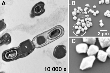
Phase-contrast microscopy of the IS5056 strain grown on NA or in peptonized milk at 48 h showed the presence of a large crystalline inclusion in sporulating cells. In most cells, this inclusion typically appeared to be cuboidal or quasicuboidal rather than bipyramidal. Because of this unique shape, sporulated cells with crystals were observed by transmission electron microscopy, and scanning electron microscopy was used to examine purified crystals. The average length of each axis of crystals in sporulated cells examined by transmission electron microscopy was 1.05 μm (standard deviation, 0.126 μm) (Fig. 1A). Scanning electron microscopy of purified crystals showed that the length of each axis ranged from approximately 0.8 to 1.4 μm, and the crystals were typically bipyramidal inclusions with a long axis shorter than the long axes characteristic for bipryramidal crystals formed by most Cry1 proteins (Fig. 1B and C). The shorter long axis accounted for the quasicuboidal appearance of IS5056 crystals.
FIG. 1.
Morphology of insecticidal crystals produced by the IS5056 isolate of B. thuringiensis subsp. thuringiensis. (A) Transmission electron micrograph of sporulated cells. (B) Scanning electron micrograph of purified crystals. (C) Higher magnification (×3.6) of crystals in the area circled in panel B. sp, spore; c, crystal.
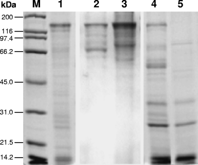
SDS-PAGE analysis of cultures grown to sporulation and lysis in peptonized milk (72 h) showed that IS5056 synthesized a protein that migrated at ∼130 kDa, a molecular mass that corresponded to that of the Cry1 δ-endotoxin of the reference HD1 isolate of B. thuringiensis subsp. kurstaki (130 kDa) (Fig. 2, lanes 1 and 2). The protein in purified IS5056 crystalline inclusions also migrated at ∼130 kDa (Fig. 2, lane 3). In IS5056, no protein corresponding to the HD1 65-kDa Cry2A protein was observed.
FIG. 2.
SDS-PAGE of the parasporal crystals produced by B. thuringiensis subsp. thuringiensis isolate IS5056. Lane 1, spore-crystal preparation of IS5056 grown for 72 h in peptonized milk; lane 2, purified crystals of IS5056; lane 3, purified crystals of the HD1 isolate of B. thuringiensis subsp. kurstaki; lane 4, B. thuringiensis subsp. israelensis strain 4Q7 transformed to express the IS5056 cry1 gene; lane 5, parental strain 4Q7 used as a control. Proteins were fractionated in a 12% gel and stained with Coomassie blue. Lane M contained protein molecular mass markers.
The results of MALDI-TOF MS peptide sequencing of the IS5056 130-kDa protein (Fig. 2, lane 3) suggested that it contained a 13-amino-acid sequence (IEDPQDLEIYLIR), a 9-amino-acid sequence (GNLEFLEEK), and two 8-amino-acid sequences (VDALFVNS and NLEFLEEE). BLAST searches using these sequences and the GenBank sequence database revealed high levels of identity with sequences of Cry1 δ-endotoxins. For example, the 9-amino-acid peptide was identical to a sequence of Cry1A (GenBank accession number ABB76664), Cry1Ba (GenBank accession number POA374), Cry1C (GenBank accession number AAX53094), and Cry1Da (GenBank accession number P19415). Also, both 8-amino-acid peptides showed 100% identity with Cry1 endotoxin sequences (GenBank accession numbers AAQ88259, ABL60921, and AAD04732). Comparisons using the 13-amino-acid peptide revealed 92% identity with Cry1 proteins, in which only one residue, proline, was found to be different at the corresponding position in these proteins (GenBank accession numbers ABL60921, AAW31761, and AAK63251).
As the results of the SDS-PAGE and MALDI-TOF MS analyses suggested that the IS5056 crystal protein belonged to the Cry1 family, PCR using primers (see above) based on the B. thuringiensis serovar aizawai cry1Ab1 gene sequence (accession number DQ991096) was performed to amplify the homologue in IS5056 for heterologous gene expression in the acrystalliferous B. thuringiensis subsp. israelensis 4Q7 strain and for comparative sequence analyses. When the IS5056 cry1 gene was expressed in the 4Q7 strain using the pHT3101 vector, crystalline inclusions that were similar in shape although smaller than those in IS5056 were observed during sporulation (data not shown). In addition, a novel protein band migrating at ∼130 kDa in the recombinant 4Q7 strain but not in parental strain 4Q7 was observed by SDS-PAGE (Fig. 2, lanes 4 and 5). The molecular mass of this protein was identical to that of the protein in the parental IS5056 strain (Fig. 2, lanes 1 and 3). In the recombinant 4Q7 strain, apparent degradation products, presumably from IS5056 Cry1, whose molecular masses ranged from ∼55 to 80 kDa were observed; peptides with molecular masses in this mass range were absent in the 4Q7 control grown under identical conditions (Fig. 2, lanes 4 and 5).
The nucleotide sequence of the amplicon revealed that IS5056 contained a cry1 gene with 3,468-nucleotide open reading frame. The gene encoded a 1,155-amino-acid protein with a predicted molecular mass of 130,511 Da corresponding to the protein observed by SDS-PAGE (Fig. 2, lanes 1, 3, and 4).
In comparative analyses, the nucleotide sequence of IS5056 cry1 was 99% identical to the nucleotide sequences of cry1Ab homologues and differed by only 5 to 17 nucleotides (Fig. 3). The predicted protein encoded by IS5056 cry1 showed 99% identity to known Cry1Ab δ-endotoxins, whereas lower levels of identity (86 to 92%) were observed in comparisons with other Cry1A subgroups (Table 1). Only one amino acid residue, Pro262, in IS5056 Cry1 varied from the corresponding amino acid in Cry1Ab in the accession number AAO13302 and ABK35130 sequences. Five differences in Cry1Ab (accession number AAW31761) were observed at corresponding sites in IS5056 Cry1 (Pro262, Gly385, Ser631, Ala905, and Glu1151), whereas the accession number AF375608 Cry1Ab16 homologue differed by 8 amino acids at corresponding sites in the IS5056 Cry1 protein (Gln154, Ala165, Ser176, Pro262, Leu885, Gly1013, Asp1029, and Lys1121).
FIG. 3.
Positional differences in nucleotide sequences in the cry1 gene of B. thuringiensis subsp. thuringiensis isolate IS5056 and selected cry1Ab homologues. Identical nucleotides are indicated by dots.
TABLE 1.
Relatedness of B. thuringiensis subsp. thuringiensis isolate IS5056 Cry1Ab21 to other Cry1A proteins
| δ-Endotoxin | PubMed accession no. | No. of replacementsa | % Identity |
|---|---|---|---|
| Cry1Ab | AAO13302, ABK35130 | 1 | 99 |
| Cry1Ab | AAW31761 | 5 | 99 |
| Cry1Ab16 | AF375608 | 8 | 99 |
| Cry1Ae | AAA22410 | 58 | 92 |
| Cry1Aa | AAP40639 | 69 | 91 |
| Cry1Ag | AAD46137 | 117 | 86 |
| Cry1Ad | AAA22340 | 128 | 86 |
| Cry1Ac | AAA22339 | 128 | 86 |
Amino acid replacements in selected proteins representing the Cry1A family.
Based on sequence comparisons, the IS5056 Cry1 protein was designated Cry1Ab21, in accordance with B. thuringiensis crystal nomenclature (http://www.lifesci.sussex.ac.uk/home/Neil_Crickmore/Bt/toxins2.html).
Plasmid location of the IS5056 cry1Ab gene.
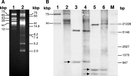
A profile of purified plasmids from IS5056 showed that this strain contained at least five plasmids (approximately 75, 65, 43, 35, and 12 kbp) (Fig. 4A, lane 1). This profile was significantly different than that of reference strain HD1 (Fig. 4A, lane 2). In Southern blot hybridization with undigested IS5056 and HD1 plasmids, two bands, presumably relaxed circular (∼75 kbp) and supercoiled (∼21 kbp) forms, hybridized to the cry1Ab probe (Fig. 4, lanes 1 and 2). In addition, when IS5056 and HD1 plasmids were digested with HindIII, the probe hybridized to predicted 1.0-kbp and ∼6-kb restriction fragments from each of these strains (Fig. 4B, lanes 3 and 4). A different hybridization pattern was observed for IS5056 and HD1 when plasmid DNAs were digested with EcoRI. Whereas two predicted EcoRI fragments (726 and 583 bp) from the Cry1Ab gene of both IS5056 and HD1 were observed, a IS5056 plasmid fragment with a molecular mass (∼19 kbp) lower that that of a fragment from HD1 (∼20 kbp) hybridized with the probe (Fig. 4B, lanes 5 and 6), showing that there was variation between the IS5056 and HD1 plasmids harboring the cry1Ab gene homologues and that IS5056 harbored a single copy of the cry1Ab gene.
FIG. 4.
Plasmid location of the cry1Ab21 gene of B. thuringiensis subsp. thuringiensis isolate IS5056. (A) Plasmid profiles of IS5056 and B. thuringiensis serovar kurstaki HD1 (lanes 1 and 2, respectively). The numbers on the left indicate approximate plasmid sizes. (B) Southern blot hybridization of IS5056 and HD1 with the cry1A probe. Lanes 1 and 2, undigested plasmids from IS5056 and HD1, respectively; lanes 3 and 4, IS5056 and HD1 plasmids digested with HindIII, respectively; lanes 5 and 6, IS5056 and HD1 plasmids digested with EcoRI, respectively. The numbers on the right indicate the sizes of DIG-labeled molecular markers (lane M). The solid arrow indicates the 1,052 bp HindIII fragment, whereas the dotted arrows indicate the 394- and 583-bp EcoRI fragments predicted from intact cry1Ab gene sequences in IS5056 and HD1, respectively. The potential location of a supercoiled plasmid (sc) harboring cry1Ab21, based on hybridization patterns observed in the panel, is also indicated.
IS5056 contains a cry2 gene homologue.
A 4-kbp amplicon was obtained following PCR amplification with the Cry2OPF and Cry2OPR primers, indicating that IS5056 contained a cry2A gene homologue. However, cuboidal crystals typical of Cry2 were not observed by bright-field or electron microscopy, and an additional crystalline band was not observed in sucrose gradients. Whether the protein bands migrating at ∼67 kDa contained a Cry2 homologue is unknown, although it is unlikely (Fig. 2, lanes 1 and 2). Nevertheless, further studies are required to determine whether the gene amplified is a cryptic homologue of cry2.
Phylogenetic relationship of IS5056 gyrB and 16S rRNA sequences to other B. thuringiensis strains.
To evaluate the phylogenetic relationship between IS5056 and other B. thuringiensis strains, partial sequences of the IS5056 gyrB gene that encoded the subunit B protein of DNA gyrase and the 16S rRNA gene were amplified, cloned, and sequenced. Comparative analyses of the IS5056 1,170-bp gyrB sequence showed that the levels of nucleotide identity among B. thuringiensis homologues ranged from 87 to 99% (Table 2). The similarity between the IS5056 16S rRNA sequence (1,348 nucleotides) and homologues in B. thuringiensis strains was much higher (99%) than that observed with gyrB, and differences of only one to nine base substitutions were observed (Table 3).
TABLE 2.
Comparison of B. thuringiensis subsp. thuringiensis isolate IS5056 gyrB with homologues in other Bacillus species
| GenBank accession no. | Bacterium | No. of substitutionsa |
|---|---|---|
| EF210275 | B. thuringiensis subsp. ostriniae | 3 |
| F210276 | B. thuringiensis subsp. tohokuensis | 6 |
| EF210259 | B. thuringiensis subsp. sotto | 10 |
| AY461780 | B. thuringiensis subsp. israelensis | 12 |
| EF210264 | B. thuringiensis subsp. alesti | 11 |
| EF210279 | B. thuringiensis subsp. fukokaensis | 19 |
| EF210261 | B. thuringiensis subsp. wratislaviensis | 33 |
| EF210268 | B. thuringiensis subsp. shandongiensis | 37 |
| EF210267 | B. thuringiensis subsp. roskildiensis | 73 |
| EF210269 | B. thuringiensis subsp. vazensis | 95 |
| AY461778 | B. thuringiensis subsp. aizawai | 104 |
| AE017355 | B. thuringiensis subsp. konkukian | 141 |
| EF210265 | B. thuringiensis subsp. oswaldocruzi | 143 |
| CP000485 | B. thuringiensis strain Al Hakam | 144 |
| CP000001 | B. cereus strain E33L | 73 |
| EF210273 | B. weihenstephanensis strain CMM4966 | 97 |
| AB190227 | B. anthracis strain H0001 | 140 |
Based on 1,170 nucleotide bases of homologous Bacillus gyrB genes.
TABLE 3.
Comparison of B. thuringiensis susbsp. thuringiensis isolate IS5056 16S rRNA with homologues in other Bacillus species
| GenBank accession no. | Bacterium | No. of substitutionsa |
|---|---|---|
| AF290545 | B. thuringiensis strain ATCC 10792 | 1 |
| AF155954 | B. thuringiensis strain 4Q281 | 1 |
| AY138290 | B. thuringiensis strain 2000031482 | 5 |
| AY138288 | B. thuringiensis strain 2000031494 | 5 |
| AB116122 | B. thuringiensis | 5 |
| AE017355 | B. thuringiensis subsp. konkukian 97-27 | 6 |
| AF155955 | B. thuringiensis strain B8 | 6 |
| CP000485 | B. thuringiensis strain Al Hakam | 7 |
| AJ310100 | B. cereus biovar toyoi strain CNCM I-1012/NCIB | 1 |
| AY138271 | B. cereus strain G8639 | 1 |
| EF126181 | B. cereus strain HCY02 | 3 |
| AE016877 | B. cereus strain ATCC 14579 | 4 |
| CP000001 | B. cereus strain E33L | 5 |
| AE017225 | B. anthracis strain Sterne | 5 |
| AE016879 | B. anthracis strain Ames | 5 |
Based on 1,348 nucleotide bases of the homologous Bacillus 16S rRNA gene.
Toxicity of IS5056 for T. ni and S. exigua larvae.
Standard bioassays showed that the crystal-spore mixture of the IS5056 strain was toxic to T. ni larvae. The LC50s for second- and fourth-instar larvae were calculated to be 16.9 μg ml−1 (statistical range, 14.05 to 20.09 μg ml−1) and 29.7 μg ml−1 (statistical range, 24.28 to 38.39 μg ml−1), respectively, whereas the LC90s were 53.6 μg ml−1 (statistical range, 41.09 to 80.58 μg ml−1) and 114.3 μg ml−1 (statistical range, 75.36 to 238.16 μg ml−1), respectively. When spore-crystal preparations were autoclaved (121°C for 20 min) to test for β-exotoxin activity, mortality was not observed with second- and fourth-instar T. ni larvae, even when the spore-crystal concentration was 400 μg ml−1. Finally, similar assays with S. exigua showed that the IS5056 crystal-spore mixture significantly inhibited larval growth and molting, even at concentrations as low as 25 μg ml−1, but no mortality was observed after 7 days of exposure to the mixture.
Strain pathogenesis.
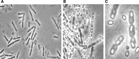
In contrast to the results reported for L. dispar (5), we found that the IS5056 isolate of B. thuringiensis invaded larvae as they died or shortly after death and rapidly colonized the larval cadaver (Fig. 5). The mortality for second-instar T. ni larvae fed a suspension of 10,000 spores and associated crystals was 23.5% (4/17) at 24 h. No mortality was observed for the remaining 13 larvae, all of which progressed to pupation. For third-instar larvae fed 20,000 spores and associated crystals, the mortality was 8% (2/25) at 24 h and 40% (10/25) at 48 h. The remaining 13 larvae progressed to pupation. Vegetative and sporulating cells were found in the cadavers of second- and third-instar larvae (Fig. 5) within 24 to 48 h of death. The NA cultures without antibiotics prepared from cadavers 7 days after death consisted of opaque white colonies, and the average colony count for second-instar larvae was 7.7 × 10⁶ colonies, a 770-fold increase over the infecting dose; the average colony count for the third-instar larvae was 1.5 × 10⁷ colonies, a 750-fold increase. During the course of this study, we examined many larvae that died from consuming the IS5056 spore-crystal mixture, and these larvae always contained numerous sporulated cells with crystals characteristic of this isolate. For further confirmation that the vegetative cells and spores observed in cadavers of treated T. ni larvae were the result of reproduction, microscopic examination of 20 colonies chosen randomly from the cultures described above showed that they were all composed of vegetative, sporulating, and sporulated cells of IS5056, as determined by the presence of the large quasicuboidal inclusions characteristic of this isolate (Fig. 1 and 5). These results demonstrated that T. ni was an excellent resource for reproduction of this isolate of B. thuringiensis. No other type of colony was observed, indicating that the cadavers were virtually pure, if not completely pure, cultures of IS5056. In contrast to this, the cultures prepared from control larvae, none of which had died after 5 days, were dominated by colonies that were translucent rather than opaque white and contained rod-shaped bacteria that never sporulated and which were much smaller than cells of IS5056.
FIG. 5.
Phase-contrast microscopy showing that B. thuringiensis subsp. thuringiensis IS5056 colonizes and sporulates in dead larvae of T. ni. Third-instar larvae were fed a spore-crystal mixture of IS5056, after which larval tissues were examined by phase-contrast microscopy at 48 h (A) and 7 days (B and C). (A and B) Magnification, ×400. (C) Magnification, ×1,000.
DISCUSSION
In the present study, we identified and characterized a new entomopathogenic soil isolate (IS5056) of B. thuringiensis subsp. thuringiensis that produces an unusual parasporal inclusion that typically appears to be cuboidal or quasicuboidal and is composed of a new Cry endotoxin, Cry1Ab21. In addition to the large crystalline inclusions and unique plasmid profile, the genetic evidence based on sequence analyses of the gyrB and 16S rRNA genes supports our classification of IS5056 as a novel isolate of B. thuringiensis subsp. thuringiensis (3).
The naturally occurring crystals in B. thuringiensis IS5056 were observed to be very large (0.8 by 1.4 μm) compared to the typical Cry1A crystals, which are approximately 0.5 by 1.0 μm. The shape of the IS5056 CryAb21 inclusion is atypical for Cry1-type protein crystals (2, 14, 27), and this enabled us to use it as a marker for our experiments in which we examined the reproduction of B. thuringiensis in T. ni. With respect to the atypical shape of the IS5056 crystal, it is known that the native shapes of Cry proteins are largely maintained by interchain disulfide bridges, which can be distorted by physical and chemical treatment (6). Interestingly, Du et al. (6) reported that native and autoclaved crystals of B. thuringiensis subsp. kurstaki HD1 are indistinguishable; they are bipyramidal, and the structure and shape presumably are stabilized by disulfide bonds. When the IS5056 crystalline inclusions were autoclaved and examined microscopically, predominantly spherical inclusions, rather than the native cuboidal, quasicuboidal, and bipyramidal inclusions, were observed (unpublished results). This suggests that there are differences in the stabilizing properties of disulfide bridges in crystals of strain HD1 and the IS5056 isolate.
Interestingly, heterologous expression of the IS5056 cryAb21 gene in the acrystalliferous B. thuringiensis subsp. israelensis 4Q7 strain showed that this strain produced a crystal that was approximately one-third the size of the crystal in wild-type strain IS5056. This observation suggests that the molecular basis for synthesis of the large crystalline inclusion in IS5056 may not be intrinsic to the CryAb21 amino acid sequence, particularly since it has few amino acid replacements compared to other Cry1Ab proteins, or that the plasmid-cured strain 4Q7 lacked the genetic background to facilitate synthesis of large crystals. In this regard, several factors are known to affect the level of crystal toxin synthesis and crystallization in B. thuringiensis. These factors include mechanisms that influence transcription efficiency and mRNA stability, translation efficiency, and posttranslational events mediated by chaperone-like accessory proteins, such as the 20-kDa and ORF2 helper proteins encoded by the Cry11A and Cry2A operons, respectively (1, 7, 24, 33, 34, 35). Whether these factors play crucial roles at the corresponding levels in enhancing Cry1Ab21 yields and crystallization remains to be investigated and is the focus of current studies. However, the apparent extensive degradation of Cry1Ab21 when it was synthesized in 4Q7 (Fig. 2, lane 4) suggests that a mechanism(s) involving molecular chaperones or helper proteins that stabilize the Cry1Ab21 protoxin by protecting it from proteolytic cleavage or by optimizing its crystallization is present in IS5056. Further studies should resolve these mechanisms.
We also demonstrated here that the IS5056 isolate was toxic to T. ni, with LC50s of 16.9 and 29.7 μg ml−1 and LC90s of 53.6 and 114.3 μg ml−1 for second- and fourth-instar larvae, respectively. The observed lethality is attributed to Cry1Ab21, as we have shown that this strain does not produce detectable levels of β-exotoxin (8). It is difficult to directly compare this level of toxicity to the levels of toxicity of other isolates and strains of B. thuringiensis that have been described previously because the methods used by other investigators vary. However, comparing the toxicity of IS5056 to, for example, the toxicity of the HD1 strain of B. thuringiensis indicated they have similar toxicities (21). Moar et al. (21) observed a toxicity of 1.23 μg ml−1 (LC50) for neonate larvae of T. ni in bioassays similar to our bioassay. Neonate larvae are the most sensitive stage, and progressively higher concentrations of toxin are required with each successive instar to obtain a similar mortality. Thus, we evaluated our new isolate with second- and fourth-instar larvae, and based on the results described above, we estimated that the levels of toxicity of HD1 and IS5053 are similar.
In contrast to the results reported for the HD1 strain of B. thuringiensis and L. dispar (5), when the pathogenesis of strain IS5053 was investigated, it was clear that this new strain is fully capable of invading, colonizing, and sporulating in cadavers of T. ni. In terms of the biology of this isolate, Cry1Ab21 intoxicates the larvae, after which spores apparently germinate in the midgut and invade the moribund and/or dead larvae, reproducing more than 700-fold in third-instar larvae. Thus, the toxin is an important virulence factor that contributes to the reproductive potential of this isolate when sufficient quantities of spores and crystals are consumed by a susceptible host.
Although IS5056 can clearly reproduce and sporulate in cadavers of T. ni killed by this isolate, the process is likely considerably different than what occurs in larvae of the Japanese beetle, Popillia japonica, a type of scarab beetle, infected with Paenibacillus popilliae (28). In contrast to B. thuringiensis, P. popilliae has a highly obligate relationship with susceptible scarab hosts and cannot be cultured easily in vitro. Nevertheless, it is possible that these two bacterial species have similar virulence factors, such as toxins, and that B. thuringiensis isolates may be capable of invading certain susceptible hosts before they die, as P. popilliae does.
Because T. ni is among the most destructive agriculture pests in North America and has been shown to develop resistance to B. thuringiensis formulations under laboratory conditions and in commercial greenhouses in response to spray regimens (11, 12), IS5056 could potentially have applied value. For this reason, we are conducting studies to determine the basis for the large Cry1Ab21 crystalline inclusion and are using molecular approaches to introduce additional toxin genes into IS5056 to increase its toxicity and expand its host range.
Acknowledgments
We thank Jeffrey J. Johnson (University of California, Riverside) for technical assistance during this study, Kumiko Kagoshima and Michio Ohba (Graduate School of Agriculture, Kyushu University, Fukuoka, Japan) for performing the serological identification of isolate IS5056, and Neil Crickmore (University of Sussex, Falmer, Brighton, United Kingdom) for providing the designation (Cry1Ab21) for the IS5056 crystal protein.
I. Swiecicka thanks the Fulbright Commission for a fellowship that supported the research conducted at University of California, Riverside. This research was also supported in part by grant 2PO4C 094 30 to I. Swiecicka from the Ministry of Science and Higher Education in Poland.
Footnotes
Published ahead of print on 14 December 2007.
REFERENCES
- 1.Agaisse, H., and D. Lereclus. 1996. STAB-SD: a Shine-Delgarno sequence in the 5′-untranslated region is a determinant of mRNA stability. Mol. Microbiol. 20:633-643. [DOI] [PubMed] [Google Scholar]
- 2.Aguiar, R. W. S., É. S Martins, F. H. Valicente, N. P. Carneiro, A. C. Batista, V. M. Melatti, R. G. Monnerat, and B. M. Ribeiro. 2006. A recombinant truncated Cry1Ca protein is toxic to lepidopteran insects and forms large cuboidal crystals in insect cells. Curr. Microbiol. 53:287-292. [DOI] [PubMed] [Google Scholar]
- 3.Bavykin, S. G., Y. P. Lysov, V. Zakhariev, J. J. Kelly, J. Jackman, D. A. Stahl, and A. Cherni. 2004. Use of 16S rRNA, 23S rRNA, and gyrG gene sequence analysis to determine phylogenetic relationships of Bacillus cereus group microorganisms. J. Clin. Microbiol. 42:3711-3730. [DOI] [PMC free article] [PubMed] [Google Scholar]
- 4.Bonk, T., and A. Humeny. 2001. MALDI-TOF-MS analysis of protein and DNA. Neuroscientist 7:6-12. [DOI] [PubMed] [Google Scholar]
- 5.Broderick, N. A., K. F. Raffa, and J. Handelsman. 2006. Midgut bacteria required for Bacillus thuringiensis insecticidal activity. Proc. Natl. Acad. Sci. USA 103:15196-15199. [DOI] [PMC free article] [PubMed] [Google Scholar]
- 6.Du, C., P. A. W. Martin, and K. W. Nickerson. 1994. Comparison of disulfide contents and solubility at alkaline pH of insecticidal and noninsecticidal Bacillus thuringiensis protein crystal. Appl. Environ. Microbiol. 60:3847-3853. [DOI] [PMC free article] [PubMed] [Google Scholar]
- 7.Ge, B., D. K. Bideshi, W. J. Moar, and B. A. Federici. 1998. Differential effects of helper proteins encoded by the cry2A and cry11A operons on formation of Cry2A inclusions in Bacillus thuringiensis. FEMS Microbiol. Lett. 165:35-41. [DOI] [PubMed] [Google Scholar]
- 8.Gough, J. M., D. H. Kemp, R. J. Akhurst, R. D. Pearson, and K. Kongsuwan. 2005. Identification and characterization of proteins from Bacillus thuringiensis with high toxic activity against the sheep blowfly, Lucilia cuprina. J. Invertebr. Pathol. 90:39-46. [DOI] [PubMed] [Google Scholar]
- 9.Gunning, R. V., H. T. Dang, F. C. Kemp, I. C Nicholson, and G. D. Moores. 2005. New resistance mechanism in Helicoverpa armigera threatens transgenic crops expressing Bacillus thuringiensis Cry1Ac toxin. Appl. Environ. Microbiol. 71:2558-2563. [DOI] [PMC free article] [PubMed] [Google Scholar]
- 10.Ichimatsu, T., E. Mizuki, K. Nishimura, T. Akao, and H. Saitoh. 2000. Occurrence of Bacillus thuringiensis in fresh water of Japan. Curr. Microbiol. 40:217-220. [DOI] [PubMed] [Google Scholar]
- 11.Janmaat, A. F., and J. Myers. 2003. Rapid evolution and the cost of resistance to Bacillus thuringiensis in greenhouse populations of cabbage loopers, Trichoplusia ni. Proc. R. Soc. Lond. B 270:2263-2270. [DOI] [PMC free article] [PubMed] [Google Scholar]
- 12.Janmaat, A. F., P. Wang, W. Kain, J.-Z. Zhuo, and J. Myers. 2004. Inheritance of resistance to Bacillus thuringiensis subsp. kurstaki in Trichoplusia ni. Appl. Environ. Microbiol. 70:5859-5867. [DOI] [PMC free article] [PubMed] [Google Scholar]
- 13.Jurat-Fuentes, J. L., F. L. Gould, and M. J. Adang. 2003. Dual resistance to Bacillus thuringiensis Cry1Ac and Cry2Aa toxins in Heliothis virescens suggests multiply mechanism of resistance. Appl. Environ. Microbiol. 69:5898-5906. [DOI] [PMC free article] [PubMed] [Google Scholar]
- 14.Kaelin, P., and F. Gadani. 2000. Occurrence of Bacillus thuringiensis on cured tobacco leaves. Curr. Microbiol. 40:205-209. [DOI] [PubMed] [Google Scholar]
- 15.Kronstad, J. W., H. E. Schnepf, and H. R. Whiteley. 1983. Diversity of location for Bacillus thuringiensis crystal protein genes. J. Bacteriol. 154:419-428. [DOI] [PMC free article] [PubMed] [Google Scholar]
- 16.Laemmli, U. K. 1970. Cleavage of structural proteins during the assembly of the head of bacteriophage T4. Nature 227:680-685. [DOI] [PubMed] [Google Scholar]
- 17.Lecadet, M.-M., E. Frachon, V. Cosmao Dumanoir, H. Ripoutea, S. Hamon, P. Laurent, and I. Thiery. 1999. Updating the H-antigen classification of Bacillus thuringiensis. J. Appl. Microbiol. 86:660-672. [DOI] [PubMed] [Google Scholar]
- 18.Lereclus, D., O. Arantes, J. Chhaufaux, and M. M. Lecadet. 1989. Transformation and expression of a cloned δ-endotoxin gene in Bacillus thuringiensis. FEMS Microbiol. Lett. 60:211-218. [DOI] [PubMed] [Google Scholar]
- 19.Lonc, E., M.-M. Lecadet, T. M. Lachowicz, and E. Panek. 1997. Description of Bacillus thuringiensis wratislaviensis (H-47), a new serotype originating from Wroclaw (Poland), and other Bt soil isolates from the same area. Lett. Appl. Microbiol. 24:467-473. [Google Scholar]
- 20.McGaughey, W. H., and M. E. Whalon. 1992. Managing insect resistance to Bacillus thuringiensis toxins. Science 258:1451-1455. [DOI] [PubMed] [Google Scholar]
- 21.Moar, W. J., L. Masson, R. Brousseau, and J. T. Trumble. 1990. Toxicity to Spodoptera exigua and Trichoplusia ni of individual P1 protoxins and sporulated cultures of Bacillus thuringiensis subsp. kurstaki HD-1 and NRD-12. Appl. Environ. Microbiol. 56:2480-2483. [DOI] [PMC free article] [PubMed] [Google Scholar]
- 22.Nester, E. W., L. S. Thomashow, M. Metz, and M. Gordon. 2002. 100 years of Bacillus thuringiensis: a critical scientific assessment. American Society for Microbiology, Washington, DC. http://www.asm.org. [PubMed]
- 23.Ohba, M., and K. Aizawa. 1978. Serological identification of Bacillus thuringiensis and related bacteria in Japan. J. Invertebr. Pathol. 32:303-309. [Google Scholar]
- 24.Park, H.-W., B. Ge, L. S. Bauer, and B. A. Federici. 1998. Optimization of Cry3A yields in Bacillus thuringiensis by use of sporulation-dependent promoters in combination with the STAB-SD mRNA sequence. Appl. Environ. Microbiol. 64:3932-3938. [DOI] [PMC free article] [PubMed] [Google Scholar]
- 25.Sambrook, J., and D. W. Russell. 2001. Molecular cloning: a laboratory manual, 3rd ed. Cold Spring Harbor Laboratory Press, Cold Spring Harbor, NY.
- 26.Sayyed, A. H., B. Raymond, M. Sales Ibiza-Palacios, B. Escriche, and D. J. Wright. 2004. Genetic and biochemical characterization of field-evolved resistance to Bacillus thuringiensis toxin Cry1Ac in the diamondback moth, Plutella xylostella. Appl. Environ. Microbiol. 70:7010-7017. [DOI] [PMC free article] [PubMed] [Google Scholar]
- 27.Schnepf, E., N. Crickmore, J. Van Rie, D. Lereclus, J. Baum, J. Feitelson., D. R. Zeigler, and D. H. Dean. 1998. Bacillus thuringiensis and its pesticidal crystal proteins. Microbiol. Mol. Biol. Rev. 62:775-806. [DOI] [PMC free article] [PubMed] [Google Scholar]
- 28.Splittstoesser, C. M., C. Y. Kawanishi, and H. Tashiro. 1978. Infection of the European chafer, Amphimallon majalis, by Bacillus popilliae: light and electron microscope observations. J. Invertebr. Pathol. 31:84-90. [Google Scholar]
- 29.Swiecicka, I., K. Fiedoruk, and G. Bednarz. 2002. The occurrence and properties of Bacillus thuringiensis isolated from free-living animals. Lett. Appl. Microbiol. 34:194-198. [DOI] [PubMed] [Google Scholar]
- 30.Swiecicka, I., and P. De Vos. 2003. Properties of Bacillus thuringiensis isolated from bank voles. J. Appl. Microbiol. 94:60-64. [DOI] [PubMed] [Google Scholar]
- 31.Tabashnik, B. E., Y. Carriere, T. J. Dennehy, S. Morin, M. S. Sisterson, R. T. Roush, A. M. Shelton, and J. Z. Zhao. 2003. Insect resistance to transgenic Bt crops: lessons from the laboratory and field. J. Econ. Entomol. 96:1031-1038. [DOI] [PubMed] [Google Scholar]
- 32.Tamez-Guerra, P., G. Damas, M. M. Iracheta, B. Oppert, R. Gomez-Flores, and C. Rodriguez-Padilla. 2006. Differences in susceptibility and physiological fitness of Mexican field Trichoplusia ni strains exposed to Bacillus thuringiensis. J. Econ. Entomol. 99:937-945. [DOI] [PubMed] [Google Scholar]
- 33.Tang, M., D. K. Bideshi, H.-W. Park, and B. A. Federici. 2006. Minireplicon from pBtoxis of Bacillus thuringiensis subsp. israelensis. Appl. Environ. Microbiol. 72:6948-6954. [DOI] [PMC free article] [PubMed] [Google Scholar]
- 34.Wu, D., and B. A. Federici. 1993. A 20-kilodalton protein preserves cell viability and promotes CytA crystal formation during sporulation in Bacillus thuringiensis. J. Bacteriol. 175:5276-5280. [DOI] [PMC free article] [PubMed] [Google Scholar]
- 35.Wu, D., and B. A. Federici. 1995. Improved production of the insecticidal CryIVD protein in Bacillus thuringiensis using cry1A(c) promoter to express the gene for an associated 20-kDa protein. Appl. Microbiol. Biotechnol. 42:697-702. [DOI] [PubMed] [Google Scholar]


