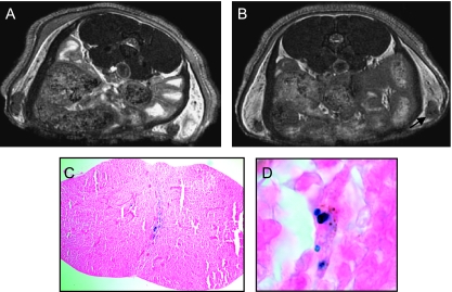
Figure 5.
Axial in vivo FIESTA images of the inguinal lymph nodes of a mouse acquired with 100 x 100 x 100 µm3 spatial resolution in 68 minutes. The mouse was injected with 100 MPIO-labeled cells, and the images were acquired on (A) days 4 and (B) 22 postinjection. Small focal regions of signal loss within the node persist over this time course. No tumor growth was evident at dissection. (C) PPB staining (original magnification, x40) of this node at day 22 shows the existence of iron-positive cells. (D) At higher magnification (x100), cells that are PPB-positive and have brown pigmentation, which indicates melanin, can be identified.

