Abstract
This paper reports a study to find small peptide substrates for the important virulence factor of Yersinia pestis, plasminogen activator, Pla. The method used to find small substrates for this protease is reported along with studies examining the ability of these peptides to inhibit activity of the enzyme. Through the use of parallel synthesis and positional scanning, small tripeptides were identified that are viable substrates for the protease.
Keywords: Yersinia pestis; plasminogen activator; protease substrate; peptide inhibitor; positional scan, parallel synthesis
Yersinia pestis, the agent of plague, has been recognized as one of the most devastating, epidemic-causing bacteria experienced by mankind.1 Typically, Y. pestis is transmitted to humans via a bite from a flea that previously fed on an infected rodent. From the initial site of infection, bacteria disseminate to the draining lymph node, causing swelling of this lymph node to form a bubo (bubonic plague).
Y. pestis contains a unique, 9.5-kb plasmid pPCP that expresses plague plasminogen activator (Pla) which is responsible for fibrinolytic and coagulase activities.2 Pla expression is associated with the marked ability of Y. pestis to colonize the vicera and thus cause lethal infection upon administration by peripheral (i.e. intradermal (i.d.), subcutaneous (s.c.) or intraperitoneal) routes of infection.3 The importance of Pla for plague pathogenesis was verified with isogenic Pla mutants of epidemic Y. pestis strains KIM and CO92, which showed an up to 106 fold reduced virulence by the s.c. routes.3,4 Recently, it was confirmed that bubonic plague, but not septicemic, depends on Pla to facilitate the rapid dissemination of bacteria from the site of i.d. infection.5 Moreover, it was shown that Pla allows Y. pestis to replicate rapidly in the airways, thus playing the essential role in causing pneumonic plague.6 The invasive properties of Pla which is a cell surface-located protease are likely due to the ability of this enzyme to induce fibrinolysis and degrade extracellular matrix and basement membranes.7,8 The activation of plasminogen to plasmin while degrading the plasmin inhibitor α2-antiplasmin by Pla is the mechanism used by Y. pestis to progress from the peripheral sites into the circulation and systemic infection.9,10 This paper reports work to identify inhibitors of Pla through parallel synthesis of small peptidic substrates for the enzyme.
One of the classic approaches for the development of protease inhibitors has been to determine the substrate specificity of the protease of interest and then use that information for the design of inhibitors.11 Such an approach is particularly useful with membrane bound proteases, such as Pla, where there is limited structural information. An approach was undertaken where ensembles of peptides were screened for their ability to act as substrates for Pla. Knowledge of the small peptide substrates accepted by the enzyme can often serve as a starting point for the development of enzyme inhibitors.
The original approach undertaken was to synthesize, on solid support, ensembles of fluorogenic peptides based on loop 5 of Pla, a region of the enzyme that is known to undergo self cleavage,10 and then screen those libraries for fluorescence (Fig. 1). Six-residue peptides, overlapping by one amino acid were synthesized with the DABCYL/EDANS quencher/fluorophor system chosen for this work.12 In theory, peptides in the mixture that are substrates for Pla would be hydrolyzed resulting in the separation of the quencher (DABCYL) from the fluorophor (EDANS) and fluorescence of the bead. The targeted libraries synthesized on the PEGA1900 support resulted in no fluorescence when submitted to the purified enzyme. Additionally, targeted libraries based on the region of plasminogen that is known to be cleaved by Pla were also screened, resulting in no fluorescence. Some potential reasons for the lack of a signal are: the enzyme and the solid support the peptides were attached to may not be compatible in terms of porosity or hydrophobicity; the enzyme may not accept small substrates or substrates that do not have a particular secondary structure.
Figure 1.
Loop 5 sequence was analyzed as overlapping 6 residue peptides
To determine if the enzyme would accept small peptide substrates in general, mixtures of “all possible” combinations of trimers, tetramers, pentamers and hexamers were generated in solution and incubated with the enzyme. These mixtures were generated using a modified version of the isokinetic approach of Houghton13,14 and consisted of all the natural amino acids, excluding cysteine and tryptophan. Upon treatment of Pla with a given volume of each peptide mixture, the solutions of pentamers and hexamers were found to fluoresce. This indicated that the problem with the initial solid supported screen was probability due to incompatibility of the enzyme with the support. Rather than attempt to remedy this problem an effort was undertaken to identify active peptides in solution. The positive results with pentamers and hexamers, versus the lack of fluorescence with trimers and tetramers, were explained through an assumption that Pla needs at least 5 amino acids to recognize its substrate. This assumption later proven to be incorrect and will be discussed later.
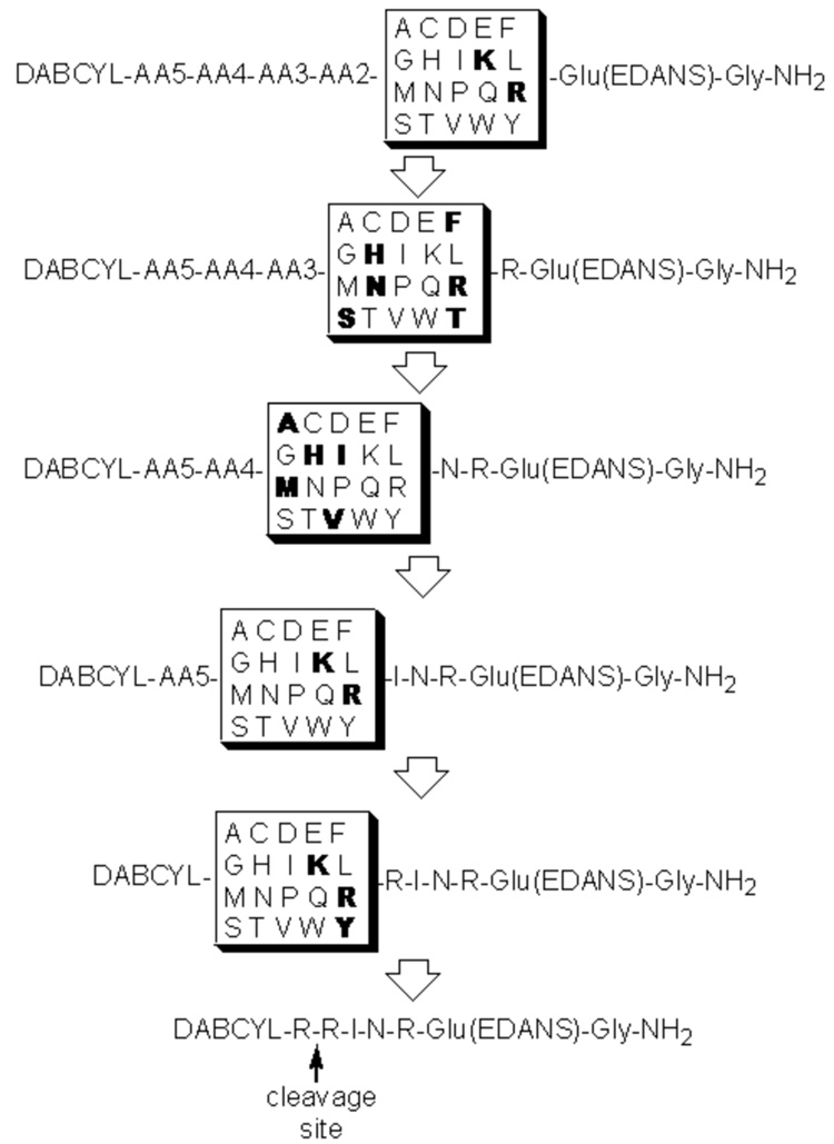
Having established a “minimal” length of the substrate a positional scan approach, similar to the work of Houghten, was used to identify individual peptides.15 In this approach one amino acid position is held constant while the others are varied. The Fmoc-Glu(EDANS)-Gly-Wang or Fmoc-Glu(EDANS)-Ala-Wang resin was divided into 20 equal portions, and each portion was coupled to an individual amino acid followed by coupling with an isokinetic mixture of eighteen amino acids. This provides each vessel with a mixture of dimers with the known amino acid at position 1 and the other eighteen amino acids represented at position 2. This process was then repeated for positions 3, 4 and 5 and then the final introduction of DABCYL. Each peptide mixture was cleaved from the support and then tested in a reaction with Pla to determine which amino acid in the first position resulted in the hydrolysis. Fluorescence was detected using a plate reader at excitation and emission wavelengths of 360 and 460 nm, respectively. The amino acid in position 1 that resulted in fluorescence was then held constant and the second position is scanned in the same manner as the first. This process is then repeated for the third, fourth and fifth positions. (It should be noted that while the original paper reports isokinetic ratios for Boc-protected amino acids and usage of 10 eq, we used Fmoc-based strategy and 5 eq during coupling.) Despite the limitation of a positional scan approach, the method provided substrates for Pla enzyme, with a sequence Arg-Arg-Ile-Asn-Arg being selected as the “best”. The substrate based on this sequence was resynthesized on Sieber amide resin to eliminate influence of Gly/Ala, and the resulting DABCYL-Arg-Arg-Ile-Asn-Arg-Glu(EDANS)-NH2 was confirmed to be a fluorogenic substrate for Pla.
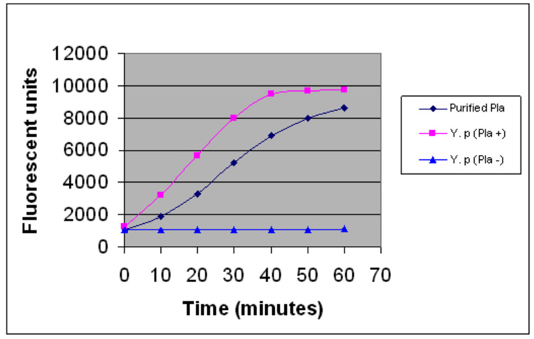
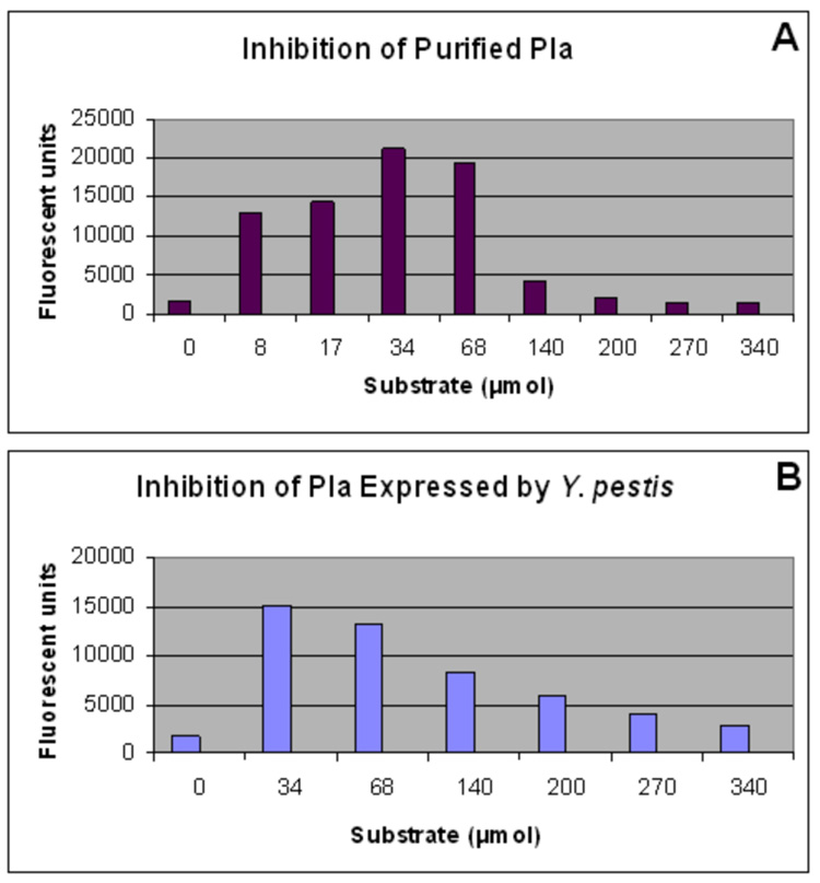
Once a small peptide substrate was identified, the Pla activity was measured in a fluorimetric assay using substrate DABCYL-Arg-Arg-Ile-Asn-Arg-Glu(EDANS)-NH2. In Figure 2, increase in fluorescence is given as a function of time for both purified recombinant Pla and an isogenic pair of Y. pestis strains either expressing or not the Pla protease on the cell surface. Both purified and cell-associated Pla enzyme cleaved the substrate in a time-dependent manner, reaching a plateau in about 60 min. The Planegative mutant showed low background fluorescence indicating substrate specificity towards Pla. At high concentrations of the fluorogenic peptide, inhibition was observed with both purified Pla as well as the enzyme expressed on the surface of Y. pestis (Fig. 3). The exact origin of this inhibition is not know but it was confirmed by LC-MS analysis of the reaction mixtures.
Figure 2.
Kinetic of cleavage of identified fluorescent substrate by Pla. The activity was measured with purified recombinant Pla, Y. pestis cells expressing Pla and Y. pestis cells lacking Pla gene.
Figure 3.
Fluorimetric assay of Pla activity at various substrate concentrations. The activity was measured at the end point of the reaction with purified recombinant Pla (A) and Y. pestis cells expressing Pla on their surface (B).
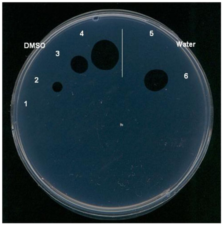
In addition to cleavage of the flurogenic substrate, a functional assay was performed. Pla activates plasminogen to plasmin by limited proteolysis. The inhibitory activity of the substrate was tested by examining its ability to inhibit the conversion of plasminogen to plasmin in a fibrin plate assay.16 Upon plasminogen’s conversion to plasmin the resulting proteolytic dissolution of fibrin is seen as a clear liquid spot on an opalescent fibrin film. Pre-incubation of purified Pla with the substrate, DABCYL-Arg-Arg-Ile-Asn-Arg-Glu(EDANS)-NH2, followed by spotting the samples on the fibrin film resulted in a concentration-dependent inhibition of Pla-mediated fibrinolysis (Fig. 4). It was also observed that the fluorogenic substrate can inhibit Pla expressed by Y. pestis cells in a similar manner, but did not prevent the action of mammalian plasminogen activator urokinase (data not shown).
Figure 4.
Fibrinolytic assay of Pla activity at various substrate concentrations. The substrate is taken in 2-fold dilutions. Spots 4 and 6 represent the samples with no substrate added. Spots 1, 2, 3 and 5 have different substrate concentrations. The substrate was dissolved either in DMSO or in water.
Following hydrolysis of the fluorogenic substrate the reaction products were identified by LC-MS analysis. The site of hydrolysis was at a basic site, in this case between the two arginines. This observation correlates with the substrate specificity of the omptin family of proteases which have a preference to cleave between two basic amino acids.17
Truncated versions of the initial six-residue substrate were examined to probe the length of peptide necessary, as well as to examine the role of the fluorogenic and quencher molecules. Versions of the hexapeptide substrate with and without the EDANS group were evaluated as substrates and as inhibitors in the fibrinolytic assay. A derivative of the peptide without the fluorophore EDANS but with the quencher DABCYL did not inhibit the activation of plasminogen at the concentration in the range of 100-350 µM but was still a substrate for the enzyme. Every truncated version except the dipeptide DABCYL-Arg-Arg-OH, were substrates for Pla (Table 1). Apparently EDANS plays a role in increasing inhibitory activity. We are in the process of examining the origin of this effect through the incorporation of molecules with structures similar to EDANS. This effect was observed when EDANS was attached by either the side chain or main chain carboxyl of the peptide (1 and 6, Table 1). Synthesis of the hexapeptide omitting DABCYL also resulted in the loss of the inhibitory activity, indicating that both fluorophore and quencher play a role in the inhibition of Pla. As a control, both free EDANS and DABCYL where examined and found not to inhibit the enzyme. As shown in Table 1 the length of the peptide can be reduced to a trimer DABCYL-Arg-Arg-Ile-EDANS and maintain activity.
Table 1.
Peptide sequences tested as substrates and functional inhibitors
| Cpda | Sequence | Sb | Ic |
|---|---|---|---|
| 1 | DABCYL-RRINR-Glu(EDANS)-NH2 | + | + |
| 2 | DABCYL-RRINR-OH | + | − |
| 3 | DABCYL-RRIN-OH | + | − |
| 4 | DABCYL-RRI-OH | + | − |
| 5 | DABCYL-RR-OH | − | − |
| 6 | DABCYL-RRINR-Glu-EDANS | + | + |
| 7 | DABCYL-RRIN-EDANS | + | + |
| 8 | DABCYL-RRI-EDANS | + | + |
Compounds 2–5 and DABCYL-Arg-OH have been synthesized and used as LC-MS standards to determine cleavage fragments. Based on retention time and mass detection DABCYL-Arg-OH was one of products in all cleavage mixtures except 5.
Substrate: the vertical line designates that the peptides were cleaved by Pla (LC-MS tested).
Inhibitor: the inhibitory activity of the peptides was determined using functional assay on fibrin plates.
Previously, in the case of mixtures of peptides, it was found that only hexamer and pentamer mixtures resulted in fluorescence. Since our initial experiments were conducted with the same amount of total peptide resulted from the isokinetic approach, the difference between the experiment discussed here and the results with mixtures is likely due to the fact that in a given amount of the mixtures there is significantly more of each individual trimer than each pentamer (up to 182 fold). Consequently under the conditions of the initial assay, substrate inhibition was observed with the trimer and tetramer mixtures. However when discrete tetra-and tri- peptides were synthesized and tested they displayed substrate and inhibitory properties similar to those of the lead hexamer substrate.
Since a tripeptide DABCYL-Arg-Arg-Ile-EDANS was found to be a substrate similar to the hexapeptide DABCYL-Arg-Arg-Ile-Asn-Arg-Glu(EDANS)-NH2, its optimization was under taken. A focused library of tripeptide substrates with Arg-Arg held constant, and the third position varied was screened. Profiling of Pla with this DABCYL-Arg-Arg-X-EDANS library revealed preference for hydrophobic aliphatic (Ala, Ile and Val), neutral-polar side chains (Thr, Ser, and Cys) and small Gly amino acids at that position (Fig. 5).
Figure 5.
Profiling the tripeptide DABCYL-Arg-Arg-X-EDANS library. The X represents one of the twenty amino acids residues. The y axis is end point fluorescent signal after 2 h of incubation of the substrate with purified Pla. The x axis provides the spatial address of the amino acid as represented by the one-letter code. Each substrate was examined at concentration of ~35 µmol.
In addition to providing information about the substrate specificity of the enzyme this work has provided a series of small fluorogenic peptides that are currently being used as leads for the development of inhibitors of the protease Pla. Additionally such fluorogenic substrates are being used to develop a high-throughput screen for small molecule inhibitors of Pla.
Supplementary Material
Scheme 1.
The highlighted amino acids are amino acids that resulted in preferred cleavage of the substrate. Ultimately, based on qualitative differences in the rate of cleavage between different active sequences, we selected DABCYL-Arg-Arg-Ile-Asn-Arg-Glu(EDANS)-NH2 as the substrate candidate for Pla.
Acknowledgements
This work was supported by the Region VI “Western” RCE (NIH award 1-U54-AI-057156) grant, an award from the Sealy Center for Vaccine Development of the University of Texas Medical Branch, and the Robert A. Welch Foundation.
We thank Dr. Robert R. Brubaker for providing the strains of Y. pestis and helpful discussions.
Footnotes
Publisher's Disclaimer: This is a PDF file of an unedited manuscript that has been accepted for publication. As a service to our customers we are providing this early version of the manuscript. The manuscript will undergo copyediting, typesetting, and review of the resulting proof before it is published in its final citable form. Please note that during the production process errors may be discovered which could affect the content, and all legal disclaimers that apply to the journal pertain.
References
- 1.Perry RD, Fetherston JD. Clin. Microbiol. Rev. 1997;10:35. doi: 10.1128/cmr.10.1.35. [DOI] [PMC free article] [PubMed] [Google Scholar]
- 2.Sodeinde OA, Goguen JD. Infect. Immun. 1988;56:2743. doi: 10.1128/iai.56.10.2743-2748.1988. [DOI] [PMC free article] [PubMed] [Google Scholar]
- 3.Sodeinde OA, Subrahmanyam YVBK, Stark K, Quan T, Bao Y, Goguen JD. Science. 1992;258:1004. doi: 10.1126/science.1439793. [DOI] [PubMed] [Google Scholar]
- 4.Welkos SL, Friedlander AM, Davis KJ. Microb. Pathogen. 1997;23:221. doi: 10.1006/mpat.1997.0154. [DOI] [PubMed] [Google Scholar]
- 5.Sebbane F, Jarrett CO, Gardner D, Long D, Hinnebusch BJ. Proc. Natl. Acad. Sci. U.S.A. 2006;103:5526. doi: 10.1073/pnas.0509544103. [DOI] [PMC free article] [PubMed] [Google Scholar]
- 6.Lathem WW, Price PA, Miller VL, Goldman WE. Science. 2007;315:509. doi: 10.1126/science.1137195. [DOI] [PubMed] [Google Scholar]
- 7.Goguen JD, Bugge T, Degen JL. Methods. 2000;21:179. doi: 10.1006/meth.2000.0989. [DOI] [PubMed] [Google Scholar]
- 8.Lähteenmäki K, Virkola R, Sarén A, Emödy E, Korhonen TK. Infect. Immun. 1998;66:5755. doi: 10.1128/iai.66.12.5755-5762.1998. [DOI] [PMC free article] [PubMed] [Google Scholar]
- 9.Lahteenmaki K, Kuusela P, Korhonen TK. FEMS Microbiol. Rev. 2001;25:531. doi: 10.1111/j.1574-6976.2001.tb00590.x. [DOI] [PubMed] [Google Scholar]
- 10.Kukkonen M, Lähteenmäki K, Suomalainen M, Kalkkinen N, Emödy L, Lång H, Korhonen TK. Mol. Microbiol. 2001;40:1. doi: 10.1046/j.1365-2958.2001.02451.x. [DOI] [PubMed] [Google Scholar]
- 11.Leung D, Abbenante G, Fairlie DP. J. Med. Chem. 2000;43:305. doi: 10.1021/jm990412m. [DOI] [PubMed] [Google Scholar]
- 12.Matayoshi ED, Wang GT, Krafft GA, Erickson J. Science. 1990;247:954. doi: 10.1126/science.2106161. [DOI] [PubMed] [Google Scholar]
- 13.Ostresh JM, Winkle JH, Hamashin VT, Houghten RA. Biopolymers. 1994;34:1681. doi: 10.1002/bip.360341212. [DOI] [PubMed] [Google Scholar]
- 14.Harris JL, Backes BJ, Leonetti F, Mahrus S, Ellman JA, Craik CS. Proc. Natl. Acad. Sci. U.S.A. 2000;97:7754. doi: 10.1073/pnas.140132697. [DOI] [PMC free article] [PubMed] [Google Scholar]
- 15.Houghten RA, Pinilla C, Blondelle SE, Appel JR, Dooley CT, Cuervo JH. Nature. 1991;354:84. doi: 10.1038/354084a0. [DOI] [PubMed] [Google Scholar]
- 16.Beesley ED, Brubaker RR, Janssen WA, Surgalla MJ. J. Bacteriol. 1967;94:19. doi: 10.1128/jb.94.1.19-26.1967. [DOI] [PMC free article] [PubMed] [Google Scholar]
- 17.Vandeputte-Rutten L, Kramer RA, Kroon J, Dekker N, Egmond MR, Gros P. Embo J. 2001;20:5033. doi: 10.1093/emboj/20.18.5033. [DOI] [PMC free article] [PubMed] [Google Scholar]
Associated Data
This section collects any data citations, data availability statements, or supplementary materials included in this article.


