Abstract
Objective
Studies comparing people suffering from depression who commited suicide with control subjects have yielded inconsistent results regarding serotonin (5-HT) involvement in pathology, possibly owing to procedural factors. Our objective was to investigate which 5-HT receptor subtypes might be associated with depression and suicide, whether receptor differences vary across brain regions and whether they are moderated by sex.
Methods
We assessed messenger ribonucleic acid (mRNA) expression of several 5-HT receptor subtypes and that of p11, a protein involved in the functional expression of 5-HT1B, in several stress-relevant brain regions. Tissue was obtained soon after death, and RNA integrity and pH was confirmed to be appropriate. Brain tissue from suicide subjects suffering from depression and from control subjects who had died from other causes (10 men and 10 women in each condition) was obtained within 6.5 hours postmortem. Quantitative polymerase chain reaction analyses determined mRNA expression of 5-HT receptor subtypes and p11 within the frontopolar cortex, orbitofrontal cortex, hippocampus, amygdala and paraventricular nucleus. The 5-HT transporter (5-HTT) was also assessed in the raphe nucleus.
Results
Differences of 5-HT1A, 5-HT1B and p11 mRNA expression between people who committed suicide and control subjects were relatively widespread, whereas 5-HT2A and 5-HT2C variations were restricted to the frontopolar cortex and amygdala. Within the dorsal raphe nucleus, neither 5-HT1A nor 5-HTT mRNA expression differed between those who committed suicide and control subjects.
Conclusion
Several 5-HT receptor subtypes are associated with depression and suicide, but these receptor differences vary across brain regions and are moderated by sex.
Medical subject headings: suicide; depression; receptors, serotonin
Abstract
Objectif
Des études de comparaison entre des personnes atteintes de dépression qui se sont suicidées et des sujets témoins ont produit des résultats incohérents quant au rôle de la sérotonine (5-HT) dans la pathologie, peut-être à cause de facteurs liés à la façon de procéder. Nous voulions savoir quels sous-types de récepteurs de 5-HT pourraient être associés à la dépression et au suicide, si les différences au niveau des récepteurs varient entre les régions du cerveau et si elles sont modérées selon le sexe.
Méthodes
Nous avons évalué l'expression de l'acide ribonucléique messager (ARNm) de plusieurs sous-types de récepteurs de la 5-HT et de celui de la p11, protéine qui joue un rôle dans l'expression fonctionnelle de la 5-HT1B dans plusieurs régions du cerveau reliées au stress. Nous avons prélevé des tissus peu après le décès et confirmé l'intégrité de l'ARN et du pH. Moins de 6,5 h après la mort, on a prélevé du tissu cérébral de personnes suicidées atteintes de dépression et de sujets témoins morts d'autres causes (10 hommes et 10 femmes dans chaque cas). Des analyses quantitatives basées sur la réaction en chaîne de la polymérase ont déterminé l'expression de l'ARNm de sous-types de récepteurs de la 5-HT et de la p11 dans le cortex frontopolaire, le cortex orbitofrontal, l'hippocampe, les amygdales et le noyau paraventriculaire. On a aussi évalué les transporteurs de la 5-HT (5-HTT) dans le noyau de raphé.
Résultats
Les différences au niveau de l'expression de l'ARNm de la 5-HT1A, de la 5-HT1B et de la p11 entre les personnes qui se sont suicidées et les sujets témoins étaient relativement généralisées, tandis que les variations de la 5-HT2A et de la 5-HT2C étaient limitées au cortex frontopolaire et aux amygdales. Dans le noyau de raphé dorsal, il n'y a pas eu de différences au niveau de l'expression de l'ARNm de la 5-HT1A ou du 5-HTT entre les sujets qui se sont suicidés et les sujets témoins.
Conclusion
On associe plusieurs sous-types de récepteurs de la 5-HT à la dépression et au suicide, mais ces différences entre les récepteurs varient selon les régions du cerveau et sont modérées par le sexe.
Introduction
Imaging and receptor binding studies have identified a diverse set of morphological and neurochemical disturbances that may contribute to the emergence of major depressive illness and suicide.1–6 Of these, particular attention has been devoted to serotonergic changes, owing in part to the positive effects of agents that act on serotonin (5-HT) in managing depressive illness.7 In contrast to the pharmacologic evidence implicating 5-HT functioning in depression and the evidence derived from imaging studies showing altered 5-HT receptor binding within the frontal cortex or hippocampus,8,9 there have also been reports indicating that depression with suicide was not associated with altered 5-HT receptors.10–12 Moreover, DNA microarray analyses did not detect molecular genetic differences within the dorsolateral and ventral prefrontal cortex (PFC) of subjects who committed suicide and control subjects.13
In contrast to these negative reports, differences between suicide and control subjects have been observed with respect to 5-HT receptor subtypes, the 5-HT transporter (5-HTT) and genes coding for tryptophan hydroxylase-2.14 For instance, among subjects suffering from depression who committed suicide, reduced 5-HT1A (auto)receptors were found in the dorsal raphe nucleus (DRN),15 which potentially influences forebrain 5-HT availability. As well, suicide among people with depression was associated with increased 5-HT1A receptors in the PFC and a decrease of 5-HT transporter sites.16–18 Beyond these changes, it was reported that 5-HT2A receptors in the PFC and the gene for this receptor were elevated among persons who had committed suicide.19,20 However, suicide was associated with fewer 5-HT2A binding sites in the hippocampus and lower binding affinity when compared with control subjects.21 Interestingly, inositol triphosphate (IP3), a second messenger of 5-HT, was elevated in the hippocampus of suicide subjects who suffered from depression, suggesting that depression (or suicide) was related to hypersensitivity of 5-HT2A receptors.22
The available data suggest that there are dynamic feedback and perhaps feedforward regulatory elements controlling gene expression, ribonucleic acid (RNA) processing and protein abundance. In the case of depression and suicidal behaviours, these regulatory processes may involve alterations of the expression or functioning of various aspects of the 5-HT system, although the source for some of the diverse outcomes that have been reported is uncertain. It may be that the contribution of these receptors varies across brain regions, and it may also be sex-dependent. To partially explore this possibility and to better understand the potential dynamic regulation of the 5-HT system's genome, the present investigation determined frank changes in the abundance of each transcript and how each of these might be altered with respect to one another. We describe differences that exist regarding messenger RNA (mRNA) expression of several 5-HT receptor subtypes (5-HT1A, 5-HT1B, 5-HT2A, 5-HT2C) in brain regions that have been implicated in depression (the frontopolar cortex [FPC], orbitofrontal cortex [OFC] and hippocampus), stressor reactivity and anxiety (the amygdala) and hypothalamic-pituitary-adrenal (HPA) functioning (the paraventricular nucleus [PVN] of the hypothalamus). As well, we assessed mRNA coding for p11, a protein involved in the functional expression of 5-HT1B receptors (also known as calpactin light chain, calpactin I and S100A10). Its link to depression comes from findings that in p11 knockout mice the behavioural profile observed was reminiscent of that seen in human depression and that the expression of this protein was diminished in the brains of people suffering from depression.23,24 Thus, in addition to assessing several 5-HT receptor types, we also determined whether the relation between p11 and 5-HT1B would be evident across brain regions and whether p11 was related to other 5-HT receptor subtypes. Although several other 5-HT receptor subtypes, including 5-HT6 and 5-HT7, have been implicated in depression, the present report was restricted to those most commonly assessed.25,26
Methods
Subjects
Brains from those who committed suicide (10 men and 10 women) and from control subjects (10 men and 10 women) who died from causes not directly involving any diseases of the central nervous system were obtained at autopsy at the Department of Forensic Medicine of the Semmelweis University Medical School, Budapest; at the Department of Neuropathology, National Institute of Psychiatry and Neurology, Budapest; and at the Department of Pathology of Saint George Hospital, Székesfehérvár, Hungary. The complete complement of tissue samples was available for the FPC, the OFC and the hippocampus. However, for the PVN and the amygdala, tissue was available from fewer female suicide subjects (4 and 4, respectively). The raphe nucleus was obtained from an independent sample of 12 suicide subjects (9 men and 3 women) and an equal number of control subjects (9 men and 3 women). The brains were microdissected and stored in the Human Brain Tissue Bank, Budapest. Of the 40 brains represented in the present analysis, 10 brains from subjects who had committed suicide and 10 brains from control subjects had also been used in an earlier study assessing corticotropin-releasing hormone (CRH) and γ-aminobutyric acidA subunit expression.5
All control and suicide subjects were white, from Hungary and of comparable age (Table 1). Causes of death by suicide are shown in Table 1. Death in all instances was sudden and did not involve a prolonged agonal state. Medical, psychiatric and drug histories of the suicide subjects were obtained through chart review coupled with interviews with the attending physician or psychiatrist and family members. The interviews were semistructured and included questions pertaining to family history of psychiatric illness, recent major stressors or life events encountered and previous incidents of major depression or suicide attempts, or both. In each instance, a psychiatric diagnosis of major depressive disorder was on record. The postmortem psychological autopsy and the diagnoses were conducted by an experienced psychiatrist on the basis of DSM-IV criteria.27 Insofar as could be determined, the participants had not used antidepressant medication for at least 2 months before death and did not have a history of either drug or alcohol abuse. Toxicological analyses were used to determine the presence of alcohol or illicit drugs in blood samples. The presence of a positive toxicological screen was used as an exclusion criterion in cases of death by hanging or jumping from a height. In contrast to suicide subjects, interviews with family members coupled with examination of medical records indicated that control subjects had never been treated for depression or any other psychiatric disorder and did not have a history of drug or alcohol abuse during the last 10 years.
Table 1
Tissue harvesting occurred after written informed consent was obtained from next of kin, which also included the request to consult the medical chart and to conduct neurochemical or biochemical analyses, or both. The local Ethics Committee (Semmelweis) also approved tissue harvesting. Likewise, the local Ethics Committee of the Department of Psychology approved analyses of tissue samples at Carleton University, Ottawa.
Tissue collection, dissection and storage
With the exception of 1 sample, brain tissue was obtained less than 6.5 hours after death. Table 1 shows the time from death to the time that tissue samples were frozen. Overall, tissue samples from those who died by suicide were obtained significantly later than from control subjects (F1,36 = 3.85, p < 0.01). However, as will be described shortly, mRNA expression for each of the 5-HT family members within each of the brain regions was not significantly correlated with the time at which the tissue samples were harvested. The absence of a correlation is not surprising, given that samples were obtained soon after death and within a restricted time window.
The brain samples used in the present investigation were obtained very soon after death, and thus a comment is warranted. Ethical rules for dissecting human brains vary across countries, and in some of the European countries, as in Hungary, once death is confirmed by 3 physicians or pathologists, the brain removal may proceed. Ordinarily, those who died by suicide or who died in a motor vehicle collision are defined as “medicolegal cases,” and pathological sectioning is obligatory. These brains may be removed from the skull as soon as 1–2 hours postmortem, frozen and stored until the pathological sectioning is carried out. The dissection (microdissection) of the brain can be performed after a pathological diagnosis has been obtained (including tests for HIV, tuberculosis, syphilis, hepatitis, alcohol and other drugs).
After removal from the skull, the brains were cut into 6 major pieces (4 cortical lobes, basal ganglia–diencephalon and lower brain stem–cerebellum), rapidly frozen on dry ice and stored at –70°C until microdissection (2 d–2 mo later). At the time of the dissection, the brain samples were sliced into coronal sections that were 1.0–1.5 mm thick at a temperature of 0–10°C. Two prefrontal cortical areas were cut out of the sections by a fine microdissecting (Graefe's) knife, namely, the FPC (Brodmann's area 10), which was dissected at the most polar portion of the frontal lobe below the intermediate frontal sulcus, and the OFC. The OFC included the anterior orbital gyrus and the anterior parts of the medial and lateral orbital gyri. These corresponded to Brodmann's area 11 and the ventral part of the Brodmann's area 12. The tissue samples did not contain any parts of the gyrus rectus, the posterior orbital gyrus or the neighbouring Brodmann's areas 45 and 47. Cortical samples were always taken from the right hemisphere. In addition to these cortical regions, samples from the hippocampus were taken from 2 consecutive coronal sections of the frontal portion of the temporal lobe immediately posterior to the amygdala. Two side-by-side tissue pellets were punched from each hippocampal section with a 1.5-mm needle on each side. The tissue pellets included the dentate gyrus, all the hippocampal areas (CA1–CA3) and a portion of the subiculum, but not the presubiculum or the parahippocampal gyrus. Amygdala samples were removed from coronal sections throughout the rostral pole of the temporal lobe, at the middle portion of the amygdala just ventral to the section profile of the optic tract. For microdissection, a punch needle with 1.5-mm inside diameter was used. From 2 consecutive sections, punches were taken from each side of the amygdala. The tissue pellets included the central, medial and basal, but not the lateral, amygdaloid nuclei. Finally, microdissection samples were obtained from the PVN of the hypothalamus. A punch needle with 1.0-mm inside diameter was used on the 2 sides of the hypothalamus between the third ventricle and the fornix. The microdissected tissue pellets contained both the parvo-and magnocellular subdivisions of the nucleus.
The DRN was removed from the ventral portion of the periaqueductal grey matter with a punch needle having a 0.5-mm inside diameter. The micropunches were located on the 2 sides of the midline between the medial longitudinal fascicle and the aqueduct throughout the posterior half of the midbrain, as caudal as the beginning of the fourth ventricle.
Tissue analyses
We undertook a reverse transcription–quantitative polymerase chain reaction analysis (RT-QPCR) of 5-HT1A, 5-HT1B, 5-HT2A, 5-HT2C and p11 mRNA expression.
The samples were stored in airtight containers at –70°C over a period spanning 2000–2005. After they were thawed, guanidinium thiocyanate-phenol-chloroform extraction (Trizol) was used to isolate total cellular RNA from cellular protein and genomic DNA, as described by the manufacturer's protocol. The samples were verified as free of contaminating DNA because no signal originated from genomic DNA among the no-reverse-transcription control subject samples. We checked isolated RNA for purity by ensuring that the optical density 260/280 ratio was greater than 1.8. An analysis of the RNA quality using Agilent 2100 Bioanalyzer (Agilent Technologies, Santa Clara, Calif.) showed little degradation of the 18- and 20-second bands of the mRNA. No high-molecular-weight nucleic acid was detected in any sample, further indicating that contaminating genomic DNA was undetectable. With regard to the RNA integrity number (RIN) for control subjects and those who committed suicide, we found 2 samples with low values. When these samples were removed, the mean RIN values for control and suicide subjects were 5.67 (standard deviation [SD] 0.30) and 5.78 (SD 0.15), respectively, and all scores exceeded 5. It will be noted that the outcomes were the same regardless of whether these 2 samples were or were not included in the analyses. The correlation between the RIN and the cycle threshold (Ct) of synaptophysin was –0.31 for control subjects and and –0.13 for suicide subjects, suggesting that RNA integrity was not related to the ability to detect mRNA. As well, Brainbank Budapest, as a member of the European Brain Bank Consortium (BrainNet II Europe) had 2 member institutions (Imperial College, London, and Universitat de Barcelona) perform DNA stability tests on 50 of its tissue samples (in 16 brains), including 5 brains represented in the present study. Of these, 48 samples were rated “good quality,” and only 2 samples (from a single brain not represented in this study) were unacceptable. Finally, analyses of pH from FPC samples of 20 male and 20 female brains indicated that the pH was in a reasonable range and did not differ between those who died by suicide and control subjects, with a mean of 6.58 (standard error of the mean [SEM] 0.06) for suicide subjects and a mean of 6.45 (SEM 0.06) for control subjects). The values in the present investigation fell between those reported by Li and colleagues28 and Torrey and colleagues.29 Importantly as well, the pH values were very similar in the suicide and the control populations. Given the acceptable pH values and the fact that the RIN was unrelated to the Ct values, we included all the samples in the analyses.
We prepared samples for QPCR analyses by reverse transcribing with an anchored oligo(dT) primer 5.0 μg of total RNA, using Superscript II reverse transcriptase (Invitrogen; Burlington, Ontario). Aliquots of this reaction were then used in simultaneous QPCR reactions. For QPCR, SYBR Green I detection was used according to the manufacturer's protocol (Stratagene Brilliant QPCR kit, Statagene, Cedar Creek, Tex.). A Stratagene MX-4000 Real Time thermocycler was used to collect the data. All PCR primer pairs used generated amplicons between 114 and 194 base pairs. No primer dimers were formed. This was verified in 2 ways, by running products in agarose gels and by performing a melt curve after the PCR run. Products all had melting temperature (Tm) values greater than 80°C. Amplicon identity was checked by restriction analysis. All primers were designed with the use of GeneRunner Software (Hastings Software, V. 3.05, Hudson, New York, 1994) and we assessed PCR primer efficiency empirically by determining the relation between the Ct and a dilution series of reverse-transcribed RNA. The slope of this relation was used to calculate efficiency with the Stratagene MX-4000 software. Efficiency for all primer pairs was greater than 93%. The primer sequences were as follows: synaptophysin sense, CAG ACA GGG AAC ACA TGC AAG G; antisense, GGC CCA GCC TGT CTC CTT AAA C; 5-HT1A sense, ACA GGT ACT GGG CCA TCA CG; antisense, GCG GGA TAG AGA TGA GGA AGC; 5-HT1B sense, TCA CCG ACC TGC TTG TGT CC; antisense, TCCAGGGCGATGACACAGAG; 5-HT2A sense, TCG TCA TCA TGG CAG TGT CCC; antisense, AAA GCT TGC TCG GCA GAG GC; 5-HT2C sense, TCC CTA GCC ATT GCT GAT ATG C; antisense, CGC AGA GGT GCA TGA TGG AC; 5-HTT sense, TGG ACC TGG GCA ATG TCT GG; antisense, TCC ATT TCG GTG GTA CTG TCC C; p11 sense, ACG CCA TGG AAA CCA TGA TG; antisense, GCT CTG GAA GCC CAC TTT GC.
Primers that amplify synaptophysin mRNA were used as a control to normalize the data because this mRNA species is a stably expressed housekeeping gene even under extreme perturbations (static epilepsy).30 For the OFC and FPC, 2 housekeeping genes were used, namely, synaptophysin and cyclophilin. The correlation of their Cts was found to be high (r = 0.90 and 0.93, n = 40), supporting the reliability of using synaptophysin as a single housekeeping gene5 for the remaining brain regions. Predictably, given the high correlations between synaptophysin and cyclophilin, the analyses of variance (ANOVAs) of 5-HT receptor subtypes in the PFC and OFC were comparable. Although there was intersubject variability in the Ct for synaptophysin, the average Cts for the suicide and control tissues were comparable across brain regions and did not differ between control subjects and suicide victims. To control for individual variability that ordinarily exists within the assay, the expression of each species was normalized (denoted as Ctn) by subtracting its Ct from the synaptophysin Ct. Because synaptophysin is usually a more abundant gene, this gives a negative Ctn. Thus more negative Ctns mean less expression in comparison with a Ctn that is closer to 0 (equal synaptophysin expression). Thus the difference between Ctns for an mRNA species represents the fold change (as a power of 2) in abundance. A difference of 1 cycle represents doubling, a difference of 2 cycles reflects quadrupling, and so on. For the QPCR analysis, aliquots of the same RT reaction were set up from the same PCR reaction master mix run in parallel. The QPCR analyses for the various 5-HT family members were run concurrently so that the panel of mRNA species was always run simultaneously for each subject. The abundance of 5-HTT was low in each of the terminal regions, and hence was not analyzed further because potential differences would not have been considered meaningful.
The synaptophysin Cts were unrelated to the postmortem harvest time (across brain regions, these correlations ranged from –0.01 to 0.09 for FPC, –0.26 to 0.04 for OFC, 0.06 to 0.36 for amygdala, –0.21 to 0.18 for hippocampus and –0.14 to 0.23 for PVN). Similarly, the subject's age was not correlated with 5-HT receptor variations, with the exception of the OFC. Correlations across brain regions ranged as follows: –0.2 to 0.19 for FPC,–0.10 to 0.34 for amygdala, –0.21 to 0.23 for hippocampus and –0.12 to 0.02 for PVN. Within the OFC, the age at which death occurred was related to 5-HT1A (r = 0.60, p < 0.001), 5-HT1B (r = 0.43, p < 0.001) and p11 (r = 0.61, p < 0.001).
Statistical analyses
The Ctn values for each of the 5-HT receptor subtypes, as well as p11, were analyzed by 2 (cause of death, suicide v. control subject) × 2 (sex) ANOVAs. Follow-up comparisons of the simple effects of significant interactions were analyzed by t tests with a Bonferroni correction to maintain the α level at 0.05. For each analysis, η2 is provided as an index of the variance accounted for. In the case of the amygdala and the dorsal raphe, where there were fewer female suicide samples, we conducted separate analyses. In the first, the data for men and women were pooled, and hence, effects related to sex or the sex × suicide interaction were not determined; in the second, only the data from men were included in the analysis. In each instance, the data analyses yielded consistent results.
We conducted Pearson product correlations independently for male and female tissue samples to determine the relations between specific 5-HT family members. As already indicated, correlations were also conducted to determine whether the subject's age was related to mRNA expression and whether postmortem time to harvest tissue was correlated with mRNA expression. Although time of tissue harvesting was unrelated to mRNA expression, as already indicated, subject age was related to several measures within the OFC. For these analyses, age was included as a covariate in the ANOVA of the mRNA expression. Finally, analyses of tissue from subjects who died through drug overdose could not be distinguished from those who had died by hanging or jumping from a height. To be sure, the number of subjects was small, which likely precluded detetection of significant effects, but insofar as this factor was concerned, the amount of variance accounted for (η2) was small.
Results
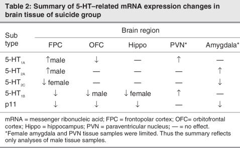
Owing to the multiple brain regions and receptor transcripts determined, the complete data set is somewhat complex. Accordingly, we provide an overview of the findings in Table 2.
Table 2
With regard to the FPC, Figure 1 shows the mRNA expression for each of the 5-HT receptors within the FPC. Within this region, 5-HT1A mRNA expression was significantly higher among women than among men (F1,36 = 31.16, p < 0.01, η2 = 0.41) and higher in suicide than in control subjects (F1,36 = 7.05, p = 0.01, η2 = 0.09). Although the interaction between these variables was not significant, the increase of 5-HT1A in the suicide group was only significant in men. In the case of 5-HT2A, mRNA expression varied as a function of the cause of death × sex interaction (F1,36 = 6.31, p = 0.01, η2 = 0.10). Follow-up tests indicated that 5-HT2A mRNA expression in the brains of male control subjects was lower than in female samples. As in the case of 5-HT1A, the 5-HT2A mRNA expression was elevated among male suicide subjects relative to control subjects, but a similar effect was not apparent among female subjects. The analysis of 5-HT2C expression yielded a significant cause of death × sex interaction, F 1,36 = 5.68, p = 0.02, η2 = 0.13. The follow up tests confirmed that among men 5-HT2C mRNA expression did not differ as a function of the cause of death, whereas among women 5-HT2C expression was lower in suicide subjects than in control subjects. Finally, mRNA expression of 5-HT1B receptors was lower in suicide subjects than in control subjects (F1,36 = 8.06, p < 0.05, η2 = 0.10) as was p11 expression (F1,36 = 6.74, p = 0.01, η2 = 0.15).
Fig. 1: Mean (and standard error of the mean) messenger ribonucleic acid (mRNA) expression of 5-HT1A, 5-HT2A, 5-HT2C, 5-HT1B and p11 in the frontopolar cortex of suicide victims who suffered from depression and of control subjects. The left panel shows the data for men, and the right panel presents the data for women. Data are presented as normalized cycle thresholds (Ctn) wherein the expression of each species was normalized by subtracting its cycle threshold (Ct) from the synaptophysin Ct. Thus a negative Ctn indicates that the mRNA species was less abundant than that of synaptophysin. A change of 1 Ctn is equivalent to a 2-fold difference in the abundance of that species. *p < 0.05 relative to control subjects of the same sex.
With regard to the OFC, given its presumed involvement in cognitive processes31 and modulation of emotional behaviours,2,32 we were particularly interested to establish whether 5-HT variations would be apparent in the brains of those who died by suicide. In contrast to the FPC, the mRNA expression of 5-HT1A within the OFC was lower in the suicide group than in control subjects (F1,29 = 6.37, p < 0.001, η2 = 0.31) (Fig. 2). The expression of 5-HT2A was relatively variable and did not differ between male and female samples or as a function of suicide subject compared with control subject. The expression of 5-HT2C was likewise found not to differ as a function of either the cause of death or of sex, although there was a modest trend for lower 5-HT2C expression in those who committed suicide when compared with control subjects (p = 0.11).
Fig. 2: Mean (and standard error of the mean) messenger ribonucleic acid expression of 5-HT1A, 5-HT2A, 5-HT2C, 5-HT1B and p11 in the orbital frontal cortex of of suicide victims who suffered from depression and of control subjects. The left panel shows the data for men, and the right panel presents the data for women. Data are presented as normalized cycle thresholds (Ctn) wherein the expression of each species was normalized by subtracting its cycle threshold (Ct) from the synaptophysin Ct. *p < 0.05 relative to control subjects of the same sex.
The expression of 5-HT1B receptors varied as a function of the cause of death × sex interaction (F1,29 = 4.54, p < 0.05, η2 = 0.09). The follow-up tests indicated that mRNA expression did not differ between male suicide subjects and control subjects. The female control subjects displayed higher 5-HT1B mRNA expression relative to male control subjects. Unlike the lack of effect seen among male subjects, in female subjects 5-HT1B mRNA expression was lower in the suicide group than in the control subjects. The mRNA expression of p11 was lower in men than in women (F1,29 = 6.20, p < 0.02, η2 = 0.13) and was appreciably lower in the suicide samples when compared with control samples (F1,29 = 14.89, p < 0.01, η2 = 0.31).
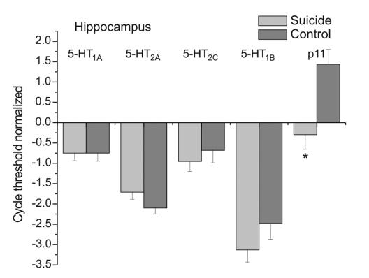
With regard to the hippocampus, hippocampal 5-HT1A, 5-HT2A and 5-HT2C mRNA expression did not differ as a function of sex, cause of death or the interaction between these conditions (Fig. 3). In contrast, 5-HT1B and p11 mRNA expression varied as a function of the cause of death × sex interaction (F1,32 = 6.23 and 8.29, p < 0.02 and < 0.001, η2 = 0.15 and 0.15, respectively). The follow-up tests of the simple effects indicated that, among women, 5-HT1B receptor expression and p11 were lower in suicide subjects than in control subjects, whereas among men, there was no difference between these conditions (see Fig. 3).
Fig. 3: Mean (and standard error of the mean) messenger ribonucleic acid expression of 5-HT1A, 5-HT2A, 5-HT2C, 5-HT1B and p11 in the hippocampus of suicide victims who suffered from depression and of control subjects. The left panel shows the data for men, and the right panel presents the data for women. Data are presented as normalized cycle thresholds (Ctn) wherein the expression of each species was normalized by subtracting its cycle threshold (Ct) from the synaptophysin Ct.*p < 0.05 relative to control subjects of the same sex.
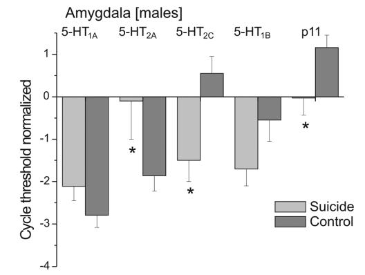
For the amygdala, tissue was only available for 4 female suicide samples, and the analysis was therefore conducted only on the male samples, for which there were 10 control and 10 suicide samples available. These analyses confirmed that 5-HT1A did not differ between suicide subjects and control subjects. In contrast, 5-HT2C mRNA expression was lower in suicide samples than in control samples (F1,18 = 7.80, p < 0.01, η2 = 0.30), whereas 5-HT2A mRNA expression was elevated in brain tissue of those who committed suicide (F1,18 = 7.31, p 0.02, η2 = 0.16) (Fig. 4). The expression of 5-HT1B did not differ as a function of the cause of death, although there was a tendency for mRNA expression to be lower in the brain tissue of suicide subjects, compared with control subjects (F1,18 = 2.97, p = 0.09, η2 = 0.14). The mRNA expression of p11, however, was significantly diminished in the amygdala of suicide subjects (F1,18 = 5.74, p 0.05, η2 = 0.18).
Fig. 4: Mean (and standard error of the mean) messenger ribonucleic acid expression of 5-HT1A, 5-HT2A, 5-HT2C, 5-HT1B and p11 in the amygdala of suicide victims who suffered from depression and of control subjects. Because there were only a few samples from women, only the data from men are presented. Data are presented as normalized cycle thresholds (Ctn) wherein the expression of each species was normalized by subtracting its cycle threshold (Ct) from the synaptophysin Ct. *p < 0.05 relative to control subjects of the same sex.
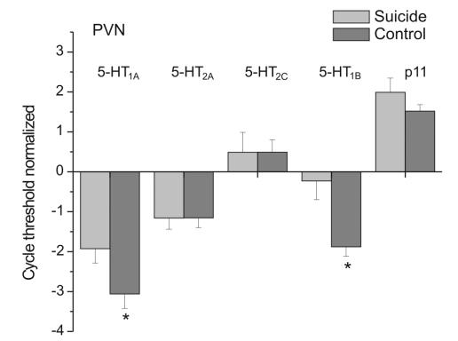
With regard to the PVN of the hypothalamus, the number of samples from women was relatively small (5 control and 4 suicide subjects), and we therefore conducted separate analyses that included or did not include samples from women. Because the number of female and male samples were roughly the same, and because the statistical outcomes were comparable irrespective of whether sex was included as an independent variable, the data are presented with sex included as a variable. As depicted in Figure 5, both 5-HT1A and 5-HT1B mRNA expression was significantly higher in suicide samples than in control subject samples (F1,29 = 5.62 and 9.76, p < 0.05 and 0.001, η2 = 0.17 and 0.27, respectively). In contrast to the mRNA variations for these receptor subtypes, mRNA expression for 5-HT2A, 5-HT2C and p11 did not reach significance.
Fig. 5: Mean (and standard error of the mean) messenger ribonucleic acid expression of 5-HT1A, 5-HT2A, 5-HT2C, 5-HT1B and p11 in the paraventricular nucleus of the hypothalamus of suicide victims who suffered from depression and control subjects. The left panel shows the data for men, and the right panel presents the data for women. Data are presented as normalized cycle thresholds (Ctn) wherein the expression of each species was normalized by subtracting its cycle threshold (Ct) from the synaptophysin Ct. *p < 0.05 relative to control subjects of the same sex.
With regard to the dorsal raphe nucleus, in contrast to most of the other brain regions examined, mRNA expression of 5-HTT, 5-HT1A, 5-HT1B, p11 and tryptophan hydroxylase were relatively variable and did not vary as a function of suicide. The number of female samples was small (3 suicide and 3 control subjects), and the meaningfulness of the data might therefore be questionable. However, the absence of an effect was notable regardless of whether only male samples were assessed or whether the data from male and female samples were pooled. It was found that among women there was a trend for higher mRNA levels (especially for 5-HT1A and 5-HTT, but a greater sample size will clearly be needed before adequate conclusions can be drawn.
Correlations between 5-HT receptors and p11
Within the FPC, OFC and hippocampus the correlations between p11 and 5-HT1B were significant among both men and women and among suicide and control subjects (r values ranged from 0.59 to 0.96). These correlations were less pronounced in the amygdala (r = 0.60 in control subjects, and only 0.40 among suicide subjects) and the PVN (r = 0.81 among suicide subjects but only 0.11 in control subjects). In the OFC and PVN, the correlations between 5-HT1A and 5-HT1B were relatively pronounced (r ranged from 0.55 to 0.71), but analyses in other brain regions did not reveal similar effects.
Discussion
The confluence of evidence implicating 5-HT processes in depression has come from pharmacologic studies,7 positron emission tomography analyses2,3,8 and assessments of specific 5-HT–related gene polymorphisms.17,33,34 As well, relevant data have come from studies that assessed postmortem 5-HT receptor binding or mRNA expression in specific brain regions of subjects who committed suicide relative to a comparison sample that died suddenly.16,18 Nevertheless, as indicated earlier, the frequent inconsistencies reported across laboratories or as a function of techniques used to assess the 5-HT variations have been disquieting. In addition to including a range of brain regions associated with depression and stressor reactivity, in the present investigation, we revisited the issue of 5-HT receptor changes associated with suicide and depression, but we used tissue collected soon after death and included both men and women in our study. Moreover, the viability of the RNA was confirmed to be adequate in both an in-house analysis and in an analysis conducted by personnel from 2 members of the Brain Bank Consortium (BrainNet II Europe), namely, the Imperial College, London, and the Universitat de Barcelona. In addition, the pH levels of samples were acceptable and were comparable in suicide and control subjects. Of course, the data in the present investigation were restricted to mRNA expression, and ultimately, the findings will need to be confirmed with respect to protein content.
Consistent with earlier reports,15–18,20,21 in the current investigation, 5-HT1A and 5-HT2A mRNA expression was elevated in the FPC of male suicide samples, whereas this difference was not significant in samples from women. It will be recalled that inconsistent results have been reported concerning 5-HT2A variations in frontal cortical regions of suicide subjects, compared with control subjects,35 and it is possible that sex differences might account for the contradictory results that have been reported.
In animal studies, chronic stressors reduced 5-HT1B binding,24,36 and altered 5-HT1B functioning has been implicated as a factor associated with depressive illness,36 although correspondence was not found between 5-HT1B receptor gene polymorphisms and suicidality.37–41 In the present investigation, 5-HT1B mRNA receptor expression was markedly reduced in the FPC of suicide subjects, as it was in the OFC and the hippocampus. Further, consistent with the perspective that p11 modulates 5-HT1B functioning,23 we observed that p11 was reduced in the FPC and that the abundance of 5-HT1B and p11 transcripts in both control and suicide subjects was highly correlated.
The function of p11 is to act as a calcium sensor that localizes other proteins with membrane phospholipids; it is part of a protein complex that includes annexin 2.42,43 In the absence of p11 (e.g., in p11 knockout mice) the localization of 5-HT1B at the cell surface is markedly reduced.24 These findings suggest tightly coupled transcriptional regulation, but it is not clear how this might occur because little is known about the regulation of 5-HT receptors at the transcriptional level. A common promoter region is unlikely because the receptor genes are not located on the same chromosomes. The p11 gene is on chromosome 1, whereas the 5-HT1B gene has been localized to chromosome 6 (see NLM GenBank www.ncbi.nlm.nih.gov/Genbank/). Given that suicide has been associated with various processes other than those related to 5-HT functioning, including brain-derived neurotrophic factor,44 as well as GABAA and CRH functioning,5 it is uncertain whether one of these factors or some other 5-HT receptor contributes to the coincident changes of 5-HT1B and p11 expression.
The OFC has been implicated as part of the neural circuitry involved in depression.2,44 Indeed, among suicide attempters, α-[(11)C]methyl-L-tryptophan trapping was reduced (relative to control subjects) in the OFC and PFC, suggesting low rates of 5-HT synthesis in these regions.45 Like other PFC regions, the OFC has been implicated in cognitive processing and possibly contributes to emotional rather than cognitive perspective taking46 as well as impulsive-aggressive behaviours.47,48 Further, 5-HT and dopamine in the medial PFC and OFC have been shown to differentially modulate impulsive decision making,48 and 5-HT2A and 5-HT2C antagonists have been shown to differentially influence impulsive responding.49 In light of these considerations, it may not be entirely surprising that 5-HT receptor differences between suicide and control subjects in the OFC did not parallel those evident the FPC. In this regard, within the OFC, the mRNA expression of 5-HT1A was lower in suicide than in control subjects (recall that 5-HT1A expression was elevated in the FPC of suicide subjects), whereas 5-HT2A and 5-HT2C mRNA expression was unaffected in this region. Contrary to these between-region differences, the changes of both 5-HT1B and p11 mRNA expression in the OFC and the FPC were comparable to one another. This raises the possibility that there is a shift in the balance of 5-HT signaling between those areas that modulate impulsivity, which may result in impaired assessment of behavioural consequences (suicidality).
Although the hippocampus has frequently been implicated as an essential conduit in the neuronal processing associated with depressive illness,50 analyses of the 5-HT receptor changes that occur in the hippocampus have not provided an encouraging or consistent profile of variations. For example, although several studies indicated that neither 5-HT1A nor 5-HT2A receptor binding differed in the hippocampus of suicide and control subjects,10,51,52 other investigators9 found that 5-HT2A receptor binding was reduced in the hippocampus of people suffering from depression. As well, Rosel and colleagues21,22 reported that hippocampal 5-HT2A binding sites were reduced in cases of violent suicide but that this was coupled with higher binding affinity between the receptor and the G protein and therefore with increased concentrations of the second messenger, IP3. Further, it was reported that variations of the 5-HT1B receptor gene at the A-161T locus were associated with impulsive-aggressive behaviours and were more prominent in suicide than in control subjects. In effect, it seems that the A-161T locus, which potentially influences 5-HT1B transcription, might contribute to suicide predisposition by influencing impulsive-aggressive behaviours.53 Adding still more complexity to the mix was the finding that 5-HT2A mRNA expression within the hippocampus was elevated in teenage suicide subjects relative to a comparison group.19 The present findings, unfortunately, do little to resolve the paradoxical findings, except to add that within the hippocampus neither 5-HT1A nor 5-HT2A mRNA receptor expression differed between suicide and control subjects. This was the case among both men and women and was observed in the context of other 5-HT receptors (notably, 5-HT1B and p11) being altered within the hippocampus and in other brain regions.
Anxiety is frequently comorbid with depression, and so neurochemical activity within the amygdala might be considered in the analysis of major depressive disorder. Although particular attention has been devoted to amygdala CRH and noradrenaline involvement in mediating anxiety,54–56 increasing evidence from both animal54 and human57 studies has also implicated amygdala 5-HT in this regard. Serotonergic changes in the amygdala have not typically been related to depression and suicide, but postmortem analyses have revealed that 5-HT utilization (reflected by 5-HIAA accumulation) was elevated in the amygdala of subjects who died by suicide relative to age-matched control subjects. Further, positron emission tomography analyses indicated lower 5-HTT binding potential in the amygdala of drug-free patients with major depressive disorder relative to comparison subjects.58 Likewise, 5-HT2 receptor abundance, reflected by [3H]ketanserin binding, has been found to be elevated in the amygdala of suicide subjects relative to control subjects,59 although others60 did not detect comparable effects. In the present investigation, 5-HT2A mRNA expression was elevated in the amygdala of suicide subjects, whereas 5-HT2C receptor expression, and to a lesser extent 5-HT1B and p11 mRNA expression, were reduced in this region. Whether these effects were related to anxiety or more closely aligned with depression is uncertain. Nonetheless, it does appear that suicide was associated with distinct 5-HT receptor changes, just as 5-HT receptor alterations were evident in cortical regions. Indeed, in most respects, the nature of changes in the amygdala paralleled those seen in the FPC.
Elevated HPA functioning has been associated with depressive illness,61,62 and it has been reported that 5-HT1A receptors within the PVN moderate hypothalamic CRH and, hence, pituitary adrenocorticotropic hormone and adrenal cortisol release.63,64 Further, 5-HT2A receptors may promote the desensitization of 5-HT1A receptors, and the interaction between these receptors has been found to influence HPA neuroendocrine activity.65 As such, it was of interest to establish whether these receptor subtypes would be altered in the PVN of suicide subjects. It was indeed found that 5-HT1A expression was elevated in the PVN of suicide subjects, as was 5-HT1B expression, and the expression of these receptor subtypes was significantly correlated. Because treatments that affect PVN 5-HT1A and 5-HT2B receptors influence neuroendocrine functioning,65 the present data are consistent with the view that neuroendocrine variations associated with depression may be related to these 5-HT processes. This view is in keeping with the proposition that multiple 5-HT receptor variations may be associated with pathology, and ultimately, that treatment strategies may be served well by concurrent tweaking of different receptor subtypes.66
It has been reported that 5-HT1A (auto)receptors were increased in the DRN of persons who suffered from depression and committed suicide,52 which might influence forebrain 5-HT availability. In the present investigation, as previously observed,15 5-HT1A variations were not evident in the DRN of suicide subjects. Similarly, unanimity has not been reached concerning 5-HTT changes in the DRN of those who died by suicide. Although the findings of Caspi and colleagues33 have focused attention on the potential role of 5-HTT gene polymorphism in depression, several studies indicated that 5-HTT ligand binding was not altered among suicide subjects.15, 67,68 However, it was reported that there was a marked reduction in the number of neurons that expressed 5-HTT mRNA.15 In the present investigation, as in these other studies, there was no evidence of altered 5-HTT or 5-HT1A receptor mRNA expression in the DRN. Thus the role of 5-HT1A and 5-HTT in supporting suicide and depression is uncertain.
In general, conclusions regarding neurotransmitter and receptor differences in the brains of suicide subjects, compared with control subjects, have been hampered by the diversity of procedures that exist across reports. In the present investigation, some of these problems were dealt with effectively (e.g., sex of subjects, verification of RNA quality, obtaining of tissue samples soon after death). However, as with most studies that have assessed the brains of those who died by suicied, it was not possible to relate biological factors to specific symptoms that each subject might have presented. Nevertheless, some of the differences that exist across studies could potentially reflect characteristics of the suicide subjects examined. In this regard, van Heeringen and colleagues69 tied frontal cortical 5-HT2A receptors in persons who attempted suicide to elevated feelings of hopelessness and harm avoidance. Moreover, Oquendo and colleagues35 indicated that lifetime aggression scores were associated with elevated 5-HT2A receptor binding in the PFC of suicide subjects. The essential point is that suicidality across people suffering from depression may involve multiple processes and that the suicidal phenotype is a complex one. Thus it should not be surprising that varied results are evident across studies, which is possibly related to the specific characteristics of those who comprised the study population. Of course, the possibility also exists that the biological effects that have been observed reflect processes related to suicide (e.g., impulsivity, disinhibition, anxiety, executive functioning) rather than depression per se.70 In fact, it has been reported that variations of the 5-HT1B receptor gene (at the A-161T locus), which has been associated with impulsive-aggressive behaviours, were more prominent in suicide subjects than in control subjects.71 Ultimately, analysis of the processes related to depression and suicide might be best served by evaluating endophenotypes that make up the disorder and by focusing on specific symptoms and specific neurochemical processes related to illness.72
Acknowledgments
This research was supported by a grant from the Canadian Institutes of Health Research (H.A., M.O.P. and Z.M.), and from the Hungarian National Research Grant (OTKA) No. 034496 (M.P.) and by EU Grant FP6, BNEII No. LSHM-CT-2004-503039.). H.A. holds a Canada Research Chair in Neuroscience and M.O.P. is A NARSAD Independent Researcher.
Footnotes
Contributors: Drs. Anisman, Palkovits, Merali and Poulter designed the study. Drs. Du, Palkovits, Faludi, Kovacs, Szontagh-Kishazi and Merali acquired the data, which Drs. Anisman, Du, Faludi and Merali analyzed. Drs. Anisman, Faludi and Poulter wrote the article, and Drs. Du, Palkovits, Kovacs, Szontagh-Kishazi and Merali revised it. All authors gave final approval for the article to be published.
Competing interests: None declared.
Correspondence to: Dr. H. Anisman, Institute of Neuroscience, Carleton University, 1125 Colonel By Dr., Ottawa ON K1S 5B6; fax 613 520-4052; hanisman@ccs.carleton.ca
References
- 1.Biegon A, Israeli M. Regionally selective increases in beta-adrenergic receptor density in the brains of suicide victims. Brain Res 1988; 442: 199-203. [DOI] [PubMed]
- 2.Drevets WC. Neuroimaging studies of mood disorders. Biol Psychiatry 2000;48:813-9. [DOI] [PubMed]
- 3.Drevets WC. Neuroimaging and neuropathological studies of depression: implications for the cognitive-emotional features of mood disorders. Curr Opin Neurobiol 2001;11:240-9. [DOI] [PubMed]
- 4.Dwivedi Y, Conley RR, Roberts RC, et al. [(3)H]cAMP binding sites and protein kinase a activity in the prefrontal cortex of suicide victims. Am J Psychiatry 2002;159:66-73. [DOI] [PubMed]
- 5.Merali Z, Du L, Hrdina P, et al. Dysregulation in the suicide brain: mRNA expression of corticotropin-releasing hormone receptors and GABA(A) receptor subunits in frontal cortical brain region. J Neurosci 2004;24:1478-85. [DOI] [PMC free article] [PubMed]
- 6.Rajkowska G. Histopathology of the prefrontal cortex in major depression: what does it tell us about dysfunctional monoaminergic circuits? Prog Brain Res 2000;126:397-412. [DOI] [PubMed]
- 7.Pineyro G, Blier P. Autoregulation of serotonin neurons: role in antidepressant drug action. Pharmacol Rev 1999;51:533-91. [PubMed]
- 8.Bhagwagar Z, Hinz R, Taylor M, et al. Increased 5-HT(2A) receptor binding in euthymic, medication-free patients recovered from depression: a positron emission study with [(11)C]MDL 100,907. Am J Psychiatry 2006;163:1580-7. [DOI] [PubMed]
- 9.Mintun MA, Sheline YI, Moerlein SM, et al. Decreased hippocampal 5-HT2A receptor binding in major depressive disorder: in vivo measurement with [superscript 1-sup-8F]altanserin positron emission tomography. Biol Psychiatry 2004;55:217-24. [DOI] [PubMed]
- 10.Lowther S, De Paermentier F, Cheetham SC, et al. 5-HT1A receptor binding sites in post-mortem brain samples from depressed suicide victims and control subjects. J Affect Disord 1997;42:199-207. [DOI] [PubMed]
- 11.Rosel P, Arranz B, Vallejo J, et al. High affinity [3H]imipramine and [3H]paroxetine binding sites in suicide brains. J Neural Transm 1997;104:921-9. [DOI] [PubMed]
- 12.Stockmeier CA, Dilley GE, Shapiro LA, et al. Serotonin receptors in suicide victims with major depression. Neuropsychopharmacology 1997;16:162-73. [DOI] [PubMed]
- 13.Sibille E, Arango V, Galfalvy HC, et al. Gene expression profiling of depression and suicide in human prefrontal cortex. Neuropsychopharmacology 2004;29:351-61. [DOI] [PubMed]
- 14.Lopez de Lara C, Brezo J, Rouleau G, et al. Effect of tryptophan hydroxylase-2 gene variants on suicide risk in major depression. Biol Psychiatry 2007;62:72-80. [DOI] [PubMed]
- 15.Arango V, Underwood MD, Boldrini M, et al. Serotonin 1A receptors, serotonin transporter binding and serotonin transporter mRNA expression in the brainstem of depressed suicide victims. Neuropsychopharmacology 2001;25:892-903. [DOI] [PubMed]
- 16.Arango V, Underwood MD, Gubbi AV, et al. Localized alterations in pre-and postsynaptic serotonin binding sites in the ventrolateral prefrontal cortex of suicide victims. Brain Res 1995;688:121-33. [DOI] [PubMed]
- 17.Arango V, Huang YY, Underwood MD, et al. Genetics of the serotonergic system in suicidal behavior. J Psychiatr Res 2003;37:375-86. [DOI] [PubMed]
- 18.Mann JJ, Huang YY, Underwood MD, et al. A serotonin transporter gene promoter polymorphism (5-HTTLPR) and prefrontal cortical binding in major depression and suicide. Arch Gen Psychiatry 2000;57:729-38. [DOI] [PubMed]
- 19.Pandey GN, Dwivedi Y, Rizavi HS, et al. Higher expression of serotonin 5-HT(2A) receptors in the postmortem brains of teenage suicide victims. Am J Psychiatry 2002;159:419-29. [DOI] [PubMed]
- 20.Turecki G, Briere R, Dewar K, et al. Prediction of level of serotonin 2A receptor binding by serotonin receptor 2A genetic variation in postmortem brain samples from subjects who did or did not commit suicide. Am J Psychiatry 1999;156:1456-8. [DOI] [PubMed]
- 21.Rosel P, Arranz B, San L, et al. Altered 5-HT(2A) binding sites and second messenger inositol trisphosphate (IP(3)) levels in hippocampus but not in frontal cortex from depressed suicide victims. Psychiatry Res 2000;99:173-81. [DOI] [PubMed]
- 22.Rosel P, Arranz B, Vallejo J, et al. Variations in [3H]imipramine and 5-HT2A but not [3H]paroxetine binding sites in suicide brains. Psychiatry Res 1998;82:161-70. [DOI] [PubMed]
- 23.Svenningsson P, Chergui K, Rachleff I, et al. Alterations in 5-HT1B receptor function by p11 in depression-like states. Science 2006;311: 77-80. [DOI] [PubMed]
- 24.Svenningsson P, Greengard P. p11 (S100A10) - an inducible adaptor protein that modulates neuronal functions. Curr Opin Pharmacol 2006;7:27-32. [DOI] [PubMed]
- 25.Svenningsson P, Tzavara ET, Qi H, et al. Biochemical and behavioral evidence for antidepressant-like effects of 5-HT6 receptor stimulation. J Neurosci 2007;27:4201-9. [DOI] [PMC free article] [PubMed]
- 26.Nandam LS, Jhaveri D, Bartlett P. 5-HT7, neurogenesis and antidepressants: a promising therapeutic axis for treating depression. Clin Exp Pharmacol Physiol 2007;34:546-51. [DOI] [PubMed]
- 27.American Psychiatric Association. Diagnostic and statistical manual of mental disorders. 4th ed. Washington: The Association; 1994.
- 28.Li JZ, Vawter MP, Walsh DM, et al. Systematic changes in gene expression in postmortem human brains associated with tissue pH and terminal medical conditions. Hum Mol Genet 2004;13:609-16. [DOI] [PubMed]
- 29.Torrey EF, Webster M, Knable M, et al. The Stanley Foundation brain collection and neuropathology consortium. Schizophr Res 2000;44:151-5. [DOI] [PubMed]
- 30.Chen J, Sochivko D, Beck H, et al. Activity-induced expression of common reference genes in individual cns neurons. Lab Invest 2001;81:913-6. [DOI] [PubMed]
- 31.Volz KG, Schubotz RI, von Cramon DY. Decision-making and the frontal lobes. Curr Opin Neurol 2006;19:401-6. [DOI] [PubMed]
- 32.Hynes CA, Baird AA, Grafton ST. Differential role of the orbital frontal lobe in emotional versus cognitive perspective-taking. Neuropsychologia 2006;44:374-83. [DOI] [PubMed]
- 33.Caspi A, Sugden K, Moffitt TE, et al. Influence of life stress on depression: moderation by a polymorphism in the 5-HTT gene. Science 2003;301:386-9. [DOI] [PubMed]
- 34.Lemonde S, Du L, Bakish D, et al. Association of the C(-1019)G 5-HT1A functional promoter polymorphism with antidepressant response. Int J Neuropsychopharmacol 2004;7:501-6. [DOI] [PubMed]
- 35.Oquendo MA, Russo SA, Underwood MD, et al. Higher postmortem prefrontal 5-HT2A receptor binding correlates with lifetime aggression in suicide. Biol Psychiatry 2006;59:235-43. [DOI] [PubMed]
- 36.Lopez JF, Vazquez DM, Chalmers DT, et al. Regulation of 5-HT receptors and the hypothalamic-pituitary-adrenal axis. Implications for the neurobiology of suicide. Ann N Y Acad Sci 1997;836:106-34. [DOI] [PubMed]
- 37.Huang YY, Grailhe R, Arango V, et al. Relationship of psychopathology to the human serotonin1B genotype and receptor binding kinetics in postmortem brain tissue. Neuropsychopharmacology 1999;21:238-46. [DOI] [PubMed]
- 38.Nis populahiguchi N, Shirakawa O, Ono H, et al. No evidence of an association between 5HT1B receptor gene polymorphism and suicide victims in a Japanesetion. Am J Med Genet 2001;105:343-5. [DOI] [PubMed]
- 39.Rujescu D, Giegling I, Sato T, et al. Lack of association between serotonin 5-HT1B receptor gene polymorphism and suicidal behavior. Am J Med Genet B Neuropsychiatr Genet 2003;116:69-71. [DOI] [PubMed]
- 40.Stefulj J, Buttner A, Skavic J, et al. Serotonin 1B (5HT-1B) receptor polymorphism (G861C) in suicide victims: association studies in German and Slavic population. Am J Med Genet B Neuropsychiatr Genet 2004;127:48-50. [DOI] [PubMed]
- 41.Tsai SJ, Hong CJ, Yu YW, et al. Association study of serotonin 1B receptor (A-161T) genetic polymorphism and suicidal behaviors and response to fluoxetine in major depressive disorder. Neuropsychobiology 2004;50:235-8. [DOI] [PubMed]
- 42.De Seranno S, Benaud C, Assard N, et al. Identification of an AHNAK binding motif specific for the Annexin2/S100A10 tetramer. J Biol Chem 2006;281:35030-8. [DOI] [PubMed]
- 43.Girard C, Tinel N, Terrenoire C, et al. Borsotto M. p11, an annexin II subunit, an auxiliary protein associated with the background K+ channel, TASK-1. EMBO J 2002;21:4439-48. [DOI] [PMC free article] [PubMed]
- 44.Duman RS, Monteggia LM. A neurotrophic model for stress-related mood disorders. Biol Psychiatry 2006;59:1116-27. [DOI] [PubMed]
- 45.Leyton M, Paquette V, Gravel P, et al. Alpha-[11C]Methyl-L-tryptophan trapping in the orbital and ventral medial prefrontal cortex of suicide attempters. Eur Neuropsychopharmacol 2006;16:220-3. [DOI] [PubMed]
- 46.Seminowicz DA, Mayberg HS, McIntosh AR, et al. Limbic-frontal circuitry in major depression: a path modeling meta analysis. Neuroimage 2004;22:409-18. [DOI] [PubMed]
- 47.Best M, Williams JM, Coccaro EF. Evidence for a dysfunctional prefrontal circuit in patients with an impulsive aggressive disorder. Proc Natl Acad Sci U S A 2002;99:8448-53. [DOI] [PMC free article] [PubMed]
- 48.Winstanley CA, Theobald DE, Dalley JW, et al. Double dissociation between serotonergic and dopaminergic modulation of medial prefrontal and orbitofrontal cortex during a test of impulsive choice. Cereb Cortex 2006;16:106-14. [DOI] [PubMed]
- 49.Winstanley CA, Theobald DE, Dalley JW, et al. 5-HT2A and 5-HT2C receptor antagonists have opposing effects on a measure of impulsivity: interactions with global 5-HT depletion. Psychopharmacology (Berl) 2004;176:376-85. [DOI] [PubMed]
- 50.Sheline YI, Mittler BL, Mintun MA. The hippocampus and depression. Eur Psychiatry 2002;17:300-5. [DOI] [PubMed]
- 51.Cheetham SC, Crompton MR, Czudek C, et al. Serotonin concentrations and turnover in brains of depressed suicide victims. Brain Res 1989;502:332-40. [DOI] [PubMed]
- 52.Stockmeier CA, Shapiro LA, Dilley GE, et al. Increase in serotonin-1A autoreceptors in the midbrain of suicide victims with major depression-postmortem evidence for decreased serotonin activity. J Neurosci 1998;18:7394-401. [DOI] [PMC free article] [PubMed]
- 53.López-Figueroa AL, Norton CS, López-Figueroa MO, et al. Serotonin 5-HT1A, 5-HT1B and 5-HT2A receptor mRNA expression in subjects with major depression, bipolar disorder and schizophrenia. Biol Psychiatry 2004;55:225-33. [DOI] [PubMed]
- 54.Davis M, Rainnie D, Cassell M. Neurotransmission in the rat amygdala related to fear and anxiety. Trends Neurosci 1994;17:208-14. [DOI] [PubMed]
- 55.LeDoux JE. Emotion circuits in the brain. Annu Rev Neurosci 2000;23:155-84. [DOI] [PubMed]
- 56.Izumi T, Inoue T, Kitaichi Y, et al. Target brain sites of the anxiolytic effect of citalopram, a selective serotonin reuptake inhibitor. Eur J Pharmacol 2006;534:129-32. [DOI] [PubMed]
- 57.Hariri AR, Holmes A. Genetics of emotional regulation: the role of the serotonin transporter in neural function. Trends Cogn Sci 2006;10:182-91. [DOI] [PubMed]
- 58.Parsey RV, Hastings RS, Oquendo MA, et al. Lower serotonin transporter binding potential in the human brain during major depressive episodes. Am J Psychiatry 2006;163:52-8. [DOI] [PubMed]
- 59.Hrdina PD, Demeter E, Vu TB, et al. 5-HT uptake sites and 5-HT2 receptors in brain of antidepressant-free suicide victims/depressives: increase in 5-HT2 sites in cortex and amygdala. Brain Res 1993;614:37-44. [DOI] [PubMed]
- 60.Cheetham SC, Crompton MR, Katona CL, et al. Brain 5-HT2 receptor binding sites in depressed suicide victims. Brain Res 1988;443:272-80. [DOI] [PubMed]
- 61.Mitchell AJ. The role of corticotropin releasing factor in depressive illness: a critical review. Neurosci Biobehav Rev 1998;22:635-51. [DOI] [PubMed]
- 62.Nemeroff CB. The corticotropin-releasing factor (CRF) hypothesis of depression: New findings and new directions. Mol Psychiatry 1996;1:336-42. [PubMed]
- 63.Pan L, Gilbert F. Activation of 5-HT1A receptor subtype in the paraventricular nuclei of the hypothalamus induces CRH and ACTH release in the rat. Neuroendocrinology 1992;56:797-802. [DOI] [PubMed]
- 64.Osei-Owusu P, James A, Crane J, et al. 5-Hydroxytryptamine 1A receptors in the paraventricular nucleus of the hypothalamus mediate oxytocin and adrenocorticotropin hormone release and some behavioral components of the serotonin syndrome. J Pharmacol Exp Ther 2005;313:1324-30. [DOI] [PubMed]
- 65.Zhang Y, Gray TS, D'Souza DN, et al. Desensitization of 5-HT1A receptors by 5-HT2A receptors in neuroendocrine neurons in vivo. J Pharmacol Exp Ther 2004;310:59-66. [DOI] [PubMed]
- 66.Millan MJ. Multi-target strategies for the improved treatment of depressive states: Conceptual foundations and neuronal substrates, drug discovery and therapeutic application. Pharmacol Ther 2006;110:135-370. [DOI] [PubMed]
- 67.Bligh-Glover W, Kolli TN, Shapiro-Kulnane L, et al. Serotonin transporter in the midbrain of suicide victims with major depression. Biol Psychiatry 2000;47:1015-24. [DOI] [PubMed]
- 68.Little KY, McLauglin DP, Ranc J, et al. Serotonin transporter binding sites and mRNA levels in depressed persons committing suicide. Biol Psychiatry 1997;41:1156-64. [DOI] [PubMed]
- 69.van Heeringen C, Audenaert K, Van Laere K, et al. Prefrontal 5-HT2a receptor binding index, hopelessness and personality characteristics in attempted suicide. J Affect Disord 2003;74:149-58. [DOI] [PubMed]
- 70.van Heeringen C. Suicide, serotonin, and the brain. Crisis 2001; 22:66-70. [DOI] [PubMed]
- 71.Zouk H, McGirr A, Lebel V, et al. The effect of genetic variation of the serotonin 1B receptor gene on impulsive aggressive behavior and suicide. Am J Med Genet B Neuropsychiatr Genet 2007;144:996-1002. [DOI] [PubMed]
- 72.Hasler G, Drevets WC, Manji HK, et al. Discovering endophenotypes for major depression. Neuropsychopharmacology 2004;29:1765-81. [DOI] [PubMed]


