
Figure 3.
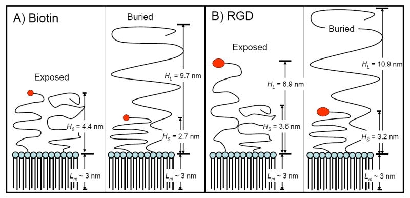
Schematic representation of the surface architectures used in this study for biotin (A) and RGD (B). Brush heights were calculated from Eqns. 1-3 using values of 0.45 nm2 for the average area per lipid molecule and 0.35 nm for the PEG monomer length. The monolayer thickness (Lm) was estimated to be ~ 3 nm from the persistence length of the stearoyl chains [1]. See text for details.
1. Israelachvili J (1992). Intermolecular and Surface Forces. 2nd ed: Academic Press.