Abstract
The study was a retrospective evaluation and comparison. Seventy-five elderly patients (>50 years) with AO type-B2 ankle fractures were treated by open reduction and internal fixation. All patients were followed up retrospectively for at least 12 months. The 75 patients were divided into two groups, based on the method of treatment. The Knowles pin (KP) group included 45 patients with an average age of 62.7 years. The tubular plate (TP) group included 30 patients with an average age of 60.0 years. The clinical results were compared between the two groups. Both of the groups were similar in respect to the injury mechanisms, fracture pattern, open fracture grade, compounding medical conditions, and ankle score (all P values <0.28). However, the KP group had significantly smaller wound incisions, shorter surgery time, shorter hospital stay, less meperidine use, less symptomatic hardware, and lower complication rates than the TP group (all P values <0.03). In conclusion, lateral fixation of AO type-B2 ankle fractures in the elderly by the Knowles pin is recommended due to its simplicity, efficacy and low complication rate.
Résumé
Etude rétrospective et comparative de 75 patients de plus de 50 ans avec une fracture malleolaire type B2 dans le système AO. Les patients, divisés en 2 groupes selon la méthode d’ostéosynthèse, avaient tous été suivis au moins un an. Le groupe traité par broche de Knowles (KP) comprenait 45 patients d’âge moyen 62.7 ans. Le groupe traité par plaque tubulaire comprenait 30 patients d’âge moyen 60 ans. Les 2 groupes étaient similaires pour le mécanisme fracturaire, le type de fracture, le degré d’ouverture, les conditions médicales, et le score fonctionnel (tous les P<0.28). Le groupe KP avait une plus petite incision cutanée, un temps opératoire plus court, une durée d’hospitalisation plus courte, moins de consommation d’antalgique, moins de gêne sur le matériel et un taux de complications plus faible que le groupe TP (tous les P<0.03). En conclusion nous recommandons la fixation des fractures de type AO B2 chez le sujet âgé par une broche de Knowles.
Introduction
Surgical treatment for ankle fractures associated with lateral displacement and rotation of the talus is generally accepted [1, 5, 6, 12]. The lateral malleolus plays the key role in the stability of the ankles because 1-mm lateral displacement of the talus decreases the tibiotalar contact surface by 42% [15]. For an OTA 44-B2 (AO type-B2) ankle fracture (a trans-syndesmotic fibular fracture with medial lesion), open reduction and internal fixation were recommended by previous studies, because the talus tends to shift in the mortise [7, 9, 13, 16].
Operative treatment of ankle fractures in elderly patients with an age of more than 50 years becomes more difficult, leading to increased risks of wound breakdown and fixation failure [2, 4, 8]. Although fibular plating in elderly patients is a popular technique, more soft tissue dissection may result in wound complication [9]. In contrast, Knowles pinning is an intramedullary technique offering an interfragmental compression effect and only needs minimal soft tissue stripping [11]. This situation inspired us to retrospectively follow up and compare the clinical outcome of AO type-B2 ankle fractures in elderly patients (>50 years) who were treated with Knowles pinning or lateral tubular plating.
Patients and methods
Between 1999 and 2005, 115 elderly patients (>50 years) with ankle fractures were treated surgically at our institution. Inclusion criteria for this study were: (1) OTA 44-B2 fractures (AO type-B2 ankle fractures); (2) acute fractures; (3) internal fixation with either a Knowles pin (KP) or a tubular plate (TP); (4) patients with the ability to walk without any assistance before injury. Exclusion criteria for this study were: (1) nonunion or pathological fractures; (2) severe open fractures (Gustilo grade III); (3) severe comminuted fractures (>75%); (4) patients with syndesmosis instability. There were 83 patients (KP, n=49; TP, n=34) who met the inclusion criteria. However, eight patients could not be followed up due to death (two cases) or relocation (six cases), and were excluded. Therefore, 75 patients with an average age of 61.2 years were followed up 12 months after discharge from the hospital and were included in this study. Thirty-five of 75 patients had medial malleolar fractures and 40 had medial tenderness associated with a medial clear space of 4 mm, consistent with a ruptured deltoid ligament. There were eight open lateral malleolar fractures, including three Gustilo type I and five type II. An open fracture was treated by irrigation, thorough debridement, and appropriate intravenous antibiotics. There were six surgeons involved in our study. Four surgeons favoured KP, and the remaining two surgeons favoured TP to treat lateral malleolar fracture.
The 75 patients were divided into two groups, based on the method of treatment. The KP group included 45 patients with an average age of 62.7 years. Thirty-seven patients (82.2%) suffered from vehicular trauma. There were five open fractures, including two type I and three type II. The TP group included 30 patients with an average age of 60.0 years. Twenty-three patients (76.7%) suffered from vehicular trauma. There were three open fractures, including one type I and two type II (Table 1).
Table 1.
The preoperative data in the two groups
| Knowles pin | Tubular plate | P value | |
|---|---|---|---|
| Injury mechanism | |||
| Vehicular trauma (*N) | 37 | 23 | 0.57 |
| High-energy falls (*N) | 2 | 2 | 1.0 |
| Sports injury or minor trauma (*N) | 4 | 4 | 0.71 |
| Other (*N) | 2 | 1 | 1.0 |
| Gustilo open type | |||
| I (*N) | 2 | 1 | 1.0 |
| II (*N) | 3 | 2 | 1.0 |
| Injury pattern | |||
| Deltoid ligament ruptures (*N) | 23 | 17 | 0.81 |
| Medial malleolar fractures (*N) | 22 | 13 | 0.81 |
| Transverse fractures (*N) | 4 | 2 | 1.0 |
| Oblique and spiral fractures (*N) | 35 | 23 | 1.0 |
| Comminuted fractures (*N) | 6 | 5 | 0.75 |
| Demographics | |||
| Gender (F/M) | 25/20 | 18/12 | 0.81 |
| Mean age (years) | 62.7 | 60.0 | 0.31 |
| Compounding medical conditions | |||
| Hypertension (*N) | 4 | 2 | 1.0 |
| Diabetes (*N) | 3 | 3 | 0.68 |
| Renal diseases (*N) | 1 | 1 | 1.0 |
| Respiratory (*N) | 1 | 1 | 1.0 |
*N= number of patients
Surgical technique
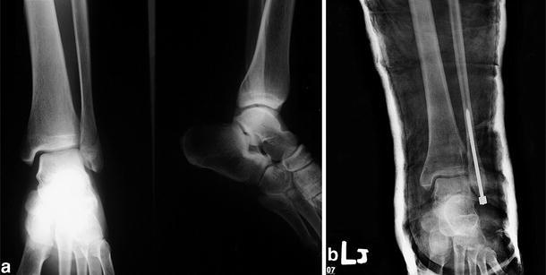
Ankle fracture operations were always performed under spinal anaesthesia. When a medial malleolar fracture was present, a separate incision was made for open reduction and internal fixation. This fixation was either by Kirschner wires augmented by a screw or by two screws. If the medial malleolus was intact and the deltoid ligament was ruptured, the deltoid ligament was not repaired. In the KP group, the operative technique has been described by Lee et al. [11]. A lateral skin incision was made 1 cm below the distal fibular tip to 1 cm above the fracture site. Soft tissue was dissected, and the fracture ends were exposed. A small bone holder or towel clip was used temporarily to hold the attained anatomical reduction. After identifying that there was no syndesmosis instability by using the stress test or intraoperative roentgenograms, a Knowles pin of 4-mm threaded diameter and 3.2-mm shaft diameter (Zimmer, Warsaw, IL), with threads capable of passing the fracture site, was directly inserted by hand drilling through the fibular tip into the medullary canal without predrilling. The entry point was 2 mm medial to the fibular tip. Ideally, the direction of the pin was parallel to the medullary canal (Fig. 1). Fluoroscopy was used to verify the pin site and post-reduced joint space. For a comminuted fracture, it was permitted to fix it by cerclage wires or sutures in addition to the Knowles pin. This procedure could hold the distal fibular alignment before the Knowles pins were inserted. In the TP group, lateral tubular plating was used to stabilise the fibula. Preoperation and postoperation, both groups received one-dose of prophylactic antibiotics.
Fig. 1.

A 58-year-old female patient who had left lateral malleolar fracture associated with the wide medial clear space was treated with a Knowles pin. a Preoperative anteroposterior and lateral radiographs showed an AO type-B2 ankle fracture. b Radiograph at the immediate postoperative period showed good fracture reduction. The entry point and pin direction were ideal
Plain films were taken immediately postoperatively to evaluate reduction, which was graded using a scale modified from that published previously [13]. Good reduction was defined as no fibula shortening, a posterior displacement less than 2 mm and a 1-mm increase in medial clear space. A fair reduction represented a fibula shortening of 2 mm, posterior displacement of 2–4 mm and a 1–3 mm increase in the medial clear space. A poor reduction was defined as a fibula shortening in excess of 2 mm, posterior displacement of over 4 mm and a greater than 3 mm increase in medial clear space.
A short leg cast with the ankle in a neutral position was applied postoperatively for 4 weeks for soft tissue healing. The postoperative rehabilitation process consisted initially of partial weight bearing. Full weight bearing was permitted 1 month after cast removal or when union was evident radiographically. Patients were reviewed at 1, 2, 3, 4 and 6 months after the fracture. AP and lateral roentgenograms were taken for all patients at each follow-up appointment for evaluations of fracture healing and the implant position. Radiographic healing was interpreted by the attending surgeon at each follow-up and was verified by all the authors of the present study during a retrospective review. Radiographic healing was defined as evidence of bridging callus across the fracture sites or the obliteration of the fracture lines on both anteroposterior and lateral views.
At the last follow-up, we evaluated the clinical results according to the ankle scoring system of Baird and Jackson [1], which was modified from Weber [17]. In this system, the subjective and objective clinical data are combined with radiographic results, with the maximum score being 100 points. The maximum clinical score is 75 points. Pain, stability of the ankle, ability to run, ability to walk, and motion of the ankle are evaluated. Maximum radiographic results contribute 25 points. An overall score of 96–100 points ranked as excellent, 91–95 points as good, 81–90 points as fair, and 0–80 as poor results. We define the excellent and good results as a satisfactory outcome. Fair and poor results were thought to be an unsatisfactory outcome. The Student’s t-test, chi-square test with Yates’ correction, and Fisher’s exact test were used to compare the two groups. The statistic software SPSS 10.0 was used to analyse the data: P values below 0.05 were considered significant.
Results
Forty-five of the original 49 (91.8%) treated with the Knowles pin and 30 of the original 34 (88.2%) treated with tubular plate had complete follow-up (P=0.71). The average period of follow-up was 34.5 months (range 12–80 months) for the KP group and 32.6 months (range 12–77 months) for the TP group (P=0.38). Both the groups were similar in respect of injury mechanisms, fracture pattern, open fracture grade, demographics and compounding medical conditions (all P values <0.31) (Table 1).
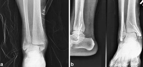
All the ankle fractures showed radiographic evidence of healing within 6 months (Fig. 2). The evaluations of immediate postoperative roentgenograms for adequacy of the reduction by one set of criteria [13] produced a good reduction in 43/45 (95%) of KP patients and 29/30 (96%) of TP patients. No difference in the good reduction rate between two groups was found (P=1.0). However, the average operative time was significantly less in the KP group (22 min) when compared to the TP group (43 min) (P<0.001). The average wound size was significantly smaller in the KP group (4.2 cm) when compared to the TP group (8.1 cm) (P<0.001). The average hospital stay was longer (P=0.03) in the TP group (5.5±0.72; range: 3–9 days) when compared to the KP group (3.4±0.42; range: 1–7 days) (Table 2).
Fig. 2.

A 60-year-old male patient with right bilateral malleolar fracture was treated with a Knowles pin (lateral malleolus) and two Kirschner wires augmented one screw (medial malleolus). a Preoperative anteroposterior radiograph showed an AO type-B2 ankle fracture. b X-rays at 2 months showed fracture healing with excellent restoration of the fracture alignment and the ankle joint
Table 2.
Clinical results in the two groups
| KP (M±SD) | TP (M±SD) | P value | |
|---|---|---|---|
| Union in 6 months | 45/45 | 30/30 | 1.0 |
| Fracture reduction | |||
| Good | 43/45 | 29/30 | 1.0 |
| Fair | 2/45 | 1/30 | 1.0 |
| Poor | 0 | 0 | 1.0 |
| Surgical data | |||
| OR time (min) | 22±4.5 | 43±6.8 | <0.001 |
| Wound size (cm) | 4.2±0.55 | 8.1±0.63 | <0.001 |
| Hospital stay (days) | 3.4±0.42 | 5.5±0.72 | 0.03 |
KP= Knowles pin; TP= tubular plate; M= mean; SD= standard deviation
No patient in the KP group complained of symptomatic hardware problems. Removal of the Knowles pin was easily accomplished by making a 1-cm skin incision along the old scar under local or spinal anesthesia. Twenty-four of 45 patients asked to have the hardware removed. In the TP group B, 12 out of 30 patients complained of feeling the plates and screws. Eighteen out of 30 patients asked to have the hardware removed. The KP group experienced fewer hardware symptoms (P<0.001). However, elective hardware removal did not significantly differ between the two groups (P=0.64)
The use of narcotic analgesics at our institution was patient-controlled meperidine (pethidine). For the first 3 days, a total value for meperidine was recorded, and the use was significantly less (P<0.001) in the KP group when compared to the TP group. There was an average of 34.4 mg for the KP group (range 0–150 mg) versus 103.3 mg for the TP group (range 0–350 mg).
The KP group experienced fewer complications than the TP group (P=0.023, Fisher’s exact test). The KP group had no complications, and the TP group had four instances of complications (13.3%), consisting of a skin necrosis, a delayed wound healing and two superficial infections. One female patient with an open type II fracture had a skin necrosis at 14 days after lateral plating. She had a secondary operation to remove the plate, and then received a split-thickness skin graft after granulation tissue surrounded the wound. Although her fracture was healed under a cast immobilisation, intermittent pain and a marked decrease in motion of the ankle was noted. The delayed wound healing occurred in a patient with renal disease. This problem was resolved after 2 months wound care. Both the superficial infections occurred in diabetic patients who were diagnosed clinically at the first follow-up visit, which was 7 days after surgery. A 1 week regimen of oral antibiotics resolved the infections.
At the final follow-up, the ankle scores of the patients were evaluated for the functional outcomes. Differences in the mean ankle score between the two groups were not significant (P=0.28). The mean ankle score was 94.2±3.2 points for the KP group and 91.6±5.1 points for the TP group.
Discussion
The AO classification system of ankle fractures amplifies from the Weber system and has further divided each of the Weber types into three groups. The AO type-B ankle fracture is the most common injury type. The AO type-B1 is an isolated lateral malleolar fracture. Type B2 is a lateral malleolar fracture with a medial lesion (deltoid ligament rupture or medial malleolar fracture). Type B3 is a lateral malleolar fracture with a medial lesion and fracture of the posterolateral tibia [6]. The treatment of AO type-B1 can be conservative. However, types B2 and B3 are most typically treated with open reduction and internal fixation (ORIF) [3, 10, 14, 16]. All the patients included in the study had AO type-B2 injury and underwent ORIF.
Although fibular plating remains a popular method to treat lateral malleolar fractures, symptomatic hardware is commonly noted [3, 9]. Brown et al. [3] reported that 31% of the 126 patients had lateral pain overlying their fracture hardware with plates and screws. In this study, 12 out of 30 patients with lateral plating complained of feeling the plates and screws. In contrast, no patient in the Knowles pin group complained of this problem. Although intramedullary fixation such as rush pinning, Kirschner wiring, etc., can also solve the symptomatic hardware problems, to date these intramedullary implants have not been widely used due to their migration and poor rotational control of the distal fragment [12]. In our study, there was neither implant migration nor loss of reduction in the Knowles pin group. We felt that the compressive force by a Knowles pin could make the contact of an oblique surface of cancellous bone stable enough to resist proximal migration or rotation of the fracture site. It could explain why a good radiographic reduction was obtained in 95% of the patients and a union rate of 100%.
Surgical treatment of ankle fractures in the elderly carries an increased risk due to osteoporosis that may preclude stable fixation and cause poor wound healing due to the quality of the soft tissue around the ankle [2, 4]. The infection rate is up to 12% in this age group [2, 10]. In this study, Knowles pinning in the elderly patients resulted in stable fixation without any complications. The interfragmental compression effect could make the fracture site stable. Less soft tissue stripping essentially decreased the wound complications. In contrast, lateral plating resulted in four complications (13%), including one skin necrosis, one delayed wound healing and two superficial infections. One of these complications in particular, involved an open grade II fracture, and the remaining three occurred in conjunction with either diabetes or renal disease. For an open fracture, the skin necrosis might be due to the severe trauma inducing poor viability of the surrounding tissue, and the lateral plating could increase skin tension. Furthermore, because the elderly patients with systemic diseases might have poor distal circulation or poor skin conditions, more soft tissue dissection could produce wound complications. Thus, if the elderly patients had open fractures or systemic diseases, lateral malleolar plating was a risk.
In our study, Knowles pinning conferred many advantages. Theoretically, elderly patients have some cognitive deficit from age. Less operation time may decrease the risk of anaesthesia. Less meperidine use is also important for an elderly patient group that may have some medical conditions. Decreased drug use and hospital stay may make the technique beneficial to both the patient and the hospital administration.
In conclusion, lateral fixation of AO type-B2 ankle fractures in the elderly by Knowles pin is a simple, safe and effective technique. Knowles pinning as opposed to tubular plating has a shorter operation time, smaller wound size, less meperidine use, shorter hospital stay, less symptomatic hardware, and a lower complication rate.
References
- 1.Baird RA, Jackson ST (1987) Fractures of the distal part of the fibula with associated disruption of the deltoid ligament. J Bone Joint Surg Am 69:1346–1352 [PubMed]
- 2.Beauchamp CG, Clay NR, Thexton PW (1983) Displaced ankle fractures in patients over 50 years of age. J Bone Joint Surg Br 65(3):322–329 [DOI] [PubMed]
- 3.Brown OL, Dirschl DR, Obremskey WT (2001) Incidence of hardware-related pain and its effect on functional outcomes after open reduction and internal fixation of ankle fractures. J Orthop Trauma 15(4):271–274 [DOI] [PubMed]
- 4.Fernandez GN (1988) Internal fixation of the oblique, osteoporotic fracture of the lateral malleolus. Injury 19(3):257–258 [DOI] [PubMed]
- 5.Franklin JL, Johnson KD, Hansen ST Jr (1984) Immediate internal fixation of ankle fractures. J Bone Joint Surg Am 66(9):1349–1356 [PubMed]
- 6.Griend RAV, Savoie FH, Hughes JL (1991) Fractures of the ankle. In: Rockwood CA Jr, Green DP, Bucholz RW (eds) Rockwood and Green’s fractures in adult: 1983–2030
- 7.Hammacher ER, Schutte PR, Bast TJ (1986) Minimal osteosynthesis of lateral malleolar fractures. Neth J Surg 38(3):87–89 [PubMed]
- 8.Koval KJ, Petraco DM, Kummer FJ et al (1997) A new technique for complex fibula fracture fixation in the elderly: a clinical and biomechanical evaluation. J Orthop Trauma 11(1):28–33 [DOI] [PubMed]
- 9.Lamontagne J, Blachut PA, Broekhuyse HM et al (2002) Surgical treatment of a displaced lateral malleolus fracture. J Orthop Trauma 16:498–502 [DOI] [PubMed]
- 10.Litchfield JC (1987) The treatment of unstable fracture of the ankle in the elderly. Injury 18:128–132 [DOI] [PubMed]
- 11.Lee YS, Huang CC, Chen CN et al (2005) Operative treatment of displaced lateral malleolar fractures. The Knowles pin technique. J Orthop Trauma 19:192–197 [DOI] [PubMed]
- 12.Mcdade WC (1975) Treatment of ankle fractures. In: AAOS Instructional Course Lectures, Ch 14. St. Louis, C.V. Mosby; 251–294
- 13.Mclennan JG, Ungersma JA (1986) A new approach to the treatment of ankle fractures. Clin Orthop 213:125–136 [PubMed]
- 14.Olerud C, Molander H (1986) Bi- and trimalleolar ankle fractures operated with nonrigid internal fixation. Clin Orthop 206:253–260 [PubMed]
- 15.Phillips WA, Schwartz HS, Keller CS et al (1985) A prospective randomized study of the management of severe ankle fractures. J Bone Joint Surg Am 67:67–78 [PubMed]
- 16.Ramasamy PR, Sherry P (2001) The role of a fibular nail in the management of Weber type B ankle fractures in elderly patients with osteoporotic bone. Injury 32:477–485 [DOI] [PubMed]
- 17.Weber BG, Simpson LA (1985) Corrective lengthening osteotomy of the fibula. Clin Orthop 199:61–67 [PubMed]


