Abstract
Purpose: To evaluate prospectively the increase in the size of the tibial and femoral bone tunnel following arthroscopic anterior cruciate ligament (ACL) reconstruction with quadrupled-hamstring autograft. Methods: Twenty-five consecutive patients underwent arthroscopic ACL reconstruction with quadrupled-hamstring autograft. Preoperative clinical evaluation was performed using the Lysholm knee score, Tegner activity level, and International Knee Documentation Committee forms and a KT-1000 arthrometer (side to side). Computed tomography (CT) of the femoral and tibial tunnel was performed on the day after operation in all cases and at mean follow-up of 10 months (range 9–11 months).Results: All of the clinical evaluation scales performed showed an overall improvement. The postoperative anterior laxity difference was <3 mm in 16 patients (70%) and 3–5 mm in seven patients (30%). The mean average femoral tunnel diameter increased significantly (3%) from 9.04±0.05 mm postoperatively to 9.3±0.8 mm at 10 months; tibial tunnel increased significantly (11%) from 9.03±0.04 mm to 10±0.8 mm. There were no statistically significant differences between tunnel enlargement, clinical results, and arthrometer evaluation. Conclusions: The rate of tunnel widening observed in this study seems to be lower than that reported in previous studies that used different techniques. We conclude that an anatomical surgical technique and a less aggressive rehabilitation process influenced the amount of tunnel enlargement after ACL reconstruction with doubled hamstrings.
Résumé
But: Évaluer prospectivement l’augmentation de taille des tunnels osseux après reconstruction arthroscopique du LCA par les ischio-jambiers. Méthode: 25 patients avaient été opérés. L’évaluation pré-opératoire avait été faite avec le score de Lysholm, les critères IKDC et de niveau d’activité Tegner et la mesure bilatérale avec l’arthromètre KT-1000. Un scanner des tunnels était réalisé dans tous les cas le jour suivant la chirurgie et à un suivi moyen de 10 mois (9–11). Résultats: Tous les scores cliniques montraint une amélioration globale. La différence de laxité antérieure après l’intervention était de moins de 3mm pour 16 patients (70%) et de 3 à 5 mm pour 7 (30%). Le diamètre moyen du tunnel fémoral augmentait significativement (3%) de 9,04±0,05 mm en post-opératoire à 9,3±0,8 mm à 10 mois ; le tunnel tibial augmentait (11%) de 9,03±0,004 à 10±0,8 mm. Il n’y avait pas de corrélation significative entre l’élargissement des tunnels, les résultats cliniques et les mesures à l’arthromètre. Conclusions: La proportion d’élargissement des tunnels observée dans cette étude semble plus faible rapportées avec des techniques différentes. Nous pensons qu’une technique très anatomique et une rééducation peu agressive influence l’élargissement des tunnels dans la reconstruction du LCA par les ischio-jambiers.
Introduction
Anterior cruciate ligament (ACL) reconstruction is a popular procedure in orthopaedic knee surgery. Among the related complications, tunnel enlargement has been reported in recent years regardless of the technique used [1, 5, 13, 23, 33].
Jackson et al. [16] noted cystic changes in the bone tunnels when the ACL was reconstructed with a bone-patellar tendon bone allograft; Linn et al. [20] reported an increase in bone tunnel size with Achilles tendon allograft, while L’Insalata et al. [19] observed this phenomenon with the use of hamstrings [26, 28, 30]. Tunnel enlargement has also been reported after ACL reconstruction with prosthetic ligaments as demonstrated in a retrospective study by Fukubayashi and Ikeda [9].
The mechanism of bone tunnel enlargement following ACL reconstruction is not yet clearly understood. Among the possible causes, mechanical factors such as graft tunnel motion, stress deprivation of bone within the tunnel wall, improper graft tunnel placement, and aggressive rehabilitation have been considered [11, 14, 22, 25, 32]. Biological factors thought to contribute to tunnel enlargement include a cytokine-mediated nonspecific inflammatory response, cell necrosis due to toxic products (ethylene oxide, metal), a foreign body (allograft) immune response, and heat necrosis as a response to drilling [18].
The purpose of our study was to evaluate prospectively the increase in size of the tibial and femoral bone tunnel following arthroscopic ACL reconstruction with quadruple-hamstring autograft and to propose a method for accurately calculating this phenomenon using computed tomography (CT) scanning.
Methods and materials
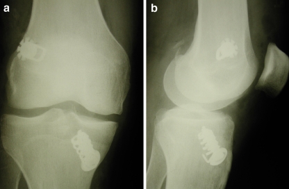
Our prospective study included 25 consecutive patients (18 men and seven women) with chronic instabilities who were treated from April 2004 to June 2004. They underwent a two-incision arthroscopic ACL reconstruction with quadrupled-hamstring autograft using a very strong fixation construction that fixed the graft close to the joint line, followed by a less aggressive postoperative rehabilitation that included postoperative immobilisation in a full extension brace for 2 weeks. The devices used were the swing-bridge (Citieffe, Bologna, Italy) for the femoral side and the Evolgate (Citieffe, Bologna, Italy) for the tibial side; we have previously described their biomechanical properties [2, 6, 7] (Fig. 1a,b). The mean patient age at the time of surgery was 30 years (range 17–44).
Fig. 1.
a Plain radiograph in anteroposterior view, b plain radiograph in lateral view
Preoperative clinical evaluation of knee function and stability was performed using the Lysholm knee score, the Tegner activity level, and the International Knee Documentation Committee (IKDC) forms and an arthrometer KT-1000 (side-to-side difference at 30 lb).
Postoperative treatment
Postoperatively, each patient’s knee was placed in a full-extension knee immobiliser. The 2nd day after operation, isometric exercises for muscular strength were begun, and patients were allowed to walk with progressive weightbearing using crutches.
Two weeks after surgery, active range-of-motion (ROM) exercises to reach 90° were started. At 4 weeks, the knee brace and crutches were removed, and patients started to practice FKT exercises to increase muscular strength and to reach full ROM. At the 3rd month after operation, patients began progressive functional activities (such as running) and isotonic and isokinetic exercises. They were allowed to return to sport activities 4–6 months after operation.
Radiographic evaluation
CT of the femoral and tibial tunnel was done the day after operation and at follow-up. All the examinations were performed using a Philips MX 8000 16-slice multislice CT (MSCT) scanner with post-processing multislab reconstructions on the sagittal and coronal planes. MSCT scanning was performed from a level just above the femoral external foramen to a level below the outer hole of the tibial tunnel in order to visualise the positioning of the autograft-fixing metallic devices. The slice thickness was 1 mm, with retro-reconstruction of 0.75mm made in all patients before postprocessing imaging with multislab views.
We measured the trans-osseous tunnel diameter at eight different levels, four each for femoral and tibial tunnels. The reference tunnel diameter measurements were taken as follows:
Femoral tunnel at the notch, axial (Fig. 2)
Femoral tunnel in the middle point, axial (Fig. 3)
Femoral tunnel in the middle point, on coronal image recons (Fig. 4)
Femoral tunnel in the middle point, on sagittal image recons (Fig. 5)
Tibial tunnel, axial at plateau (Fig. 6)
Tibial tunnel, axial at middle point (Fig. 7)
Tibial tunnel, sagittal at plateau (Fig. 8)
Tibial tunnel, sagittal at middle point (Fig. 9)
Fig. 2.
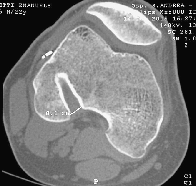
F1, femoral tunnel at the notch, axial
Fig. 3.
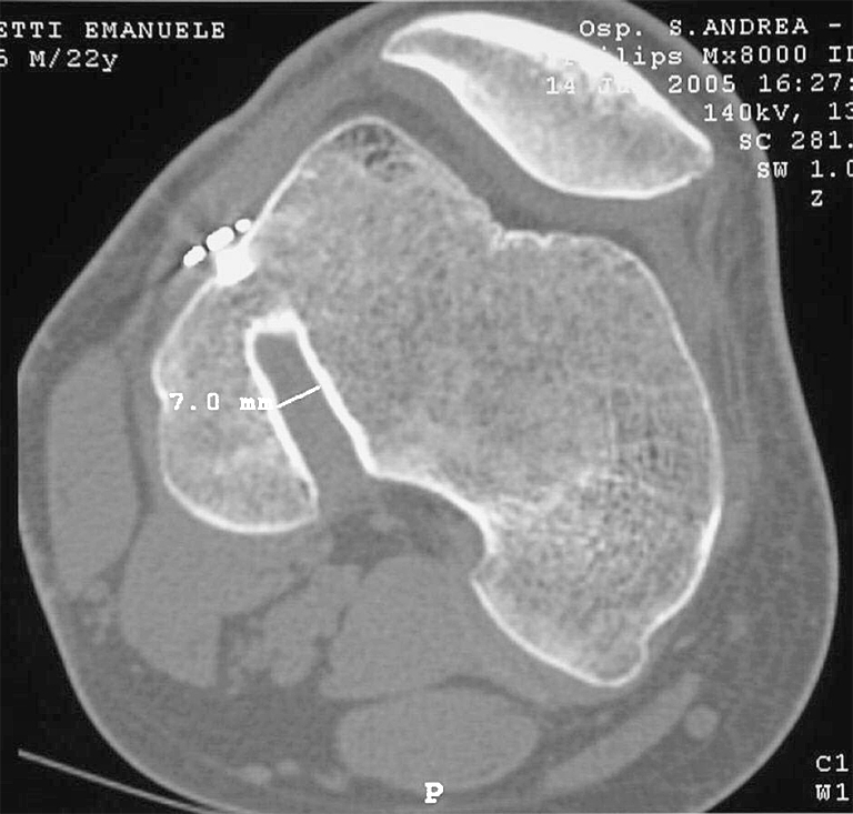
F2, femoral tunnel in the middle point, axial
Fig. 4.
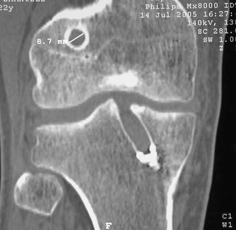
F3, femoral tunnel in the middle point, on coronal image reconstruction
Fig. 5.
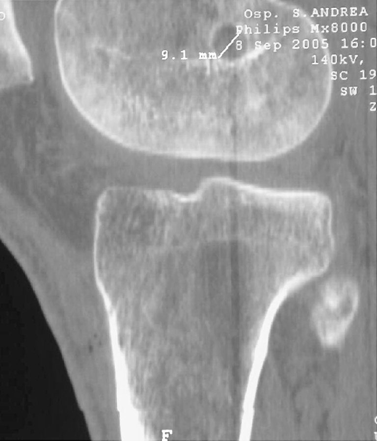
F4, femoral tunnel in the middle point, on sagittal image reconstruction
Fig. 6.
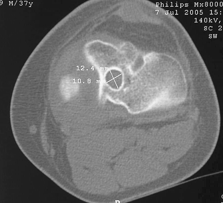
T1, tibial tunnel, axial at plateau
Fig. 7.
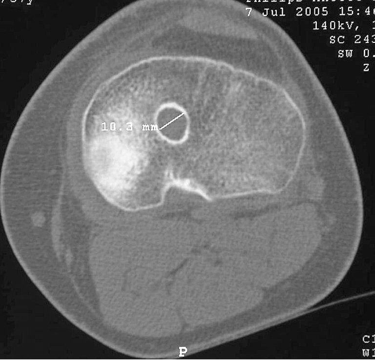
T2, tibial tunnel, axial at middle point
Fig. 8.
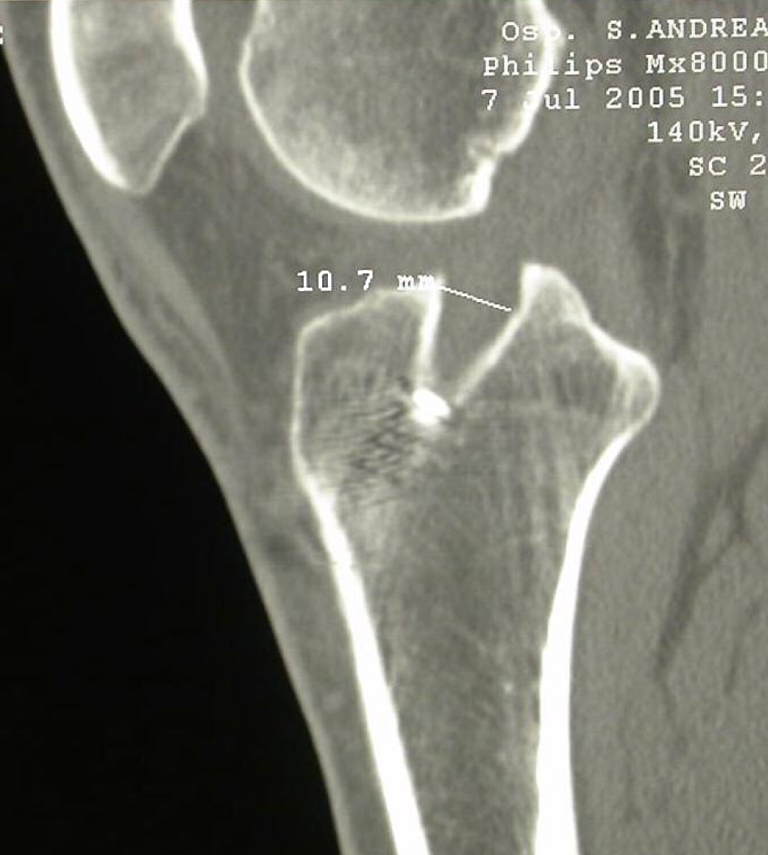
T3, tibial tunnel, sagittal at plateau
Fig. 9.
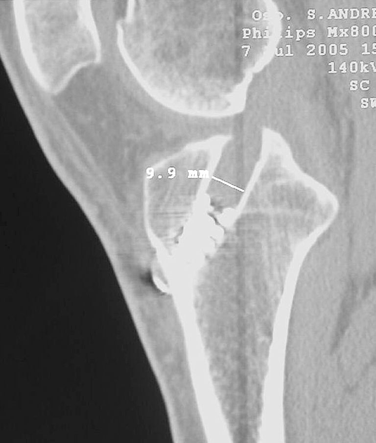
T4, tibial tunnel, sagittal at middle point
All diameters were calculated in millimetres.
Follow-up
Twenty-three patients were reviewed after a mean period of 10 months (range 9–11) with an accurate clinical examination and using a KT-1000 arthrometer, the Lysholm knee score, the Tegner activity level, and International Knee Documentation Committee (IKDC) forms. (Two patients refused to undergo radiological evaluation and were excluded from the study.) New CT scanning of the femoral and tibial tunnel was performed following the same criteria used in the immediate postoperative evaluation.
Statistical analysis of all data was performed using Student’s t-test.
Results
A meniscectomy was done in eight patients (five medial, three lateral). The clinical outcome showed no postoperative complications of infection, deep venous thromboses, or nerve injuries. Full ROM was observed in all the patients. The mean pre-injury Tegner score was 6.6 points; postoperatively it was 6.0 points (range 3–10).
The mean Lysholm score increased from 50.2±14 points pre-operatively to 94.6±16 points (range 70–100) postoperatively. The mean IKDC score preoperatively was 18.2 points and postoperatively was 86.8 points (range 70–100). On the IKDC activity scale, 14 patients (61%) were graded as level C and nine (39%) as level D preoperatively; postoperatively, 10 patients (43%) were graded as level A and 13 (57%) as level B.
The mean anterior laxity difference between the involved knee and the contralateral healthy knee dropped from 7.6±2.6 preoperatively to 2.8±1.7 mm postoperatively at a 30-lb anterior force (Table 1).
Table 1.
Preoperative and postoperative scores (IKDC International Knee Documentation Committee)
| Preoperative | Postoperative | |
|---|---|---|
| Tegner | 6.6 (preinjury) | 6 (range 3–10) |
| Lysholm | 50.2±14 | 94.6±16 |
| IKDC | 18.2: 14 (61%) group C, 9 (39%) group D | 86.8: 10 (43%) group A, 13 (57%) group B |
| KT-1000 side-to-side at 30 lb | 7.6±2.6 | 2.8±1.7 |
The mean average femoral tunnel diameter increased significantly from 9.04±0.05 mm postoperatively to 9.3±0.8 mm at 10 months; tibial tunnel increased significantly from 9.03±0.04 mm to 10±0.8 mm (Table 2).
Table 2.
Tunnel diameter measurements
| Femoral | Tibial | |||||||
|---|---|---|---|---|---|---|---|---|
| Patient | F1 | F2 | F3 | F4 | T1 | T2 | T3 | T4 |
| 1 | 9.9 | 10.4 | 12.4 | 10.6 | 10.6 | 11.6 | 10.7 | 12.2 |
| 2 | 9 | 9 | 8.8 | 10.1 | 10 | 10.4 | 11.5 | 11.1 |
| 3 | 9 | 9.6 | 10.7 | 9.4 | 10.1 | 10.2 | 10.5 | 11.4 |
| 4 | 8.3 | 9.2 | 10.3 | 9.2 | 9.8 | 9.8 | 11.1 | 11.2 |
| 5 | 9 | 9.9 | 10 | 10.8 | 10.8 | 10.3 | 10.7 | 9.9 |
| 6 | 10.6 | 9.2 | 8.7 | 8.4 | 10 | 10.6 | 10.8 | 11.5 |
| 7 | 9 | 8.5 | 9.3 | 9 | 10 | 10.7 | 9.5 | 9.7 |
| 8 | 8.8 | 9.2 | 9.3 | 8.7 | 11.4 | 11.4 | 9.8 | 11.1 |
| 10 | 10.3 | 10.7 | 11.2 | 11.9 | 10.5 | 11.4 | 11 | 12.5 |
| 11 | 8.1 | 7 | 8.7 | 8.2 | 8.6 | 8.9 | 9.1 | 9.6 |
| 12 | 7.2 | 7.4 | 8.6 | 7.5 | 9.2 | 9.3 | 9.8 | 9.6 |
| 13 | 10 | 11.5 | 12 | 12 | 10.7 | 10.1 | 10.3 | 11.1 |
| 14 | 10 | 11.5 | 12.4 | 11.7 | 10.06 | 10.4 | 10.3 | 10.9 |
| 15 | 7.7 | 8.6 | 9.7 | 6.8 | 8.8 | 9.2 | 9 | 10.2 |
| 16 | 9.2 | 9 | 10.6 | 9.2 | 9.7 | 11 | 11.2 | 11 |
| 17 | 9.7 | 8.5 | 8.5 | 7.6 | 8.7 | 8.5 | 9.8 | 9.7 |
| 18 | 8.6 | 8.8 | 9.1 | 8.8 | 10.2 | 10.4 | 9.1 | 10.2 |
| 19 | 8.9 | 8.6 | 8.6 | 9.1 | 9 | 9.5 | 9.1 | 9.1 |
| 20 | 8 | 8.9 | 9.5 | 9.1 | 9 | 9.4 | 9.1 | 10.3 |
| 21 | 11 | 10 | 10 | 9.1 | 10.5 | 10.7 | 9.6 | 9 |
| 22 | 9.1 | 9.1 | 8.5 | 8.5 | 9 | 10.3 | 9.4 | 10.4 |
| 23 | 9.1 | 9.1 | 8.5 | 9.6 | 9.5 | 10 | 9.7 | 10.1 |
| Mean | 9.113636 | 9.259091 | 9.790909 | 9.331818 | 9.387311 | 10.18636 | 10.05 | 10.53636 |
There were no statistically significant difference between tunnel enlargement, clinical results, and arthrometer evaluation.
Discussion
Before discussing the results of our study, it would be useful to briefly comment on the method of measurement used.
Previous studies [8, 31] demonstrated that measuring bone tunnels by X-ray can result in underestimating the real diameter of tunnel enlargement. CT scanning has been recommended for evaluating the dimension of the bone tunnels. We decided to use MSCT images because this technique depicts the real boundaries of the trans-osseous tunnel more exactly. Compared to plain X-ray films, MSCT has the advantage that it does not depend on geometric factors that may influence measurement, such as a small change in knee positioning or in the exposing distance from the film surface. Interestingly, all CT measurements done the day after operation closely matched the diameter of the tunnels as they were drilled at operation (9.05±0.05).
Peyrache et al. [24] reported an increase in tunnel diameter at 3 months after operation, no changes between 3 months and 2 years, and a decrease 3 years after operation. Fink et al. [8] and Harris et al. [12] reported that the enlargement occurs particularly within the first 6 weeks after operation. There is agreement that tunnel enlargement may occur within the 1st year after operation, and no further increase in bone tunnel diameter was observed thereafter up to 2–3 years after operation. In accordance with these authors, we performed a 10-month follow-up examination because this time seems to be the ideal period for investigating maximum tunnel widening.
L’Insalata et al. [19] observed an increase in tunnel diameter; in the anteroposterior (AP) radiographic view; it was 20.9%±13.4% for the tibial tunnel and 30.2%±17.2% for the femoral tunnel after ACL reconstruction with hamstring. In the lateral view the mean percentage increase was 25.5%±16.7% for the tibial tunnel and 28.1%±14.7% for the femoral tunnel.
Jansson et al. [17] noted an average femoral and tibial bone tunnel enlargement on AP-view radiography of 33% and 23%, respectively, while Fules et al. [10] noted an average tibial tunnel enlargement of 33% using magnetic resonance imaging; they both used hamstring for ACL reconstruction.
The results of this study demonstrate a slight femoral (3%) and moderate tibial (11%) bone tunnel enlargement following ACL reconstruction with hamstring grafts. These values, also calculating the cross-sectional area of the tunnels, are much lower than the suggested threshold of 50%, which is considered to be significant for tunnel enlargement [10].
Three studies have compared bone tunnel enlargement between hamstring and patellar tendon (PT) autografts. These studies showed significantly greater tunnel enlargement with the use of hamstring grafts, but they came to different conclusions with regard to cause of the enlargement process. L’Insalata et al. [19] argued for a mechanical cause due to fixation points of the hamstring graft being a greater distance from the articular surface that the fixation points of the PT graft that create a potentially larger force moment during graft motion within the tunnel (wind shield wiper effect). However, Clatworthy et al. [3, 4] found no enlargement in the femoral tunnel of the PT group despite the use of suspensory fixation. Marked enlargement was, however, observed in the hamstring group with the same method of femoral fixation. The authors concluded that graft fixation has only a subtle effect on tunnel enlargement. Webster et al. [31] proposed that the bone block used in PT autografts acts effectively as a bone graft, releasing osteoinductive bone morphogenic protein into the bone tunnel and thus reducing postoperative widening.
The cause of bone tunnel enlargement after ACL reconstruction has not been thoroughly investigated. Several potential mechanisms and causes of this phenomenon have been discussed. Biological factors associated with tunnel enlargement include foreign body immune response to allograft [29], nonspecific inflammatory response cells, necrosis due to toxic products in the tunnel, and heat necrosis as a response to drilling.
In a review article, Hoher et al. [15] discuss the theoretical concepts surrounding the aetiology as well as possible measures for preventing bone tunnel enlargement. The longitudinal graft motion of a semitendinosus and gracilis (STG) tendon Endobutton reconstruction is referred to as the “bungee effect.” They conclude that prevention of bone tunnel enlargement may be achieved by a more anatomical initial graft fixation.
This theory is supported by the findings of Schulte et al. [27], who found that tunnel expansion was greater in the lateral radiograph for BPTP grafts fixed more distally in the tibial tunnel.
In our study we used fixation devices stronger and stiffer than those used in all previous studies. Therefore, the use of a very strong and stiff femoral and tibial “intratunnel” fixation construction with a fixation point of the graft close to the joint line could contribute to minimising the tunnel enlargement [2, 6, 7].
Other authors have investigated the relationship between tunnel widening and rehabilitation. Murty et al. [21] suggested that immobilisation for 2 weeks was associated with increased tunnel enlargement. However, L’Insalata et al. [19], Clatworthy et al. [3, 4], and Yu and Paessler [32] showed that nonaggressive rehabilitation may reduce the micromotion of the graft in the tibial tunnel, thus reducing the “synovial bathing effect” and the nonspecific inflammatory response. The synovial bathing effect could also be related to tunnel widening: the synovial fluid might flow into the space between the bone graft and the wall of the tunnel, thus changing the graft’s biological fixation.
In our experience, a less aggressive rehabilitation process resulted in an acceptable rate of tunnel enlargement.
As with many other authors [8, 28], we found no statistically significant difference between tunnel enlargement and clinical results. However, like most of the previous studies, this is only a short-term review of the clinical effects of tunnel enlargement. Further studies and longer follow-up should be done to establish the role of the tunnel widening on the definitive biological fixation of the graft.
Furthermore, enlarged tunnels have the potential to cause problems with graft positioning and fixation in revision ACL surgery; therefore, whenever possible, enlargement of the bone tunnels should be prevented.
Conclusions
The rate of tunnel widening observed in this study seems to be lower than that reported in previous studies using different techniques. We conclude that using an anatomical fixation with stiff and strong fixation devices combined with a less aggressive rehabilitation programme could contribute to minimising tunnel enlargement after ACL reconstruction with doubled hamstrings.
References
- 1.Aglietti P, Zaccherotti G, Simeone AJ, Buzzi R (1998) Anatomic versus nonanatomic tibial fixation in anterior cruciate ligament reconstruction with bone-patellar tendon-bone graft. Knee Surg Sports Traumatol Arthrosc 6 (Suppl 1):S43–S48 [DOI] [PubMed]
- 2.Brown CH, Ferretti A, Conteduca F, Morelli F et al (2001) Biomechanics of the swing-bridge technique for anterior cruciate ligament reconstruction. Eur J Sports Traum Rel Res 23 (2):69–73
- 3.Clatworthy MG, Annear P, Bulow JU et al (1999) Tunnel widening in anterior cruciate ligament reconstruction: a prospective evaluation of hamstring and patella tendon grafts. Knee Surg Sports Traumatol Arthrosc 7(3):138–145 [DOI] [PubMed]
- 4.Clatworthy MG, Bartelett J, Howell S et al (1999) The effect of graft fixation techniques on tunnel widening in hamstring ACL reconstruction. Arthroscopy 15(Suppl):5
- 5.Fahey M, Indelicato PA (1994) Bone tunnel enlargement after anterior cruciate ligament replacement. Am J Sports Med 22(3):410–414 [DOI] [PubMed]
- 6.Ferretti A, Conteduca F, Labianca L et al (2005) Evolgate fixation of doubled flexor graft in ACL reconstruction: biomechanical evaluation with cyclic loading. Am J Sports Med 33(4):574–582 [DOI] [PubMed]
- 7.Ferretti A, Conteduca F, Morelli F et al (2003) The Evolgate, a method to improve the pull-out strength of interference screws in tibial fixation of anterior cruciate ligament reconstruction with doubled gracilis and semitendinosus tendons. Arthroscopy 19(9):936–940 [DOI] [PubMed]
- 8.Fink C, Zapp M, Benedetto KP et al (2001) Tibial tunnel enlargement following anterior cruciate ligament reconstruction with patellar tendon autograft. Arthroscopy 17(2):138–143 [DOI] [PubMed]
- 9.Fukubayashi T, Ikeda K (2000) Follow-up study of Gore-Tex artificial ligament: special emphasis of tunnel osteolysis. J Long Term Eff Med Implants 10(4):267–277 [PubMed]
- 10.Fules PJ, Madhav RT, Goddard RK et al (2003) Evaluation of tibial bone tunnel enlargement using MRI scan cross-sectional area measurement after autologous hamstring tendon ACL. Knee 10 (1):87–91 [DOI] [PubMed]
- 11.Hantes ME, Mastrokalos DS, Yu J, Paessler HH (2004) The effect of early motion on tibial tunnel widening after anterior cruciate ligament replacement using hamstring tendon grafts. Arthroscopy 20 (6):572–580 [DOI] [PubMed]
- 12.Harris NL, Indelicato PA, Bloomberg MS et al (2002) Radiographic and histologic analysis of the tibial tunnel after allograft anterior cruciate ligament reconstruction in goats. Am J Sports Med 30(3):368–373 [DOI] [PubMed]
- 13.Hersekli MA, Akpinar S, Ozalay M et al (2004) Tunnel enlargement after arthroscopic anterior cruciate ligament reconstruction: comparison of bone-patellar tendon-bone and hamstring autografts. Adv Ther 21(2):123–131 [DOI] [PubMed]
- 14.Hogervorst T, van der Hart CP, Pels Rijcken TH, Taconis WK (2000) Abnormal bone scans of the tibial tunnel 2 years after patella ligament anterior cruciate ligament reconstruction: correlation with tunnel enlargement and tibial graft length. Knee Surg Sports Traumatol Arthrosc 8(6):322–338 [DOI] [PubMed]
- 15.Hoher J, Moller HD, Fu FH (1998) Bone tunnel and enlargement after anterior cruciate ligament reconstruction: fact or fiction? Knee Surg Sports Traumatol Arthrosc 6(4):231–240 [DOI] [PubMed]
- 16.Jackson DW, Windler GE, Simon TM (1990) Intraarticular reaction associated with the use of freeze-fried, ethylene-oxide sterilized bone patella tendon—bone allografts in the reconstruction of the anterior cruciate ligament. Am J Sports Med 18(1):1–10 [DOI] [PubMed]
- 17.Jansson KA, Harilainen A, Sandelin J et al (1999) Bone tunnel enlargement after anterior cruciate ligament reconstruction with the hamstring autograft and endobutton fixation technique: a clinical, radiographic and magnetic resonance imaging study with 2 years follow-up. Knee Surg Sports Traumatol Arthrosc 7(5):290–295 [DOI] [PubMed]
- 18.Jo H, Jun DS, Lee DY et al (2004) Tibial tunnel area changes following arthroscopic anterior cruciate ligament reconstructions with autogenous patellar tendon graft. Knee Surg Sports Traumatol Arthrosc 12 (4):311–316 [DOI] [PubMed]
- 19.L’Insalata JC, Klatt B, Fu FH, Harner CD (1997) Tunnel expansion following anterior cruciate ligament reconstruction: a comparison of hamstring and patellar tendon autografts. Knee Surg Sports Traumatol Arthrosc 5(4):234–238 [DOI] [PubMed]
- 20.Linn RM, Fischer DA, Smith JP et al (1993) Achilles tendon allograft reconstruction of the anterior cruciate ligament-deficient knee. Am J Sports Med 21 (6):825–831 [DOI] [PubMed]
- 21.Murty AN, el Zebdeh MY, Ireland J (2001) Tibial tunnel enlargement following anterior cruciate reconstruction: does post-operative immobilisation make a difference? Knee 8 (1):39–43 [DOI] [PubMed]
- 22.Nebelung W, Becker R, Merkel M et al (1998) Bone tunnel enlargement after anterior cruciate ligament reconstruction with semitendinous tendon using Endobutton fixation on the femoral side. Arthroscopy 14(8):810–815 [DOI] [PubMed]
- 23.Otsuka H, Ishibashi Y, Tsuda E, Sasaki K et al (2003) Comparison of three techniques of anterior cruciate ligament reconstruction with bone-patellar tendon-bone graft: differences in anterior tibial translation and tunnel enlargement with each technique. Am J Sports Med 31(2):282–288 [DOI] [PubMed]
- 24.Peyrache MD, Dijan P, Christel P, Witvoet J (1996) Tibial tunnel enlargement after anterior cruciate ligament reconstruction by autogenous bone-patellar-tendon bone graft. Knee Surg Sports Traumatol Arthrosc 4(1):2–8 [DOI] [PubMed]
- 25.Romano VM, Graf BK, Keene JS et al (1993) Anterior cruciate ligament reconstruction: the effect of tibial tunnel placement on range of motion. Am J Sports Med 21(3):415–418 [DOI] [PubMed]
- 26.Sakai H, Yajima H, Hiraoka H, Fukuda A et al (2004) The influence of tibial fixation on tunnel enlargement after hamstring tendon anterior cruciate ligament reconstruction. Knee Surg Sports Traumatol Arthrosc 12 (5):364–370 [DOI] [PubMed]
- 27.Schulte K, Majewski M, Irrgang JJ et al (1995) Radiographic tunnel changes following arthroscopical reconstruction: autograft versus allograft. Arthroscopy 11:372–373
- 28.Segawa H, Omori G, Tomita S, Koga Y (2001)Bone tunnel enlargement after anterior cruciate ligament reconstruction using hamstring tendons. Knee Surg Sports Traumatol Arthrosc 9(4):206–210 [DOI] [PubMed]
- 29.Sgaglione NA, Douglas JA (2004) Allograft bone augmentation in anterior cruciate ligament reconstruction. Arthroscopy 20 (Suppl 2):171–177 [DOI] [PubMed]
- 30.Simonian PT, Erickson MS, Larson RV et al (2000) Tunnel expansion after hamstring anterior cruciate ligament reconstruction with 1-incision endobutton femoral fixation. Arthroscopy 16(7):707–714 [DOI] [PubMed]
- 31.Webster KE, Feller JA, Hameister KA (2001) Bone tunnel enlargement following anterior cruciate ligament reconstruction: a randomised comparison of hamstring and patellar tendon grafts with 2-year follow-up. Knee Surg Sports Traumatol Arthrosc 9(2):86–91 [DOI] [PubMed]
- 32.Yu JK, Paessler HH (2005) Relationship between tunnel widening and different rehabilitation procedure after cruciate ligament reconstruction with quadrupled hamstring tendons. Chin Med J (Engl) 118(4):320–326 [PubMed]
- 33.Zijl JA, Kleipool AE, Willems WJ (2000) Comparison of tibial tunnel enlargement after anterior cruciate ligament reconstruction using patellar tendon autograft or allograft. Am J Sports Med 28 (4):547–551 [DOI] [PubMed]


