Abstract
The treatment of fractures of the distal third of the tibia has evolved with the development of improved imaging and surgical techniques. The outcome of treatment using conventional fixation is poor. We report a study on indirect reduction in 26 patients. All cases achieved radiological union and full weight bearing. The good to excellent results suggest that this method should be considered in metaphyseal fractures where intra-medullary nails are not suitable.
Résumé
Le traitement des fractures de la partie distale du tibia avec troisième fragment peut être amélioré avec le développement des techniques d’imagerie et de traitements chirurgicaux. Le devenir du traitement, utilisant une fixation conventionnelle est peu satisfaisante. Nous rapportons une étude sur la réduction indirecte de cette fracture chez 26 patients. Tous les cas sont consolidés sur le plan radiographique, avec appui total. Les excellents résultats suggèrent que cette méthode peut être utilisée dans les fractures métaphysaires dans lesquelles l’enclouage centro médullaire n’est pas possible.
Introduction
The distal third tibial fractures are unique. The location of the fracture is close to the ankle joint and it is not uncommon for the fracture line to extend into the joint. The distal one third of the tibia has less muscle coverage in comparison to rest of the tibia. Often, these fractures are comminuted and are unstable. Disturbingly, they can be associated with severe closed or open soft tissue injury. Hence, complex fractures of the distal tibia are difficult to treat [2, 8].
Traditionally, these difficult fractures have been managed by open reduction and rigid internal fixation with a compression plate. A high rate of good to excellent results has been reported [17, 18]. However, this technique has not produced consistent outcomes and has a high incidence of complications, including infection, poor wound healing and non-union [3, 12, 19].
In the last 20 years, intra-medullary fixation has become the mainstay of treating tibial shaft fractures. Because of its success, the indications have been extended to those of the proximal and distal metaphyseal region. However, not all distal tibia fractures are suitable for intra-medullary rod fixation. Problems with stable fixation and malalignment have been reported [3, 6, 16].
To improve on the poor results associated with open reduction and internal fixation, minimally invasive techniques were developed to include limited internal and hybrid external fixation [11, 21]. Table 1 shows the difference between classic and minimally invasive plate fixation. Minimally invasive fixation results in minimal additional injury at surgery, emphasises meticulous soft tissue dissection, limits the stripping of fracture fragments and reduces the fracture by indirect reduction techniques [8].
Table 1.
Differences between classic and minimal fixation
| Classic open fixation | Minimally invasive fixation |
|---|---|
| 1. Direct fracture exposureDirect reduction | Untouched fracture exposure Indirect reduction |
| 2. Subperiosteal dissection | Epiperiosteal dissection |
| 3. Anatomical reduction | Anatomical alignment |
| 4. Rigid fixation | Stable but not rigid fixation |
| 5. Primary bone healing | Healing by callus formation |
Recent cadaveric studies have suggested that the circulation of the distal tibia is mainly from the extraosseous blood supply from an anastomotic network of arteries from the anterior and posterior tibial arteries, which enters the tibia from the medial surface. Open plating of the medial aspect of the distal tibia caused a statistically significant greater disruption of the extraosseous blood supply of the metaphyseal region than percutaneously applied plates [1, 7].
Compared with the traditional approach, minimally invasive techniques appear to have higher rates of union, lower rates of postoperative complications and lower incidences of bone grafting [4, 8, 12, 11]. It is with this background that the authors report the functional clinical and radiographic results of 26 distal tibial fractures that have been plated with minimally invasive techniques performed or supervised by the senior surgeon (VP) and independently assessed by an observer (GC).
Materials and methods
Between January 2000 and December 2003, a consecutive series of 26 fractures of the distal tibia were treated with a minimally invasive fixation in Health Care Hawkes Bay, Hastings, and Otago Health Care, Dunedin. The indication for fixation was distal tibial fracture that was unsuitable for conservative treatment and was not ideal for locked intra-medullary rod fixation due to the degree of communition or the distal nature of the fracture.
Of the 26 patients that were eligible, three were excluded. Two were lost to follow-up as they lived in another country and one patient refused to participate. The mean age at the time of injury was 43 years (range: 17–72). There were 15 males and eight females. The mechanisms of injury included a slip and fall in 10 patients, a fall from a significant height in three, motor vehicle accident (MVA) in six, a direct blow in two and a fall from a bike in two patients (Table 2).
Table 2.
Patients used in the study
| Age | 43 years (range: 17–72) |
| Male:female ratio | 15:8 |
| Mechanism of injury | |
| Slip and fall | 10 (40%) |
| MVA | 6 (24%) |
| Fall from height | 3 (12%) |
| Direct blow | 2 (8%) |
| Fall from bike | 2 (8%) |
| Soft tissue injury (Tscherne [22]) | |
| I | 20 |
| II | 3 |
| Fracture displacement (AP and lateral view) | |
| >30% | 7 |
| 30–50% | 13 |
| >50% | 3 |
| Fracture type (AO) | |
| A1 | 2 |
| A2 | 6 |
| A3 | 11 |
| C1 | 4 |
| Fixation | |
| Tibia only | 7 |
| Tibia and fibular | 16 |
| Type of plate | |
| AO DCP | 5 |
| AO periarticular | 2 |
| Zimmer periarticular | 16 |
| Average duration of surgery | 70 mins (range: 45–130) |
| Average radiation time | 55 s |
| Hospital stay | 4 days (range: 3–10) |
The fractures were classified radiographically according to the Muller’s AO classification [14] system. There were two in A1, six in A2, 11 in A3 and four in C1. Soft tissue injury was classified with the classification of Tscherne [22]. Twenty were of grade 1 soft tissue injuries and three were of grade 2 injuries. There were no open fractures in our group.
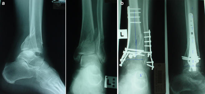
Surgery was performed on the same day (when the swelling was not bad) or as soon as the swelling allowed(i.e. when skin wrinkling occurred). A standardised technique was used for all patients in this series. A tourniquet was applied in all cases and in none was an external fixator or distracter used. When indicated, the fibula was fixed initially, by using standard AO fixation techniques. In four patients with C1 injuries, the articular fragments were fixed percutaneously or through a small incision with cannulated screws (Fig. 1). Then, the metaphyseal fragment of distal tibial was addressed through a small curved incision over the medial malleolus. An extra-periosteal or subcutaneous tunnel was created along the medial aspect of the tibia by blunt dissection using a large Bristow or a periosteal elevator. The plate could then be advanced directly beneath the soft tissue. Initial fixation was carried out with a distal screw passed parallel to the ankle joint under image intensifier guidance and the fracture was indirectly reduced onto the plate. Axial traction on the foot or application of the reduction forceps was liberally used to obtain acceptable reduction. Once the sagittal, coronal and rotational alignment appeared to be satisfactory, the proximal screws were passed percutaneously under image intensifier guidance.
Fig. 1.
a Intra-articular C1 fracture distal tibia. b Minimally invasive fixation of intra-articular fractures
Sixteen patients had both the tibia and fibula fixed, with the remaining seven cases having an intact fibula. The Zimmer medial distal periarticular plate (precontoured) was used in 18 cases, with the AO (Synthes) periarticular plate used in two and the standard AO DCP contoured by hand used in six (the intact contralateral tibia was used as a template for contouring the AO plates).
Post-operatively, all patients were treated in a resting slab for two weeks. They were then placed into a moon boot with graduated weight bearing over the next 2 months and were encouraged to move the ankle joint. Patients were assessed clinically and radiologically at regular intervals: 6 weeks, 3 months, monthly until radiological healing.
At the time of final follow-up, all patents were assessed using the criteria described by Johner and Wruhs [9]. For clinical assessment, the patients were graded into excellent, good, fair and poor, depending on pain, gait, range of movement at knee and foot, and work and hobbies. Any angulation of more than 5° of valgus-varus and 10° of the anterior-posterior angulation and over 1 cm of shortening were considered to be radiologically fair to poor results.
Results
The average duration of the operation was 70 min (range: 45–130 min), with a radiation time of 55 s. The mean hospital stay was 4 days (range: 3 to 10 days). The minimal follow-up was 12 months, with a range from 14–42 months. At 12 months, 11 patients had excellent results, nine had good, two fair and one had a poor result (Table 3). Twenty had none or slight occasional discomfort, three had moderate pain and none had severe pain.
Table 3.
Results
| a. Results clinical criteria of Johner and Wruhs [9]): | |
| Excellent | 11 (48%) |
| Good | 9 (40%) |
| Fair | 2 (9%) |
| Poor | 1 (4%) |
| b. Functional results: | |
| Pain: Minimal or no pain | 20 (87%) |
| Moderate pain | 3 |
| Mobility: Walk unlimited | 9 (39%) |
| Walk slightly limited | 7 |
| Work (at 20 weeks) | 18 (79%) |
| Hobbies | 13 (56%) |
| c. Radiological results: | |
| Valgus-varus alignment | |
| Excellent | 13 (less than 2*) |
| Good | 10 (varus within 5* in 6; valgus within 5* in 4) |
One rotational malalignment, as determined by clinical assessment
The range of ankle movement was assessed in all patients at 12 months. Fourteen had a full range of movement of their ankle and subtalar joints compared to the opposite uninjured side. Six patients had more than 80% of full movement and three had between 60% and 80% of full movement.
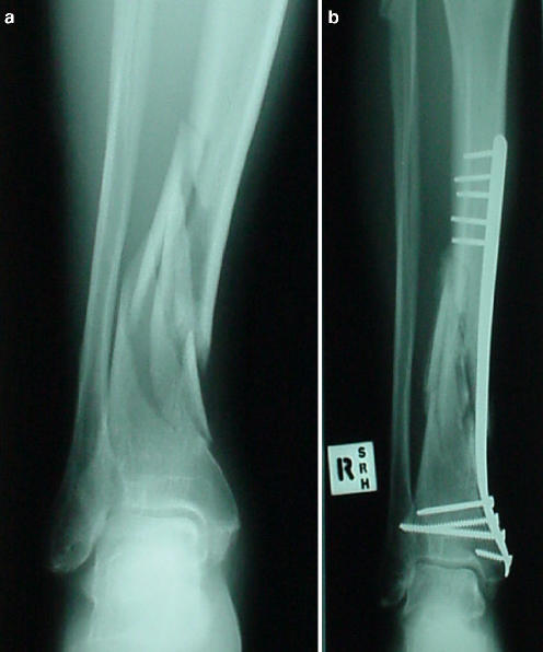
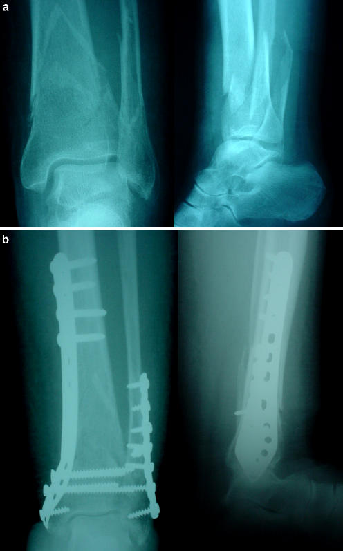
Radiological assessment revealed satisfactory alignment in all but one. Thirteen patients were within 2° of neutral alignment to the valgus-varus, six within 5° of the varus and four within 5° of the valgus alignment (Fig. 2). No patient had greater than 5° of malalignment. No patient had more than 3° of recurvatum or antecurvatum. The average length of time to radiographic healing, defined as callus seen bridging across three cortices, was 19.5 weeks (range: 14–30 weeks) (Figs. 3 and 4).
Fig. 2.
a Intra-articular C1 fracture. b Post-fixation healing in excellent alignment
Fig. 3.
a Extensive A3 fracture. b Fixation with a Zimmer plate. Callus formation is seen as early as 2 months
Fig. 4.
a Osteoporotic A3 fracture. b Healing in good alignment with some backing out of screws
Eighteen patients had returned to paid work at 24 weeks but only nine had returned to their previous employment on a full-time basis. Thirteen had returned to their previous recreational activities but none had returned to active participation in competitive sport at 12 months. All but one patient returned to their previous employment by one year.
We had one case of superficial infection treated, as an outpatient, with oral antibiotics. We also had one case of superficial saphenous nerve irritation. We had no cases of deep infection, delayed wound healing, non-union or hardware failure. There was one failure though, requiring refixation. In this patient, the initial fixation with an AO periarticular plate resulted in an external rotation deformity of more than 30°.
Discussion
The intra-medullary vessels of the tibia supply the inner two thirds of the cortex. The outer one third of the cortex receives its blood supply from the surrounding soft tissues [15]. In displaced fractures, there is disruption of the intra-medullary blood supply and the fracture fragments must rely on the surrounding soft tissues for their nutrition [7]. This soft tissue is disrupted with surgical dissection: the more extensive the dissection, the higher the chance of devascularising the bone [1, 7].
With minimally invasive plate fixation, the amount of dissection of the fracture fragments is minimised becauseof the indirect reduction techniques. It has been shown in femurs that up to 80% of the perforating arteries are disrupted in open plating, whereas no disruption in the percutaneous plated femurs occur [7]. There is no reason to think that this would not apply to the situation in the tibia. Whiteside and Lesker [23] have shown that increasing the amount of dissection in a tibial model increases healing complications. Clinical studies have confirmed that the incidence of wound healing problems, infection and non-union is high in these fractures, with the reported incidence as high as 4.5%, 22% and 8%, respectively [3, 5, 10, 12, 19].
Our study is one of the biggest reported in the literature for this type of fixation in the distal tibia, with a functional as well as a radiographic outcome. Our results are consistent with a previous report by Maffulli et al. [13]. In this study, we had one case of superficial wound infection, which was treated with oral antibiotics, and one case of revision fixation. These results are similar to those previously reported in the literature [11, 20]. The fact that all of the fractures in this study were closed injuries may have influenced the outcome. This may have been offset by the more comminuted nature of the fractures, as shown by the larger numbers of fractures classified as A3 (11 patients) in this study. Following high-energy injuries or more the comminuted nature of fractures may result in increasing complications [10].
Functionally, 73% had returned to some form of paid work at 24 weeks and 40% had returned to their previous occupation on a full-time basis. This compares favourably with results reported previously in those treated with open reduction and internal fixation [1, 13]. It still emphasises the fact that these fractures, even treated percutaneously, take a long time before full recovery can be expected.
It is encouraging to see early callus in these patients, which is consistent with a low profile plate (Zimmer). This is probably due to some micromotion occurring at the fracture site when they are fixed with a low-profile plate, as fixation is not as rigid as with a conventional AO plate. This observation allowed us to let patients partially weight bear as early as 2 weeks. In none of the patients were any hardware failure incidents observed. This is true even in osteoporotic bones, although minimal screw backing out was visible, without any significant loss of position. Probably, the locking low-profile plates in future may circumvent this problem.
Criticism against minimal fixation is its inability to achieve anatomical reduction as in the open method of fixation. The literature is not clear about the acceptable reduction in fracture tibia. Milnar et al. [14] studied 164 fracture tibias with a long-term follow-up of 30 years and concluded that there were no significant univariate associations between malunions of the tibia and the development of osteoarthritis of the knee or ankle. In no patients in this series did we observe valgus or varus malalignment more than 5°.
Percutaneous plating has a role in the management of distal tibial fractures that are not amenable to intra-medullary rod fixation either because of the distal nature of the fracture not allowing locking screw placement, intra-articular extension of the fracture or concern about varus or valgus malalignment. The functional results are no worse than those obtained with formal open reduction and internal fixation, and the complication rate is decreased.
References
- 1.Borrelli J Jr, Prickett W, Song E, Becker D, Ricci W (2002) Extraosseous blood supply of the tibia and the effects of different plating techniques: a human cadaveric study. J Orthop Trauma 16(10):691–695 [DOI] [PubMed]
- 2.Brumback RJ, McGarvey WC (1995) Fractures of the tibial plafond. Evolving treatment concepts for the pilon fracture. Orthop Clin North Am 26(2):273–285 [PubMed]
- 3.Oh C-W, Kyung H-S, Park I-H, Kim P-T, Ihn JC (2003) Distal tibia metaphyseal fractures treated by percutaneous plate osteosynthesis. Clin Orthop 408:286–291 [DOI] [PubMed]
- 4.Oh C-W, Park B-C, Kyung H-S, Kim SJ, Kim HS, Lee SM, Ihn JC (2003) Percutaneous plating for unstable tibial fractures. J Orthop Sci 8(2):166–169 [DOI] [PubMed]
- 5.Collange C, Sanders R, DiPasquale T (2000) Treatment of complex tibial periarticular fractures using percutaneous techniques. Clin Orthop 375:69–77 [DOI] [PubMed]
- 6.Dogra AS, Ruiz AL, Thompson NS, Nolan PC (2000) Dia-metaphyseal distal tibial fractures—treatment with a shortened intramedullary nail: a review of 15 cases. Injury 31(10):799–804 [DOI] [PubMed]
- 7.Farouk O, Krettek C, Miclau T, Schandelmaier P, Tscherne H (1999) The topography of the perforating vessels of the deep femoral artery. Clin Orthop 368:255–259 [PubMed]
- 8.Helfet DL, Suk M (2004) Minimally invasive percutaneous plate osteosynthesis of fractures of the distal tibia. AAOS 53:471–475 [PubMed]
- 9.Johner R, Wruhs O (1983) Classification of tibial shaft fractures and correlation with results after rigid internal fixation. Clin Orthop 178:7–25 [PubMed]
- 10.Karladani AH, Granhed H, Karrholm J, Styf J (2001) The influence of fracture etiology and type on fracture healing: a review of 104 consecutive tibial shaft fractures. Arch Orthop Trauma Surg 121(6):325–328 [DOI] [PubMed]
- 11.Kretteck C (1997) Concepts of minimally invasive plate osteosynthesis. Injury 28(Suppl 1):805–809 [DOI] [PubMed]
- 12.Leunig M, Hertel R, Siebenrock KA, Ballmer FT, Mast JW, Ganz R (2000) The evolution of indirect reduction techniques for the treatment of fractures. Clin Orthop 375:7–14 [DOI] [PubMed]
- 13.Maffulli N, Toms AD, McMurtie A, Oliva F (2004) Percutaneous plating distal tibial fractures. Int Orthop 28(3):159–162 [DOI] [PMC free article] [PubMed]
- 14.Milnar SA, Davis TR, Muir KR, Greenwood DC, Doherty M (2002) Long-term outcome after tibial shaft fracture: is malunion important? J Bone Joint Surg 84A(6):971–980 [DOI] [PubMed]
- 15.Rhinlander F (1968) The normal microcirculation of diaphyseal cortex and its response to fracture. J Bone joint Surg 50(4):784–800 [DOI] [PubMed]
- 16.Robinson CM, McLaughlan GJ, McLean IP, Court-Brown CM (1995) Distal metaphyseal fractures of the tibia with minimal involvement of the ankle. Classification and treatment by locked intramedullary nailing. J Bone Joint Surg 77(5):781–787 [PubMed]
- 17.Ruedi TP, Murphy WM (2000) AO principles of fracture management. Thieme, Stuttgart New York, p 45
- 18.Ruedi T, Webb JK, Allgower M (1976) Experience with the dynamic compression plate (DCP) in 418 recent fractures of the tibial shaft. Injury 7(4):252–257 [DOI] [PubMed]
- 19.Schutz M, Sudkamp NP (2003) Revolution in plate osteosynthesis: new internal fixator systems. J Orthop Sci 8(2):252–258 [DOI] [PubMed]
- 20.Stromsoe K, Eikvar K, Ovre S, Hvaal K (1999) Miniinvasive plate osteosynthesis of distal tibial fractures. Tidsskr Nor Laegeforen 119(29):4316–4318 [PubMed]
- 21.Tornetta P, Weiner L, Bergman M, Watnik N, Steuer J, Kelley M, Yang E (1993) Pilon fractures: treatment with combined internal and external fixation. J Orthop Trauma 7(6):489–496 [DOI] [PubMed]
- 22.Tscherne H (1983) Management of wounds in fractures with soft tissue injuries. Z Kinderchir 38(1):34–39 [DOI] [PubMed]
- 23.Whiteside L, Lesker PA (1978) The effects of periosteal and subperiosteal dissection. J Bone Joint Surg 60A:26–30 [PubMed]


