Abstract
Mycobacterium avium subsp. paratuberculosis is the causative pathogen of Johne's disease, a chronic inflammatory wasting disease in ruminants. This disease has been difficult to control because of the lack of an effective vaccine. To address this need, we adapted a specialized transduction system originally developed for M. tuberculosis and modified it to improve the efficiency of allelic exchange in order to generate site-directed mutations in preselected M. avium subsp. paratuberculosis genes. With our novel optimized method, the allelic exchange frequency was 78 to 100% and the transduction frequency was 1.1 × 10⁻⁷ to 2.9 × 10⁻⁷. Three genes were selected for mutagenesis: pknG and relA, which are genes that are known to be important virulence factors in M. tuberculosis and M. bovis, and lsr2, a gene regulating lipid biosynthesis and antibiotic resistance. Mutants were successfully generated with a virulent strain of M. avium subsp. paratuberculosis (M. avium subsp. paratuberculosis K10) and with a recombinant K10 strain expressing the green fluorescent protein gene, gfp. The improved efficiency of disruption of selected genes in M. avium subsp. paratuberculosis should accelerate development of additional mutants for vaccine testing and functional studies.
Johne's disease (paratuberculosis) is a chronic wasting disease of the intestine of ruminants caused by Mycobacterium avium subsp. paratuberculosis. It causes significant economic loss to animal producers, especially in the dairy industry, due to an increase in forage consumption, decreased milk production, and early culling due to poor health of affected animals (6, 24, 31). This disease has been difficult to control because of the lack of sensitive specific diagnostic assays and the lack of an efficacious vaccine. Available diagnostic assays, such as M. avium subsp. paratuberculosis antigen enzyme-linked immunosorbent assays and the gamma interferon assay, vary in the capacity to detect infected animals in the early stages of the disease (29). This is a major problem since infected animals begin to shed M. avium subsp. paratuberculosis in their feces early in the course of the disease before clinical signs appear. Available vaccines have been shown to reduce the severity of pathology but not to stop the shedding of bacteria (20). Consequently, there is a continuing need to develop both a better diagnostic assay and a better vaccine that, at a minimum, stops shedding of bacteria during the productive life of dairy cattle.
An important prerequisite before strategies can be developed to control this disease is a better understanding of the molecular mechanisms of M. avium subsp. paratuberculosis pathogenesis. To increase our knowledge of the genetic basis of virulence and persistence in the host and to develop efficacious potential live vaccines, an efficient method for generating targeted gene knockouts is urgently needed. In contrast to the successful gene disruption in fast-growing mycobacteria, such as M. smegmatis (8, 10, 26, 36), gene disruption in slow-growing mycobacteria has traditionally been inefficient, in part due to the high frequency of illegitimate recombination and the characteristic clumping of cells in culture (1, 25, 28).
Recent major advances in the methods used for genetic manipulation have overcome some of the difficulties encountered in attempts to disrupt genes in slow-growing mycobacteria. The ability to selectively disrupt genes of interest has improved our understanding of pathogenic mycobacterial virulence based on specific gene function. For example, allelic exchange using either linear DNA fragments or suicide vectors, insertion mutagenesis using transposons, and specialized transduction have been successful in M. tuberculosis and M. bovis (2-4, 7, 11). Although random transposon mutagenesis has been reported for M. avium subsp. paratuberculosis (12, 21, 35), directed allelic exchange mutagenesis has remained intractable. The inability to inactivate specific genes has impeded progress in the use of the recently completed genome sequence of M. avium subsp. paratuberculosis K10 (27). A new methodology or adaptation of an existing methodology to generate M. avium subsp. paratuberculosis allelic exchange mutants would provide an opportunity to gain insight into specific gene functions related to virulence and, importantly, would increase the potential for developing an effective live attenuated M. avium subsp. paratuberculosis vaccine by disrupting genes needed for in vivo survival.
Here we report the first case of targeted gene disruption and improvement in the efficiency of allelic exchange mutagenesis in a virulent clinical isolate of M. avium subsp. paratuberculosis, M. avium subsp. paratuberculosis K10, the prototype isolate used to sequence the M. avium subsp. paratuberculosis genome (13, 18, 27). The improvement in the efficiency of allelic exchange mutagenesis was demonstrated by selective disruption of orthologues of two genes known to contribute to virulence in M. tuberculosis and M. bovis (relA and pknG) (17, 38) and one gene (lsr2) known to affect colony morphology, biofilm formation, and antibiotic resistance in M. smegmatis (14, 15). In addition, we describe the use of the improved technology to generate deletion mutants expressing green fluorescent protein (GFP), using GFP-tagged M. avium subsp. paratuberculosis K10 (M. avium subsp. paratuberculosis K10-GFP) as the parental strain (22). This study established an efficient allelic exchange system for use with M. avium subsp. paratuberculosis that can be used to elucidate specific gene functions and develop novel live attenuated vaccines.
MATERIALS AND METHODS
Bacterial strains, vectors, and culture conditions.
All strains of bacteria, plasmids, and phages used in this study are listed in Table 1. The Escherichia coli Top10 strain was cultured in LB broth or on LB agar (Difco, Maryland) and was used for cloning homologous regions and for construction of allelic exchange substrates (AESs) in pYUB854. The E. coli HB101 strain was used in an in vitro λ-packaging reaction (Gigapack III; Stratagene, California). M. smegmatis mc2 155 was grown in basal Middlebrook 7H9 (Difco, Maryland) broth medium containing 0.05% Tween 80 and prepared for generation of phage lysates as previously described (9). M. avium subsp. paratuberculosis strains were grown in Middlebrook 7H9 medium containing 6.7% para-JEM GS (Trek Diagnostic Systems, Ohio) for oleic acid-albumin-dextrose-catalase supplementation, 2 μg/ml of mycobactin J (Allied Monitor, Missouri), and 0.05% Tween 80 (7H9 broth medium) or on Middlebrook 7H9 medium supplemented with 6.7% para-JEM GS, 6.7% para-JEM EYS (Trek Diagnostic Systems, Ohio) for egg yolk supplementation, 2 μg/ml of mycobactin J, and 1.5% agar base (Difco, Maryland) (7H9 agar medium). M. avium subsp. paratuberculosis liquid cultures were grown at 37°C in a shaking incubator (100 rpm), unless stated otherwise. Hygromycin (Hyg) was used at a concentration of 50 or 75 μg/ml for selection and subsequent culture of mutant colonies. Kanamycin (Kan) was used at a concentration of 25 μg/ml for subculture of GFP-tagged mutants.
TABLE 1.
Plasmid, phage, and bacterial strains used in this study
| Bacterial strain, phage, or plasmid | Description | Source or reference(s) |
|---|---|---|
| Bacterial strains | ||
| E. coli Top10 | Commercial strain used as a cloning host | Invitrogen |
| E. coli HB101 | E. coli strain without F factor | 9 |
| M. smegmatis mc2 155 | High-frequency transformation derivative of M. smegmatis mc2 6 | 36 |
| M. avium subsp. paratuberculosis K10 | Virulent clinical isolate and sequencing project strain | 13, 18 |
| M. avium subsp. paratuberculosis K10-GFP | M. avium subsp. paratuberculosis K10 containing pWES4 for GFP expression | 22 |
| Phage or plasmid | ||
| phAE87 | Conditionally replicating shuttle phasmid derivative of TM4 | 4 |
| pYUB 854 | Derivative of pYUB572; bla gene was replaced with hyg cassette | 5 |
Generation of specialized transducing mycobacteriophage containing AES.
All primers used to generate upstream and downstream homologous regions and target genes are shown in Table 2. For the relA gene, two primer sets were designed in order to compare the efficiencies of allelic exchange for small (873-bp) and large (1737-bp) sequence deletions at the same genetic locus. The primer sets used for pknG deletion and two types of relA mutants were designed to obtain in-frame deletions (replacing 1,737, 1,737, or 873 bp in the genes with a 1,915-bp insertion sequence), while the primer set used for the lsr2 mutant was designed to obtain an out-of-frame deletion (replacing 314 bp in the lsr2 locus with the 1,915-bp insertion sequence) due to the efficiency of primers for the PCR (Table 2). The lsr2 mutation introduced a stop codon at the 11th amino acid position from the start codon. Construction of each AES and subsequent delivery to the specialized transducing phage were done as previously reported (4, 9). Briefly, up- and downstream flanking fragments were amplified by PCR with primers designed to contain restriction sites corresponding to the restriction sites in the multiple cloning sites in cosmid pYUB854. Up- and downstream fragments were digested with appropriate enzymes (Table 2) and directionally cloned into pYUB854 on either side of the Hyg resistance (Hygr) gene to generate the AESs. The pYUB854 plasmids containing AESs were packaged into phasmid phAE87 using an in vitro λ-packaging solution (Gigapack III; Stratagene). The packaging solution was incubated with E. coli HB101, which was plated on LB agar containing 100 μg/ml of Hyg. The phAE87 phasmid DNA containing the AESs was prepared from the pooled Hygr colonies and electroporated into M. smegmatis mc2 155 to generate transducing mycobacteriophage. After incubation at the permissive temperature (30°C) for 3 to 4 days, each plaque was tested for the temperature-sensitive phenotype. After the correct construct for each AES was confirmed by PCR with locus-specific primers and restriction analysis, high-titer transducing mycobacteriophage (>1010 PFU/ml) were prepared in MP buffer (50 mM Tris-HCl [pH 7.6], 150 mM NaCl, 10 mM MgCl2, 2 mM CaCl2) as previously described (9).
TABLE 2.
Targeted genes and primers used for construction of allelic exchange substrates
| Targeted genea | Primerc | Oligonucleotide sequence (5′ to 3′)d | Tagged restriction enzyme site | Expected deletion size (bp)e |
|---|---|---|---|---|
| pknG | pknGU-F | GTCAGATCTTCGTGGTGTCGGTGGTCAACT | BglII | 1,737 |
| pknGU-R | GCTAAGCTTGCCCTTGCTCTTCTTGGTGGA | HindIII | ||
| pknGD-F | GTCTCTAGACACATCCTGGGCTTCCCGTTCA | XbaI | ||
| pknGD-R | TGTCTTAAGTACCTGCGGCTGCTGCTCATCG | AflII | ||
| lsr2 | lsr2U-F | CTGAGATCTTAGAAATGTACCCGTCGCTGTC | BglII | 311 |
| lsr2U-R | GTCAAGCTTTTTGCCATTGGCTTACCCTC | HindIII | ||
| lsr2D-F | GTCTCTAGACCTTCCACGCCGCAACCT | XbaI | ||
| lsr2D-R | TGTCTTAAGGGCTCAGCTCCAGCACCTTC | AflII | ||
| relASb | relASU-F | GTCAGATCTCGACCGAATCGCTCAAGACG | BglII | 873 |
| relASU-R | CTGAAGCTTGCGAACGACAGGTCCTCCAAC | HindIII | ||
| relASD-F | TCATCTAGAGCAGTGGTTCGCCAAGGAG | XbaI | ||
| relASD-R | TGACTTAAG GGGTCGCCCATCTCAAAGG | AflII | ||
| relALb | relALU-F | GTCAGATCTAAGAAGATGTACGCGGTGAGC | BglII | 1,737 |
| relALU-R | GCTAAGCTTCTTGAGCGATTCGGTCGG | HindIII | ||
| relALD-F | GTCTCTAGAATCGACCAGACCGAGGAGGAC | XbaI | ||
| relALD-R | TGACTTAAGCCACAGACCAACGGCAAGG | AflII |
GenBank accession no. AE16958.
S and L after relA indicate a relatively small sequence deletion and a large sequence deletion at the relA gene locus, respectively.
The primer designations include the designation of the gene, followed by a letter indicating the presence of an upstream (U) or downstream (D) homologous region and (after the hyphen) the direction (F, forward; R, reverse).
Restriction sites are underlined.
The sizes of inserted sequences are the same in all cases (1,915 bp).
Generation of targeted gene disruption in M. avium subsp. paratuberculosis.
The first transducing experiment with M. avium subsp. paratuberculosis K10 or M. avium subsp. paratuberculosis K10-GFP was performed using transducing phage containing AESs for pknG, relAS (S indicates that the small 873-bp sequence deletion at the relA locus was present [Table 2]), and relAL (L indicates that the large 1,737-bp sequence deletion at the relA locus was present [Table 2]), as previously described for M. tuberculosis and M. bovis BCG (4), with slight modifications (referred to as method A in this study). Briefly, M. avium subsp. paratuberculosis was cultured in 10 ml of 7H9 broth medium with 1 ml of frozen stock in a 50-ml tube at 37°C until the optical density at 600 nm (OD600) was 0.6 (approximately 6 × 10⁸ CFU/ml). The culture was centrifuged at 12,000 × g for 10 min, resuspended, and incubated in 10 ml of 7H9 broth medium without Tween 80 at 37°C for 24 h to remove any residual Tween 80 that could inhibit phage infection (9). Pelleted M. avium subsp. paratuberculosis cells were resuspended in 2 ml of 7H9 broth medium without Tween 80, and the suspension was divided into two halves. Each half of the suspension was incubated with 1 ml of MP buffer containing 1010 PFU of each phage in a 2-ml screw-cap tube at the nonpermissive temperature (37°C) for 4 h. The mixtures were added to 30 ml of 7H9 broth medium and cultured at 37°C for an additional 24 h for recovery. The cultures were centrifuged as described above and resuspended in 400 μl of 7H9 broth medium. Each half of the resuspended cultures was then plated on 7H9 agar medium containing 50 μg/ml Hyg. After 8 weeks of incubation 100 to 300 colonies were selected for analysis in each experiment. Because of the appearance of numerous spontaneous Hygr colonies in the initial plates containing transduced bacteria, the method used for preparation of the transduced bacteria and culture was modified in a second trial. In this method (referred to as method B in this study), mycobacteriophage containing AESs for relAS or relAL were transduced into M. avium subsp. paratuberculosis K10 and M. avium subsp. paratuberculosis K10-GFP. Because we hypothesized that the high level of spontaneous Hygr colonies was due in part to excessive clumping of M. avium subsp. paratuberculosis during culture, strong aggregation in the centrifugation steps, and insufficient selective pressure in the selective medium, in method B the number of bacterial clumps present in cultures was reduced by including several cycles of gravity sedimentation and by using a lower g force during centrifugation. The concentration of Hyg was also increased. Briefly, bacteria were cultured in 10 ml of 7H9 broth medium in each 50-ml tube until the OD600 was 0.6. The cultures from four tubes were mixed together and vigorously shaken. Then the culture was allowed to stand for 10 min to allow large clumps of bacteria to sediment by gravity. Twenty milliliters of the top layer of the culture was then transferred into a new 50-ml tube and vigorously vortexed. The tube was then allowed to stand for an additional 20 min without disturbance to allow further sedimentation of residual clumps. The top 10 ml of the culture was then carefully collected for use. The OD600 of the culture after sedimentation was about 0.5 (approximately 5 × 10⁸ CFU/ml). The rest of the procedures were the same as the procedures described above for method A, with three exceptions. First, the preparations were centrifuged at 3,700 × g for 30 min. Second, the amount of Hyg used in the selective agar was increased from 50 to 75 μg/ml. Third, in the experiments in which the ΔrelAS construct in M. avium subsp. paratuberculosis K10 was transduced, bacteria were washed two times with 10 ml of MP buffer to remove residual Tween 80 (9) instead of incubation in 7H9 broth medium without Tween 80, which was used in all other experiments. As a control, M. avium subsp. paratuberculosis that received no phage was plated on the same selective agar. Subsequently, the third gene, lsr2, was mutagenized using method B.
In addition, to evaluate whether the recovery time used in the experiments described above had a critical effect on the efficiency of allelic exchange, M. avium subsp. paratuberculosis receiving AES for relAL or lsr2 was directly plated onto the selective agar without the 24-h recovery time. The results were compared to those obtained in the experiment in which a recovery time was used.
Isolation and confirmation of allelic exchange mutants.
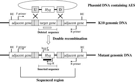
After 8 weeks of incubation on selective agar containing Hyg, each Hygr colony was recultured on new selective agar containing Hyg alone or Hyg plus Kan to expand bacterial cultures for subsequent analyses. After a colony was recultured, the correct structure of the disrupted gene in the colony was confirmed by PCR. For ΔrelAS and Δlsr2, each PCR was performed with a specific primer set binding the flanking regions of the homologous section because the sizes of the amplified fragments of the wild type and mutant are clearly distinguished by PCR (>1-kb difference) (Fig. 1 and Table 2). The following primer sets were used: for ΔrelAS, relL-3F (5′-TTCGGAGGTGAGCATCGTGG-3′) and relR-3R (5′-CCGACAACGGGTCCTGCTAC-3′); and for Δlsr2, lsrL-1F (5′-CCCCAATGTTGCAGACGC-3′) and lsrR-1R (5′TCACCCGCTCGATTTCCTT-3′). For ΔpknG and ΔrelAL, correct construction of each side was confirmed separately with site-specific primer sets because the sizes of PCR fragments obtained with a primer set binding the flanking regions of the homologous section were not well distinguished for the mutant and the wild type (178-bp difference) (Fig. 1 and Table 2). Each primer set was designed so that one primer bound within the hyg gene and the other primer bound up- or downstream of the homologous region (Fig. 1). The following primer sets were used: for the left side of ΔpknG, pknL-1F (5′-ACCAGAACTGCGACCTGACGG-3′) and hyg-R (5′-GCCCTACCTGGTGATGAGCC-3′); for the right side of ΔpknG, hyg-F (5′-CACGAAGATGTTGGTCCCGT-3′) and pknR-1R (5′-TCCACCACAACACTCGTGCC-3′); for the left side of ΔrelAL, relL-1F (5′-CAGGTGGACAACGCGATCG-3′) and hyg-R; and for the right side of ΔrelAL, hygF and relR2R (5′-TGCGTCGTTGATGAGGGTT-3′). For further confirmation, a sequencing analysis was performed with one or two isolates from the M. avium subsp. paratuberculosis K10 and M. avium subsp. paratuberculosis K10-GFP mutant groups. Transduction frequencies were calculated as follows: (X − Y)/Z, where X is the number of Hygr colonies obtained, Y is the number of spontaneous Hygr colonies from control cells which received no phage, and Z is the number of input cells for each experiment. The allelic exchange frequency was calculated by determining the percentage of allelic exchange in the population of Hygr colonies (4).
FIG. 1.
Schematic diagram of allelic exchange mutagenesis in M. avium subsp. paratuberculosis. The inserted sequence containing the Hyg gene is the same size in all mutants (1,915 bp), but the size of the deleted sequence varies in the mutants developed in this study (ΔpknG and ΔrelAL, 1,737 bp; ΔrelAS, 873 bp; Δlsr2, 311 bp [Table 2]). Arrows indicate the schematic binding sites and the directions of primers used for PCR identification. The F and R primers are the primers designed to bind outside up- and downstream homologous regions in each mutant. PCRs for ΔrelAS and Δlsr2 were performed with a primer set consisting of F and R primers, because the PCR fragments for the mutant and wild-type strains were clearly distinguished (>1-kb difference). PCRs for ΔpknG and ΔrelAL were performed with specific primer sets consisting of both F and hyg-R primers and hyg-F and R primers because the size differences between the mutant and wild-type strains with the F and R primers were not well distinguished in these cases (178-bp difference). The schematic restriction (RE) and probing (open and filled bars) sites for Southern blot analysis are also shown. Hyg, Hygr gene; U and D, up- and downstream homologous regions; RE, restriction enzyme site; open and filled bars, probes for the deleted gene and the hyg gene, respectively.
Expression analysis of disrupted M. avium subsp. paratuberculosis genes.
RNA expression of the disrupted gene was also checked by reverse transcription (RT)-PCR. Total RNA from 2 × 10⁹ cells of each strain was isolated using a FastRNA Pro Blue kit (Q-Biogene, Ohio) and treated two times with DNase I (Invitrogen, California). Five hundred nanograms of RNA was used for RT-PCR with a specific primer set for each gene using SuperScript One-Step RT-PCR systems (Invitrogen, California) according to the manufacturer's instructions. Negative (no RT) and positive (gapDH gene) (19) controls were also included in each RT-PCR.
Southern blot analyses.
Southern blot analyses were performed to show that the allelic exchange occurred at the correct position, as well as to show that illegitimate recombination at another site did not occur. DNA was extracted from each strain as previously described (23). Two micrograms of DNA from each strain was digested with an appropriate restriction enzyme (BamHI for the ΔrelAL, ΔrelAS, Δlsr2, and K10 strains; ClaI for the ΔpknG and K10 strains). One microgram of digested DNA from a mutant or the wild type was electrophoresed on a 1% agarose gel and transferred to a Hybond-N+ membrane (Amersham, New Jersey) by the capillary method. Generation of digoxigenin-labeled probes for specific binding sites (Fig. 1), hybridization, and chemiluminescence detection were performed with a DIG-High Prime DNA labeling and detection starter kit II (Roche Applied Science, Germany) used according to the manufacturer's recommendations.
RESULTS
Disruption of pknG in M. avium subsp. paratuberculosis.
Using method A, which was based on a protocol developed for M. tuberculosis and M. bovis BCG (4), M. avium subsp. paratuberculosis K10 and M. avium subsp. paratuberculosis K10-GFP were infected with a specialized transducing phage carrying AES for pknG or relAS. More than 1,000 colonies were visible after 8 weeks of incubation in each of the cultures of M. avium subsp. paratuberculosis and M. avium subsp. paratuberculosis K10-GFP transduced with the pknG or relAS AES. Screening of 300 colonies from each experiment by PCR revealed there was a high level of spontaneous Hygr. This initial trial yielded only seven ΔpknG mutants each for M. avium subsp. paratuberculosis K10 and M. avium subsp. paratuberculosis K10-GFP. No mutants were detected when cultures of M. avium subsp. paratuberculosis K10 and M. avium subsp. paratuberculosis K10-GFP transduced with the relAS AES were screened (Table 3). Furthermore, no mutants were detected in two additional experiments with the relAS AES (data not shown). Because the size of the inserted sequence was similar to the size of the deleted sequence in ΔpknG (1,915 bp versus 1,737 bp) but greater than the size of the deleted sequence in ΔrelAS (1,915 bp versus 873 bp), we performed an experiment to determine whether the sizes of inserted and deleted sequences at the recombination locus might interfere with the efficiency of allelic exchange. Another transducing phage carrying an AES for the relA deletion (relAL) was designed to delete 1,737 bp in the relA locus and tested using the same method. However, no mutants were detected when 150 colonies each of M. avium subsp. paratuberculosis K10 and M. avium subsp. paratuberculosis K10 GFP transduced with the relAL AES were screened (Table 3).
TABLE 3.
Efficiency of allelic exchange in M. avium subsp. paratuberculosis
| Host strain | Genotypea | Methodb | No. of allelic exchanges/no. of tested Hygr mutants (%)d | Total no. of Hygr mutants | Transduction frequency |
|---|---|---|---|---|---|
| M. avium subsp. paratuberculosis K10 | ΔpknG | A | 7/300 (2.3) | NAe | NA |
| ΔrelAS | A | 0/300 (0.0) | NA | NA | |
| ΔrelAL | A | 0/150 (0.0) | NA | NA | |
| ΔrelAS | Bc | 2/35 (5.7) | 35 | 1.1 × 10⁻⁸ | |
| ΔrelAL | B | 48/50 (96.0) | 291 | 1.1 × 10⁻⁷ | |
| Δlsr2 | B | 50/50 (100.0) | 738 | 2.9 × 10⁻⁷ | |
| M. avium subsp. paratuberculosis K10-GFP | ΔpknG | A | 7/300 (2.3) | NA | NA |
| ΔrelAS | A | 0/300 (0.0) | NA | NA | |
| ΔrelAL | A | 0/150 (0.0) | NA | NA | |
| ΔrelAS | B | 33/35 (94.3) | 499 | 2.0 × 10⁻⁷ | |
| ΔrelAL | B | 39/50 (78.0) | 448 | 1.8 × 10⁻⁷ | |
| Δlsr2 | B | 50/50 (100.0) | 438 | 1.7 × 10⁻⁷ |
S and L after relA indicate a small sequence deletion and a large sequence deletion at the relA gene locus, respectively.
Method A was used in the first trial, and method B was used in the second trial. For detailed information, see Materials and Methods.
The difference from other method B experiments was that M. avium subsp. paratuberculosis was washed with MP buffer to remove residual Tween 80 before absorption of phage. For detailed information, see Materials and Methods.
The values in the parentheses are the allelic exchange frequencies.
NA, not available.
Although some mutants were generated in the ΔpknG experiments, the frequency of allelic exchange compared to the frequencies for M. tuberculosis and M. bovis (4) was very low (0 to 2.3% versus 90 to 100%). These findings underscored the difficulties encountered when working with M. avium subsp. paratuberculosis and suggested that the methodology would have to be modified to use this system of transduction as a routine laboratory procedure.
Efficiency of allelic exchange mutagenesis in M. avium subsp. paratuberculosis by specialized transduction.
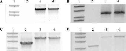
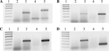
After a high rate of spontaneous Hygr was observed in the first trial with method A, the procedure was modified to determine if the efficiency of allelic exchange could be increased by modifying the method. With method B, in which the procedure was modified to decrease spontaneous Hygr as described above, 35 to 500 Hygr colonies were obtained in the relA deletion experiment after 8 weeks of incubation. Colonies having each type of targeted gene deletion were transferred onto new agar plates containing Hyg or Hyg plus Kan. The mutant colonies were first identified by PCR using locus-specific primers (Fig. 2). Then the correct position of allelic exchange was confirmed by performing a sequencing analysis of one or two mutant isolates from each mutant group (M. avium subsp. paratuberculosis K10 and M. avium subsp. paratuberculosis K10-GFP) (data not shown). In addition, the lack of RNA expression for deleted genes was also confirmed by RT-PCR using two isolates from each mutant group (M. avium subsp. paratuberculosis K10 and M. avium subsp. paratuberculosis K10-GFP) (Fig. 3). Both target genes were expressed in the control strain (M. avium subsp. paratuberculosis K10), but they were absent in the corresponding gene deletion mutants. Furthermore, these results were confirmed by Southern blot analysis, as shown in Fig. 4. Southern blotting with a hyg gene-specific probe for a mutant, as well as a target gene-specific probe for the wild type, revealed single hybridization bands at the expected sizes, which indicated that the target gene was deleted and replaced with the inserted fragment containing the hyg gene. Compared with the allelic exchange frequencies obtained in first trial using method A, the allelic exchange frequencies in the second trial using method B were greatly increased (from 0 to 2.3% to 78 to 96%) (Table 3). Compared to incubation in 7H9 broth medium without Tween 80, washing with MP buffer to remove the residual Tween 80 resulted in decreases in the allelic exchange frequency and the transduction frequency (Table 3). Contrary to our assumption about the sizes of the deletions and inserts with allelic exchange, the size of the deletions in relA did not have a significant effect on the frequency of mutants generated with method B (873-bp deletion versus 1,915-bp insertion and 1,737-bp deletion versus 1,915-bp insertion) (Tables 2 and 3) in this study.
FIG. 2.
PCR identification for specific gene construction in mutants. (A) PCR for ΔpknG; (B) PCR for ΔrelAL; (C) PCR for ΔrelAS; (D) PCR for Δlsr2. Lane 1, DNA size marker; lane 2, wild type (M. avium subsp. paratuberculosis K10); lane 3, mutant in M. avium subsp. paratuberculosis K10; lane 4, mutant in M. avium subsp. paratuberculosis K10-GFP. The primer sites for ΔpknG (A) and ΔrelAL (B) PCRs were located in the Hyg gene (inserted gene) for the forward primer and outside the downstream homologous region of each disrupted gene for the reverse primer. Note that the wild-type gene was not amplified in panels A and B because of the primer design. The primer sites for ΔrelAS (C) and Δlsr2 (D) PCRs were located outside up- and downstream homologous regions of each disrupted gene, which allowed identification of mutants based on the sizes of the amplified fragments.
FIG. 3.
RT-PCR analysis of gene expression in M. avium subsp. paratuberculosis strains. (A) RT-PCR for pknG (380 bp); (B and C) RT-PCR for relA from ΔrelAL (303 bp) (B) and ΔrelAS (303 bp) (C) mutants; (D) RT-PCR for lsr2 (145 bp). Lane 1, DNA marker; lanes 2 and 3, negative (without RT) and positive controls (gapDH) for RT-PCR, respectively; lanes 4 and 5, target gene expression in mutant and wild-type (M. avium subsp. paratuberculosis K10) strains, respectively. Negative and positive controls for RT-PCR of M. avium subsp. paratuberculosis K10 RNA were also analyzed (data not shown).
FIG. 4.
Southern blot analysis of genomic DNA from mutant and wild-type strains. (A) Southern blot characterization for ΔpknG; (B) Southern blot characterization for ΔrelAS and ΔrelAL; (C) Southern blot characterization for Δlsr2. The restriction and probing sites are shown in Fig. 1. The membrane was hybridized with the first probe (hyg probe). After chemiluminescence detection, the first probe was stripped out, and the membrane was reprobed with the second probe (probe for the deleted gene) and examined. All bands are consistent with the expected sizes.
To test whether the optimized method (method B) works well with additional gene deletions, a third gene, lsr2, was selected for disruption. Lsr2 is a cytosolic protein implicated in cell wall lipid synthesis, which has an important role in colony morphology and biofilm formation in M. smegmatis (14). The confirmation method used for lsr2 deletion was exactly the same as the method described above (Fig. 2 and 3). As shown in Table 3, generation of Δlsr2 with method B showed that there was 100% correlation of Hygr with successful allelic exchange. These data indicate that method B works equally well with additional genes.
We also compared the effects of recovery times between 0 and 24 h in three knockout experiments. For the ΔrelAL mutation in M. avium subsp. paratuberculosis K10-GFP and the Δlsr2 mutation in M. avium subsp. paratuberculosis K10, the total numbers of Hygr colonies were about twofold greater after 24 h of incubation in 7H9 broth medium before plating, which is consistent with one replication cycle of M. avium subsp. paratuberculosis (24 to 48 h). However, for the ΔrelAL mutation in M. avium subsp. paratuberculosis K10, the number of Hygr colonies decreased after 24 h of incubation. In contrast to previous findings with M. bovis BCG (4), which showed the highest allelic exchange frequency with a recovery time of 24 h, the allelic exchange frequencies in each experiment were virtually the same with and without the recovery time in the present study (Table 4).
TABLE 4.
Effect of recovery time on the efficiency of allelic exchange mutagenesis
| Recovery time (h) | ΔrelAL K10
|
ΔrelAL K10-GFP
|
Δlsr2K10
|
|||
|---|---|---|---|---|---|---|
| No. of ΔrelAL mutants/no. of Hygr mutants (%)a | Total no. of Hygr mutants | No. of ΔrelAL mutants/no. of Hygr mutants (%)a | Total no. of Hygr mutants | No. of Δlsr2 mutants/no. of Hygr mutants (%)a | Total no. of Hygr mutants | |
| 0 | 50/50 (100) | 656 | 39/50 (78) | 266 | 50/50 (100) | 402 |
| 24 | 48/50 (96) | 291 | 42/50 (84) | 448 | 50/50 (100) | 738 |
Values in parentheses are the allelic exchange frequencies.
Generation of GFP-tagged mutants of M. avium subsp. paratuberculosis.
Expression of GFP in M. avium subsp. paratuberculosis is variable, and only a few transformants express high GFP levels (32). Thus, to construct GFP-tagged mutants with equivalent high fluorescence levels, it may be useful to carry out allelic exchange directly with an M. avium subsp. paratuberculosis host with optimal GFP expression, such as our strain M. avium subsp. paratuberculosis K10-GFP. The data show that allelic exchange mutagenesis occurred in M. avium subsp. paratuberculosis K10-GFP at the same rate as it occurred in M. avium subsp. paratuberculosis K10 (Tables 3 and 4). Every 10 isolates of each mutant made from M. avium subsp. paratuberculosis K10-GFP, except ΔpknG (7 isolates), were examined by fluorescence microscopy for the presence of GFP. Even after extensive incubation without antibiotic pressure for the GFP plasmid (Kan), some mutant strains still expressed GFP. The percentages of GFP-expressing mutants in the colonies examined ranged from 20% (Δlsr2) to 100% (ΔrelAS). We also examined the potential of the GFP-tagged mutants to be a useful tool for tracing the mutants within bovine macrophages after infection. The presence of GFP-expressing mutants in macrophages was clearly detected by fluorescence microscopy (data not shown).
DISCUSSION
Allelic exchange mutagenesis using specialized transduction has been used successfully with some slow-growing mycobacteria, including M. tuberculosis, M. bovis, and M. avium (4, 30). However, successful use of this technology has not been reported previously for M. avium subsp. paratuberculosis. We explored the use of this approach to develop targeted gene disruptions in M. avium subsp. paratuberculosis, one of the slowest-growing mycobacterial species, whose generation time is 24 h or longer (34). The ability to obtain directed gene knockouts in M. avium subsp. paratuberculosis is a major breakthrough in Johne's disease research. Results from sequencing the M. avium subsp. paratuberculosis genome have shown that 41.6% of the annotated genes in the genome are unknown or hypothetical open reading frames (27). Only through specific gene disruptions can potential phenotypes be assigned to these unknown genes. Rational design and construction of attenuated mutants as possible vaccines are now within reach. Persistence within host macrophages is a key feature of mycobacterial pathogenesis that needs to be understood further. By selectively disrupting pknG and relA by allelic exchange, we took an important step in this direction as both pknG and relA have been shown to be key virulence determinants in M. tuberculosis and M. bovis (17, 38). The ability to selectively disrupt genes in M. tuberculosis has already facilitated increases in our knowledge of specific gene functions in M. tuberculosis (2, 3, 7, 11, 16, 33).
In the first trial (method A), which was similar to a previous study of M. tuberculosis and M. bovis BCG (4), we obtained a high rate of spontaneous Hygr or illegitimate recombination. A previous study with M. avium produced similar results (30). To overcome the very low efficiency of allelic exchange in that study, the authors used an M. avium leuD deletion mutant as a genetic host with the Streptomyces clelicolor ledD gene as a selective marker. In contrast, we obtained a high efficiency of allelic exchange in M. avium subsp. paratuberculosis (allelic exchange frequency, up to 100%; transduction frequency, 2.9 × 10⁻⁷) (Table 3) after modifying the original methodology (method B). Importantly, the successful optimization of the method should now allow this tool to be used routinely to generate directed gene deletions in an isogenic virulent strain of M. avium subsp. paratuberculosis. To obtain the very efficient allelic exchange in the second trial, we reduced the chance that clumped bacteria would be plated on the selective agar by consecutive gravity sedimentations and by use of a low g force in the centrifugation steps. To disrupt cell clumps, other mechanical methods might be used, such as passage through a syringe or sonication. However, these physical disruptions might cause damage to the cells, which might decrease the viability of transduced bacteria on Hyg-containing medium. In addition, we also increased the concentration of Hyg from 50 to 75 μg/ml in the second trial and did not use the drug concentration typically used for other mycobacteria (23, 30). When the concentration of Hyg was increased to 75 μg/ml, the rate of spontaneous Hygr was greatly diminished. When 50 μg/ml Hyg was used, many spontaneous Hygr mutants were generated in the experiments with M. avium (30) and M. avium subsp. paratuberculosis (Table 3). In contrast, 75 μg/ml Hyg provided excellent selective pressure for isolating mutant colonies of M. tuberculosis, M. bovis (4), and M. avium subsp. paratuberculosis in this study (Table 3). The effects of gravity sedimentation, a lower g force in centrifugation (3,700 × g versus 12,000 × g), and a higher concentration of Hyg (75 μg/ml versus 50 μg/ml), which was assumed to contribute to the high efficiency of allelic exchange in method B, were evaluated separately. Each procedure had a significant effect, reducing the number of spontaneous Hygr colonies by 50 to 90% (data not shown), thus supporting our hypothesis.
All transduction frequencies obtained with method B, except the transduction frequency for ΔrelAL in M. avium subsp. paratuberculosis K10, were calculated to be between 1.1 × 10⁻⁷ and 2.9 × 10⁻⁷ per recipient cell, and these values were similar to the values obtained in previous studies of transposon mutagenesis in M. avium subsp. paratuberculosis by specialized transduction (21). However, we estimated that the concentration of cells at an OD600 between 0.5 and 0.6 was 5 × 10⁸ to 6 × 10⁸ CFU/ml based on the results of CFU counting in our lab, while in other studies of transposon mutagenesis of M. avium subsp. paratuberculosis the workers interpreted similar optical density values as indicating that the concentration was 1.5 × 10⁸ to 2.0 × 10⁸ CFU/ml (12, 21, 35). If we had used a concentration of 2.0 × 10⁸ CFU/ml for recipient cells, the calculated transduction frequencies in this study would have been three times greater. In contrast to the previous findings for M. bovis BCG (4), the recovery time for transduced M. avium subsp. paratuberculosis with specialized transducing mycobacteriophage did not have much effect on the allelic exchange frequency in the current study (Table 4). This suggests that the recovery time is not a critical factor for achieving a high efficiency of allelic exchange.
One novel benefit of present study was the ability to create defined mutants in a GFP-expressing strain of M. avium subsp. paratuberculosis K10. This feature should enable easy tracking of mutants in a variety of downstream assays, including infection of macrophages, as shown in this study. We demonstrated that the efficiencies of allelic exchange in M. avium subsp. paratuberculosis K10-GFP mutants were similar to those in wild-type M. avium subsp. paratuberculosis strain K10 (Tables 3 and 4), and some of these mutants still expressed GFP after lengthy incubation without selective pressure for the GFP plasmid. Importantly, the pWES4 plasmid (the plasmid encoding GFP in M. avium subsp. paratuberculosis K10-GFP) introduced into M. avium did not alter bacterial virulence (32). Therefore, it is evident that M. avium subsp. paratuberculosis mutants containing GFP would have an advantage in investigations of the functions of deleted genes in host cells. In addition, GFP can be a potential antigenic marker for differentiation between wild-type and potential vaccine strains used as a live attenuated vaccine. By making GFP-expressing mutants from the parent strain M. avium subsp. paratuberculosis K10-GFP, we can save at least several months which would be required to introduce the GFP plasmid into mutant strains for this purpose. Moreover, we maximized the chance that mutants would express optimal GFP fluorescence like that in the original M. avium subsp. paratuberculosis K10-GFP host.
Lsr2 has been shown to be a cytosolic protein related to lipid biosynthesis in the cell wall and antibiotic resistance in M. smegmatis (14, 15). Deletion of the lsr2 gene in M. smegmatis resulted in an alteration in colony morphology (generation of smooth colonies) and an alteration in biofilm and pellicle formation. The lsr2 mutant of M. avium subsp. paratuberculosis generated in this study also had a distinctive smooth-colony morphology and was defective in pellicle formation (unpublished data), which indicates that the role of lsr2 in M. avium subsp. paratuberculosis is also related to lipid biosynthesis of cell wall components. The lipid-rich cell wall is a distinctive feature of mycobacteria. The cell wall components of M. tuberculosis have been shown to contribute to the virulence of this organism and modulate the host immune response (16, 37). Therefore, lsr2 in pathogenic mycobacteria may be related to virulence. Indeed, deletion of lsr2, as well as deletion of pknG and relA, resulted in a reduction in survival in bovine macrophages (unpublished data). Recently, Colangeli et al. proposed that lsr2 may be an essential gene in M. tuberculosis since they were unable to delete the gene in their study (15). Whether this is true or not, lsr2 can be deleted in M. avium subsp. paratuberculosis, one of the slow-growing mycobacterial species.
In conclusion, we established an efficient allelic exchange mutagenesis system for M. avium subsp. paratuberculosis by generating three different targeted gene disruptions, one of which involved two deletions that were different sizes (relA), in M. avium subsp. paratuberculosis K10 and M. avium subsp. paratuberculosis K10-GFP. The three genes are assumed to have important roles in the virulence of M. avium subsp. paratuberculosis, as they do in other mycobacterial species. Along with the recently completed genome sequence (27) and a random transposon mutagenesis system for M. avium subsp. paratuberculosis (12, 21, 35), this tool should allow us to gain more insight into pathogenesis and further efforts to develop an effective vaccine for M. avium subsp. paratuberculosis.
Acknowledgments
This study was supported in part by the Washington State Monoclonal Antibody Center, the Johne's Disease Integrated Program funded by the Animal Biosecurity program of USDA-CSREES National Research Initiative 2004-356051-14243, USDA-APHIS grants 03-9100-0788-GR and 03-9100-07-GR, and USDA Animal Health intramural grant WNV-00150.
Footnotes
Published ahead of print on 11 January 2008.
REFERENCES
- 1.Aldovini, A., R. N. Husson, and R. A. Young. 1993. The uraA locus and homologous recombination in Mycobacterium bovis BCG. J. Bacteriol. 175:7282-7289. [DOI] [PMC free article] [PubMed] [Google Scholar]
- 2.Azad, A. K., T. D. Sirakova, L. M. Rogers, and P. E. Kolattukudy. 1996. Targeted replacement of the mycocerosic acid synthase gene in Mycobacterium bovis BCG produces a mutant that lacks mycosides. Proc. Natl. Acad. Sci. USA 93:4787-4792. [DOI] [PMC free article] [PubMed] [Google Scholar]
- 3.Balasubramanian, V., M. S. Pavelka, Jr., S. S. Bardarov, J. Martin, T. R. Weisbrod, R. A. McAdam, B. R. Bloom, and W. R. Jacobs, Jr. 1996. Allelic exchange in Mycobacterium tuberculosis with long linear recombination substrates. J. Bacteriol. 178:273-279. [DOI] [PMC free article] [PubMed] [Google Scholar]
- 4.Bardarov, S., S. Bardarov, Jr., M. S. Pavelka, Jr., V. Sambandamurthy, M. Larsen, J. Tufariello, J. Chan, G. Hatfull, and W. R. Jacobs, Jr. 2002. Specialized transduction: an efficient method for generating marked and unmarked targeted gene disruptions in Mycobacterium tuberculosis, M. bovis BCG and M. smegmatis. Microbiology 148:3007-3017. [DOI] [PubMed] [Google Scholar]
- 5.Bardarov, S., J. Kriakov, C. Carriere, S. Yu, C. Vaamonde, R. A. McAdam, B. R. Bloom, G. F. Hatfull, and W. R. Jacobs, Jr. 1997. Conditionally replicating mycobacteriophages: a system for transposon delivery to Mycobacterium tuberculosis. Proc. Natl. Acad. Sci. USA 94:10961-10966. [DOI] [PMC free article] [PubMed] [Google Scholar]
- 6.Benedictus, G., A. A. Dijkhuizen, and J. Stelwagen. 1987. Economic losses due to paratuberculosis in dairy cattle. Vet. Rec. 121:142-146. [DOI] [PubMed] [Google Scholar]
- 7.Berthet, F. X., M. Lagranderie, P. Gounon, C. Laurent-Winter, D. Ensergueix, P. Chavarot, F. Thouron, E. Maranghi, V. Pelicic, D. Portnoi, G. Marchal, and B. Gicquel. 1998. Attenuation of virulence by disruption of the Mycobacterium tuberculosis erp gene. Science 282:759-762. [DOI] [PubMed] [Google Scholar]
- 8.Boshoff, H. I., and V. Mizrahi. 2000. Expression of Mycobacterium smegmatis pyrazinamidase in Mycobacterium tuberculosis confers hypersensitivity to pyrazinamide and related amides. J. Bacteriol. 182:5479-5485. [DOI] [PMC free article] [PubMed] [Google Scholar]
- 9.Braunstein, M., S. S. Bardarov, and W. R. Jacobs, Jr. 2002. Genetic methods for deciphering virulence determinants of Mycobacterium tuberculosis. Methods Enzymol. 358:67-99. [DOI] [PubMed] [Google Scholar]
- 10.Braunstein, M., A. M. Brown, S. Kurtz, and W. R. Jacobs, Jr. 2001. Two nonredundant SecA homologues function in mycobacteria. J. Bacteriol. 183:6979-6990. [DOI] [PMC free article] [PubMed] [Google Scholar]
- 11.Camacho, L. R., D. Ensergueix, E. Perez, B. Gicquel, and C. Guilhot. 1999. Identification of a virulence gene cluster of Mycobacterium tuberculosis by signature-tagged transposon mutagenesis. Mol. Microbiol. 34:257-267. [DOI] [PubMed] [Google Scholar]
- 12.Cavaignac, S. M., S. J. White, G. W. de Lisle, and D. M. Collins. 2000. Construction and screening of Mycobacterium paratuberculosis insertional mutant libraries. Arch. Microbiol. 173:229-231. [DOI] [PubMed] [Google Scholar]
- 13.Chacon, O., L. E. Bermudez, and R. G. Barletta. 2004. Johne's disease, inflammatory bowel disease, and Mycobacterium paratuberculosis. Annu. Rev. Microbiol. 58:329-363. [DOI] [PubMed] [Google Scholar]
- 14.Chen, J. M., G. J. German, D. C. Alexander, H. Ren, T. Tan, and J. Liu. 2006. Roles of Lsr2 in colony morphology and biofilm formation of Mycobacterium smegmatis. J. Bacteriol. 188:633-641. [DOI] [PMC free article] [PubMed] [Google Scholar]
- 15.Colangeli, R., D. Helb, C. Vilcheze, M. H. Hazbon, C. G. Lee, H. Safi, B. Sayers, I. Sardone, M. B. Jones, R. D. Fleischmann, S. N. Peterson, W. R. Jacobs, Jr., and D. Alland. 2007. Transcriptional regulation of multi-drug tolerance and antibiotic-induced responses by the histone-like protein Lsr2 in M. tuberculosis. PLoS Pathog. 3:e87. [DOI] [PMC free article] [PubMed] [Google Scholar]
- 16.Cox, J. S., B. Chen, M. McNeil, and W. R. Jacobs, Jr. 1999. Complex lipid determines tissue-specific replication of Mycobacterium tuberculosis in mice. Nature 402:79-83. [DOI] [PubMed] [Google Scholar]
- 17.Dahl, J. L., C. N. Kraus, H. I. Boshoff, B. Doan, K. Foley, D. Avarbock, G. Kaplan, V. Mizrahi, H. Rubin, and C. E. Barry III. 2003. The role of RelMtb-mediated adaptation to stationary phase in long-term persistence of Mycobacterium tuberculosis in mice. Proc. Natl. Acad. Sci. USA 100:10026-10031. [DOI] [PMC free article] [PubMed] [Google Scholar]
- 18.Foley-Thomas, E. M., D. L. Whipple, L. E. Bermudez, and R. G. Barletta. 1995. Phage infection, transfection and transformation of Mycobacterium avium complex and Mycobacterium paratuberculosis. Microbiology 141:1173-1181. [DOI] [PubMed] [Google Scholar]
- 19.Granger, K., R. J. Moore, J. K. Davies, J. A. Vaughan, P. L. Stiles, D. J. Stewart, and M. L. Tizard. 2004. Recovery of Mycobacterium avium subspecies paratuberculosis from the natural host for the extraction and analysis in vivo-derived RNA. J. Microbiol. Methods 57:241-249. [DOI] [PubMed] [Google Scholar]
- 20.Harris, N. B., and R. G. Barletta. 2001. Mycobacterium avium subsp. paratuberculosis in veterinary medicine. Clin. Microbiol. Rev. 14:489-512. [DOI] [PMC free article] [PubMed] [Google Scholar]
- 21.Harris, N. B., Z. Feng, X. Liu, S. L. Cirillo, J. D. Cirillo, and R. G. Barletta. 1999. Development of a transposon mutagenesis system for Mycobacterium avium subsp. paratuberculosis. FEMS Microbiol. Lett. 175:21-26. [DOI] [PubMed] [Google Scholar]
- 22.Harris, N. B., D. K. Zinniel, M. K. Hsieh, J. D. Cirillo, and R. G. Barletta. 2002. Cell sorting of formalin-treated pathogenic Mycobacterium paratuberculosis expressing GFP. BioTechniques 32:522. 4:526-527. [DOI] [PubMed] [Google Scholar]
- 23.Hatfull, G. F., and W. R. Jacobs. 2000. Molecular genetics of mycobacteria. ASM Press, Washington, DC.
- 24.Johnson-Ifearulundu, Y. J., J. B. Kaneene, D. J. Sprecher, J. C. Gardiner, and J. W. Lloyd. 2000. The effect of subclinical Mycobacterium paratuberculosis infection on days open in Michigan, USA, dairy cows. Prev. Vet. Med. 46:171-181. [DOI] [PubMed] [Google Scholar]
- 25.Kalpana, G. V., B. R. Bloom, and W. R. Jacobs, Jr. 1991. Insertional mutagenesis and illegitimate recombination in mycobacteria. Proc. Natl. Acad. Sci. USA 88:5433-5437. [DOI] [PMC free article] [PubMed] [Google Scholar]
- 26.Knipfer, N., A. Seth, and T. E. Shrader. 1997. Unmarked gene integration into the chromosome of Mycobacterium smegmatis via precise replacement of the pyrF gene. Plasmid 37:129-140. [DOI] [PubMed] [Google Scholar]
- 27.Li, L., J. P. Bannantine, Q. Zhang, A. Amonsin, B. J. May, D. Alt, N. Banerji, S. Kanjilal, and V. Kapur. 2005. The complete genome sequence of Mycobacterium avium subspecies paratuberculosis. Proc. Natl. Acad. Sci. USA 102:12344-12349. [DOI] [PMC free article] [PubMed] [Google Scholar]
- 28.McFadden, J. 1996. Recombination in mycobacteria. Mol. Microbiol. 21:205-211. [DOI] [PubMed] [Google Scholar]
- 29.National Research Council (U.S.) Committee on Diagnosis and Control of Johne's Disease. 2003. Diagnosis and control of Johne's disease. National Academies Press, Washington, DC.
- 30.Otero, J., W. R. Jacobs, Jr., and M. S. Glickman. 2003. Efficient allelic exchange and transposon mutagenesis in Mycobacterium avium by specialized transduction. Appl. Environ. Microbiol. 69:5039-5044. [DOI] [PMC free article] [PubMed] [Google Scholar]
- 31.Ott, S. L., S. J. Wells, and B. A. Wagner. 1999. Herd-level economic losses associated with Johne's disease on US dairy operations. Prev. Vet. Med. 40:179-192. [DOI] [PubMed] [Google Scholar]
- 32.Parker, A. E., and L. E. Bermudez. 1997. Expression of the green fluorescent protein (GFP) in Mycobacterium avium as a tool to study the interaction between mycobacteria and host cells. Microb. Pathog. 22:193-198. [DOI] [PubMed] [Google Scholar]
- 33.Pavelka, M. S., Jr., and W. R. Jacobs, Jr. 1999. Comparison of the construction of unmarked deletion mutations in Mycobacterium smegmatis, Mycobacterium bovis bacillus Calmette-Guerin, and Mycobacterium tuberculosis H37Rv by allelic exchange. J. Bacteriol. 181:4780-4789. [DOI] [PMC free article] [PubMed] [Google Scholar]
- 34.Shin, S. J., J. H. Han, E. J. Manning, and M. T. Collins. 2007. Rapid and reliable method for quantification of Mycobacterium paratuberculosis by use of the BACTEC MGIT 960 system. J. Clin. Microbiol. 45:1941-1948. [DOI] [PMC free article] [PubMed] [Google Scholar]
- 35.Shin, S. J., C. W. Wu, H. Steinberg, and A. M. Talaat. 2006. Identification of novel virulence determinants in Mycobacterium paratuberculosis by screening a library of insertional mutants. Infect. Immun. 74:3825-3833. [DOI] [PMC free article] [PubMed] [Google Scholar]
- 36.Snapper, S. B., R. E. Melton, S. Mustafa, T. Kieser, and W. R. Jacobs, Jr. 1990. Isolation and characterization of efficient plasmid transformation mutants of Mycobacterium smegmatis. Mol. Microbiol. 4:1911-1919. [DOI] [PubMed] [Google Scholar]
- 37.Strohmeier, G. R., and M. J. Fenton. 1999. Roles of lipoarabinomannan in the pathogenesis of tuberculosis. Microbes Infect. 1:709-717. [DOI] [PubMed] [Google Scholar]
- 38.Walburger, A., A. Koul, G. Ferrari, L. Nguyen, C. Prescianotto-Baschong, K. Huygen, B. Klebl, C. Thompson, G. Bacher, and J. Pieters. 2004. Protein kinase G from pathogenic mycobacteria promotes survival within macrophages. Science 304:1800-1804. [DOI] [PubMed] [Google Scholar]


