Abstract
Mechanical transmission of porcine reproductive and respiratory syndrome virus (PRRSV) throughout a coordinated sequence of events that replicated common farm worker behavior during warm weather (10°C to 16°C) was assessed using a field-based model. The model involved fomites (boots and containers), vehicle sanitation, transport, and personnel movement. In a previous study, the model successfully demonstrated mechanical transmission of PRRSV in 8 out of 10 replicates during cold weather. A field strain of PRRSV was inoculated into carriers consisting of soil samples, which were adhered to the undercarriage of a vehicle. The vehicle was driven approximately 50 km to a commercial truck washing facility where the driver's boots contacted the carriers during washing, introducing the virus to the vehicle interior. The vehicle was then driven 50 km to a simulated farm site, and the driver's boots mechanically spread virus into the farm anteroom. Types of containers frequently employed in swine farms contacted drippings from the footwear on the anteroom floor. The truck wash floor, vehicle cab floor mats, boot soles, anteroom floor, and the ventral surface of containers were sampled to track the virus throughout the model. Ten replicates were conducted, along with sham-inoculated controls, and control replicates. In 2 replicates, infectious PRRSV was detected on the anteroom floor and in 1 replicate, infectious PRRSV was detected on the surface of the container by swine bioassay. All sham-inoculated controls and protocol controls were negative. These results indicate that mechanical transmission of PRRSV throughout a coordinated sequence of events in warm weather can occur, but in contrast to data from studies conducted during cold weather, it appears to be a relatively infrequent event.
Introduction
A thorough understanding of the routes of transmission of porcine reproductive and respiratory syndrome virus (PRRSV) is critical for the successful control and eradication of the disease. Known routes of PRRSV transmission between swine farms include infected pigs and semen, contaminated fomites (boots, coveralls, needles), mosquitoes, and houseflies (1,2,3,4,5,6). Recently, a field model was developed to test the mechanical transmission of PRRSV onto a simulated farm setting through a coordinated sequence of events during cold weather (7). Under the conditions of the study, mechanical transmission of PRRSV during cold weather was a frequent event and occurred in 8 out of 10 replicates (7). Furthermore, viable PRRSV was detected at multiple sampling points throughout the model; including the floor of a vehicle washing facility, transport vehicle interior, soles of boots of study personnel, the anteroom floor of a simulated farm, and the ventral surfaces of containers frequently observed in swine farms (7). Therefore, it appeared that mechanical transmission of PRRSV during cold weather is a frequent event.
However, not all farms are located in regions of the world that have a winter season. Furthermore, areas such as the midwest USA experience the warm, wet weather of the season of spring. In the west central Minnesota, during the months of March, April, and May, the mean daytime temperatures during the spring season range from −8°C to 19°C with relative humidity levels between 75 and 95% (National Weather Service, personal communication, April 17, 2002). While PRRSV is readily inactivated by drying and exposure to temperatures of 56°C, it can remain infectious for 30 d at 4°C, 1 to 6 d at temperatures of 20°C, and PRRSV can remain viable for 9 to 11 d when kept moist (8,9). It was speculated that because the environmental conditions during these 3 mo were not excessively hot or dry, PRRSV could survive outside of the host for a short period of time. Therefore, the purpose of the present study was to modify the existing field model to assess mechanical transmission of PRRSV throughout a coordinated sequence of events during periods of warm weather, and compare these findings to those previously reported in the cold weather study. For the purpose of this study, warm weather was defined as an environmental temperature ranging between 10°C and 20°C, during the time the replicates were conducted. It was hypothesized that while mechanical transmission of PRRSV may occur during warm weather, it was not a frequent event.
Materials and methods
Assumptions and observations
As in the cold weather study, the model was based on a set of assumptions and field observations that were modified for warm weather (7). The principal investigator had made the observations when visiting modern commercial swine enterprises during the period of 1987 to 2001. The assumptions were as follows: 1) during periods of warm weather (10°C to 20°C) PRRSV can survive outside of the host for extended periods, enhancing mechanical transmission from site to site; 2) during periods of warm weather, livestock transport vehicles, veterinary vehicles, and other fomites (such as boots), can contact PRRSV at potentially contaminated points, such as infected farms, commercial truck washes, or slaughterhouses; and 3) the introduction of PRRSV-contaminated fomites into the farm office results in infection of the animal population.
The observations were as follows: 1) during periods of warm weather, the floor surface in the entryway (anteroom) to farms that employ shower-in and shower-out facilities is frequently dirty, due to accumulation of soil from the footwear of personnel and visitors; 2) miscellaneous shipments (animal health products, doses of semen, tools, food items for farm personnel) enter swine farms on a daily basis and temporarily reside on the soiled anteroom floor prior to introduction into the animal airspace; and 3) contaminated items frequently enter the animal airspace without being disinfected.
Terminology
The specific components of the model were defined as follows:
Carrier — A carrier was defined as a medium that enhanced the survivability of PRRSV outside of the host and assisted in its mechanical spread between sites. Since the study was to be conducted during the springtime, soil samples were selected as the material of choice for the construction of carriers.
Vehicle — To transport the carrier between sites, a motorized vehicle (Ford Explorer XLT 1997) was used. This vehicle had served as the principal investigator's mode of transportation to swine farms over the previous 3 y. The chosen point to attach the carrier to the vehicle was the ventral surface of the fender immediately dorsal to the vehicle's wheels, hereafter known as the “wheel well.”
Contamination point — The purpose of the contamination site was to serve as the point that the study personnel contacted the contaminated carriers during the process of cleaning the vehicle. A commercial truck wash located in rural Minnesota was selected to serve in this capacity. The site had the capability of providing hot water (46°C) at a rate of 15 L/min and soap detergent (Envirox G; Dorsey Lever, Coon Rapids, Minnesota, USA).
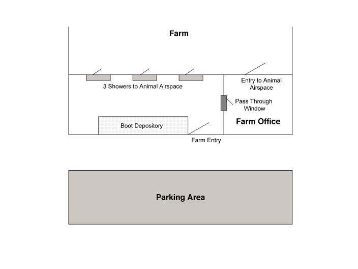
Anteroom — The anteroom was defined as the area encountered immediately upon entering the front doorway of a swine farm that employs a shower-in and shower-out procedure. The purpose of the anteroom is to provide personnel and visitors with a place to store coats and footwear prior to entering the shower-in facility (Figure 1). Since this type of study was far too risky to conduct on a commercial swine operation, it was necessary to simulate the entryway or “anteroom” of a farm. To enhance the safety of the study, the personal residence of the principal investigator was used. Specifically, the study employed the garage and a lavatory facility within the investigator's residence. The garage contained a 5-metre concrete walkway that led to the lavatory. The lavatory was previously used as a sanitation area for the principal investigator following visits to swine farms. Its design was similar to an actual farm anteroom, and included a shower, a sink, and linoleum floor covering.
Figure 1. Layout of anteroom in a swine farm employing a shower-in and shower-out facility.
Transfer point — The transfer point was defined as a designated area (0.5 m2 in size) of floor space located in the anteroom, immediately to the left of the doorway. During the study, the dimensions of the transfer point were clearly delineated using blue tape, providing a defined area for the placement of boots following entry to the anteroom.
Fomites — Two sets of inanimate objects were selected as fomites, including boots used by personnel during the study, and a series of packages, hereafter defined as “containers.” The purpose of the boots (men's 25.4 cm outdoor pull-up boot, IC-820020; Cabela's, Sydney, New England, USA) was to serve as fomites for the potential mechanical transfer of PRRSV from the contamination site into the cab of the vehicle, and from the cab of the vehicle into the anteroom. Containers were defined as boxes or shipping parcels that were destined for entry into swine farms. Four different types of containers were selected for use in the study, including cardboard (representing shipments of swine pharmaceuticals or biologics), styrofoam (representing semen deliveries), metal (representing electrician's or plumber's toolboxes), and plastic (representing lunch pails of farm personnel).
Experimental design
Study personnel and construction of carriers — The study consisted of 10 replicates, all conducted by the principal investigator. All laboratory testing took place at the University of Minnesota Veterinary Diagnostic Laboratory. For construction of the carriers, soil was obtained from the garden at the principal investigator's primary residence at the start of each sampling day. The soil was manually compressed into the shape of a sphere, averaging 21.5 cm in circumference, weighing approximately 225 g, with a core temperature of 9°C. On each day that a series of replicates were conducted, a 450 g sample of soil used to construct carriers was submitted for textural analysis, calculation of pH, and percent moisture at the University of Minnesota Soil Analysis Laboratory in St. Paul, Minnesota, USA (10,11).
Inoculation and attachment of carriers — A field strain of PRRSV (MN-30100) was used throughout the study to inoculate carriers. This PRRSV isolate had been previously recovered from a chronically infected sow following an acute outbreak of PRRSV within a commercial swine system. Prior to use in this study, the isolate had been passaged 1 time in a PRRSV-naïve pig, and recovered from lymphoid tissues that were placed on MARC-145 cells (12). For inoculation of carriers, 1 mL (104.4 TCID 50/mL) of PRRSV MN-30100 was injected into the center of each carrier using a 3-mL syringe (Monoject, St Louis, Missouri, USA). Sediment present in the soil samples did not allow for the use of a needle and direct injection of the study isolate into the intact carrier. Therefore, the carrier was manually divided into half, the inoculum administered onto the inner surface of 1 section of the carrier, and the 2 halves manually compressed together to restore its original shape. Two PRRSV-inoculated carriers (virus positive carriers) and 2 sham-inoculated carriers (virus negative carriers) were used for each replicate, the sham-inoculated carriers receiving 1-mL of sterile minimum essential medium (MEM).
Attachment of both sets of carriers to the vehicle occurred at a neutral location, 50 km from the contamination site. The virus positive carriers were attached to the left front and left rear wheel wells, while the sham-inoculated carriers were attached to the right front and right rear wheel wells. Holding a carrier in a gloved hand, it was attached to the wheel well using gentle pressure in order to secure the carrier to a specific site on the vehicle. Sham-inoculated carriers were attached first, followed by virus positive carriers. For standardized placement of the carriers, a measurement was taken, originating at the most ventral point of the cranial edge of the left front or right front wheel wells extending 15.25-cm dorsally along the cranial section of the rim to the designated attachment point. The left rear and right rear carriers were attached in a similar manner; however, the measurement initiated from the ventral most point of the caudal edge of the rear wheel well, extending 15.25-cm dorsally along the caudal edge of the rim to the designated attachment point.
Transport of carrier to contamination site and the contact of carriers with boots — Following carrier attachment, the vehicle was driven 50 km to the contamination point, where it underwent the washing process. Personnel manually washed the vehicle, using a hand-held instrument, “washing wand,” that allowed for water to be directed at high pressure (15 L/min) to specific points on the vehicle's exterior. The washing process consisted of a 30-minute period in which the external surface of the vehicle was initially rinsed with 46°C water for 5 min. The washing wand was also extended manually to contact the undercarriage area and wheel wells. The top and all sides of the vehicle (including the wheel wells) were then covered with soap and hot water, using a 0.5% concentration of soap detergent, and the vehicle was allowed to soak for 15 min. It was then rinsed again with 46°C water (15 L/min) for a 10-minute period. During the washing process, water was directed on all 4 carriers displacing them onto the cement floor of the truck wash. The water and the carriers mixed together, liquefying the carrier, resulting in a pool of mud on the floor. At the end of the washing process, the principle investigator stepped into the mud pools and crushed any residual carrier material in the pool underfoot, allowing the ventral surface of the boots contact with the contaminated area. The principle investigator then immediately entered the cab of the vehicle and the boots contacted the rubber floor mat on the driver's side of the vehicle. Prior to leaving the contamination site, the truck wash floor was cleaned to avoid the buildup of residual PRRSV in the facility, not only for future replicates, but to minimize risk to other producers using the facility. To do this, the principle investigator removed the contaminated boots, placed them on the driver's side floor mat, donned a clean pair of shoes, exited the vehicle, and poured 1 L of 100% bleach on the truck wash floor. The floor was then washed with hot water and soap and all visible soil was removed via a flush gutter. To avoid contamination of controls, a sample (approximately 25%) of each sham-inoculated carrier was collected using a gloved hand. Personnel then removed their shoes, placed them in a plastic bag, donned their original boots, and traveled 50 km to the simulated farm site.
Mechanical transmission of PRRSV into anteroom and contact with containers — Upon arrival to the simulated farm, personnel exited the vehicle, walked 5 m across the concrete floor of the garage, and entered the anteroom. Upon contacting the transfer area, the principle investigator forcefully “stomped” both feet 2 times to displace soil from the soles of the boots and to allow soil to accumulate on the floor. The boots were then placed in the transfer area for 15 min, removed and the ventral surfaces of the 4 types of containers were placed in an upright position, allowing the ventral surface of the container to contact the soil for 5 s. The containers were then removed, the transfer point area disinfected using ammonia spray (Lysol; Reckitt Benckiser Wayne, New Jersey, USA), and dried with paper towels. The rubber floor mat from the driver's side of the vehicle cab was disinfected in a similar manner and allowed to air dry.
Sampling and diagnostic analysis
During each replicate, specific sampling points were identified as follows: 1) the concrete floor of the contamination site, directly beneath the residual virus positive carrier located in the carrier-water (mud) pool; 2) the driver's side floor mat of the vehicle and the ventral surface of the boots of the study personnel immediately upon entry into the vehicle after the washing process; 3) the driver's side floor mat and ventral surface of the boots of the study personnel immediately upon arrival at the simulated farm site; 4) the transfer point area, accumulated boot soil, and the ventral surface of the boots of the study personnel following the completion of the 15-minute period; and 5) the ventral surfaces of the 4 containers following completion of the 5-second contact period with boot soil in the transfer point area.
Sterile swabs (Dacron swabs; Fisher Scientific, Hanover Park, Illinois, USA) were used for sampling all surfaces, floor mats, and fomites. Prior to sampling, swabs were moistened with MEM. Items were swabbed in a horizontal (left-to-right zigzag) manner over the entire surface, starting at the top and moving downwards until reaching the bottom of the sampling area. For sampling boots, swabs were drawn from the toe region down to the heel, again in a left-to-right zigzag pattern, allowing the swab to contact the entire ventral surface of both boots. The ventral surface of the containers was swabbed using the same pattern. To monitor the PRRSV status of the truck wash floor following completion of the sanitation program, swabs were drawn over the floor surface where virus-positive carriers had resided, using the identical swabbing pattern. All swabs were placed in plastic tubes (Falcon, Franklin Lakes, New Jersey, USA) containing 3-mL of MEM, stored on ice, and delivered to the Minnesota Veterinary Diagnostic Laboratory for testing. Upon arrival to the laboratory, samples were centrifuged at 1500 g for 15 min, and the supernatants were tested for PRRSV nucleic acid by polymerase chain reaction (PCR) and for viable PRRSV by virus isolation using MARC-145 and porcine alveolar macrophage (PAM) cell lines (13). For PCR testing, the TaqMan PCR assay was used (Perkin-Elmer Applied Biosystems Foster City, California, USA) (14). Representative isolates were nucleic acid sequenced to confirm the degree of homology with the original PRRSV isolate used to inoculate the virus positive carriers (15).
Swine bioassay
Samples from transfer points and containers found to be PCR positive and virus isolation (VI) negative were tested by swine bioassay to verify the presence of infectious PRRSV (16). The protocol of swine bioassay involved the inoculation of PRRSV-naïve pigs housed in isolation facilities at the University of Minnesota College of Veterinary Medicine. These facilities consisted of a series of rooms with separate ventilation systems and individual slurry pits. Entry to the facility required a shower and entry to rooms required wearing sterile coveralls and boots that were changed between rooms. Personnel also wore disposable rubber gloves, surgical facemasks, and hairnets within rooms. Bioassay pigs were obtained from a PRRSV-naïve farm, previously verified by 5 y of diagnostic data and the absence of clinical signs of PRRS. Supernatants from PCR-positive and VI negative swab samples collected from the transfer points and containers were injected intramuscularly in the cervical region of a 4-week old pig, using an 18-gauge 3.81 cm needle, and the pigs were isolated and tested over a 14-day period. A negative control pig was sham-inoculated using 1-mL of MEM administered in an identical manner. On day 7 and 14 post-inoculation (pi), all pigs were blood tested, and the sera were analyzed for the presence of PRRSV-nucleic acid by PCR, PRRSV by VI, and PRRSV-antibodies by the IDEXX HerdCheck ELISA (IDEXX Laboratories, Westbrook, Maine, USA) (17).
Controls
Porcine reproductive and respiratory syndrome virus (PRRSV) viability over time — Prior to initiating the first replicate, a pilot study was conducted to determine the viability of the study PRRSV isolate on representative surfaces at 2 different temperatures (10°C and 20°C), in the presence or absence of soil, over time (1 to 8 h pi). Surfaces that were inoculated and sampled at 10°C included concrete, cardboard, styrofoam, rubber, plastic, and metal, while those inoculated and sampled at 20°C included cardboard, plastic, styrofoam, metal, rubber, and linoleum. Four sections of each surface (5.0 cm2 in size) were inoculated with 0.5 mL of PRRSV MN-30100 (104.4 TCID 50/mL) using syringes (Redi-Tip syringes; Fisher Scientific). Following inoculation, the 0.5-mL drop of PRRSV was spread out using a sterile Dacron swab to a diameter of 2.54 cm. At each temperature, 1 inoculated point on each surface was covered with approximately 1 g of soil, while the corresponding inoculation point on each surface remained free of soil. Individual surfaces were spaced 0.5 m apart. All surfaces were sampled at 1, 2, 4, and 8 h pi.
Positive controls — To serve as positive controls, 5.0 cm2 sections of concrete, rubber, and linoleum were inoculated with 0.5 mL of the study isolate. These surfaces were representative of the floor of the contamination site, anteroom, and vehicle floor mat. Duplicate surfaces were established as described; one set held at 20°C, and the other set held at the respective external environmental temperature of the sampling day. Samples were collected as described at 1, 2, 4, and 8 h pi. As above, duplicate samples were established, one receiving 1 g of soil cover, the other remaining free of soil. Also, a 1-mL sample of the study PRRSV isolate (non-diluted) held at 20°C was included as a positive control to insure viability of the study isolate and that the diagnostic tests were functioning properly during each replicate.
Negative controls — Sham-inoculated negative controls used during each replicate included the carriers, passenger's side floor mat in the vehicle, ventral surfaces of an identical style of boots, an area of linoleum flooring in the anteroom, and ventral surfaces of duplicate containers. All negative controls were inoculated with 1-mL MEM prior to sampling. Sham-inoculated sections of concrete, rubber, and linoleum were tested at times and temperatures similar to the positive controls. For the purpose of sham-inoculating these 3 surfaces, 0.5-mL of MEM was used. In addition, a protocol control was conducted prior to each replicate. A protocol control consisted of an exact duplicate of an actual replicate, except for the fact that all the “virus positive” carriers were inoculated with 1-mL MEM instead of PRRSV. During a protocol control, all methods of a virus positive replicate were duplicated and all sampling points tested as described.
Results
Transport and environmental data
A total of 10 replicates were conducted in Minnesota over a 5-day period during the month of April. Two replicates and 2 protocol control replicates were conducted on each sampling day and each replicate required 2 to 2.5 h to complete. The external environmental temperature recorded during each sampling day was day 1 and 2: 10°C to 12°C, day 3: 12°C to 14°C, and day 4 and 5: 15°C to 16°C. Across all replicates, the temperature recorded in the cab of the vehicle was maintained between 14°C to 15°C. The relative humidity on each sampling day was day 1: 71%, day 2: 75%, day 3: 80%, day 4: 78%, and day 5: 71%. No rainfall was recorded during any of the sampling days. Vehicle speed recorded during the 100-km roundtrip required for each replicate ranged from 48 to 112 km/h, and the road surface traveled during each replicate was 93% asphalt (93 km) and 7% (7 km) crushed rock (gravel).
Analysis of soil samples
Textural analysis of the 5 samples of soil collected on each sampling day were as follows: percent sand (mean 38.8%, range 36.2% to 43.2%), percent silt (mean 44.5%, range 41.9% to 45.8%), and percent clay (mean 16.7%, range 14.9% to 18.6%). The mean percent moisture across all samples tested was 1.54% (range 1.02% to 2.03%), with a mean pH of 6.6 (range 6.2 to 6.9).
Porcine reproductive and respiratory syndrome virus (PRRSV) viability over time
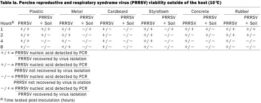
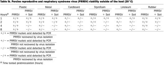
Results are summarized in Tables Ia and Ib. The PRRSV nucleic acid was detected by PCR on all soil-free surfaces held at 10°C and 20°C for up to 8 h pi. The PRRSV RNA was detected by PCR at 1 and 2 h pi on soil-covered concrete, plastic, and rubber at 10°C. All soil-covered surfaces were PCR positive 1-hour pi at 20°C; however, only plastic, linoleum, and rubber were positive 2 h pi. All other samples from soil-covered surfaces at either temperature were PCR negative throughout the remainder of the testing period. Porcine reproductive and respiratory syndrome virus was isolated from a number of soil-free surfaces; including plastic (1, 2, and 4 h pi at 10°C and 20°C), metal (1 and 2 h pi at 20°C), styrofoam (1 and 2 h at 20°C, and 1, 2, and 4 h at 10°C), concrete (1 h at 10°C), and rubber (1, 2, and 4 h at 20°C, and 1 and 2 h at 10°C). As for surfaces with soil cover, PRRSV was isolated from plastic (1 and 2 h pi), rubber (1 h pi), linoleum (1 h pi), and styrofoam (1 h pi) at 20°C, and plastic, rubber, metal, and styrofoam (1 h pi) at 10°C.
Table Ia.
Table Ib.
Controls
Porcine reproductive and respiratory syndrome virus nucleic acid was detected by PCR up to 8 h pi in soil-free positive control samples, and up to 2 h pi in soil-covered controls at both temperatures. Infectious PRRSV was isolated from all soil-free positive controls ranging from 1 to 2 h pi, from soil-covered plastic (1 and 2 h pi), and from rubber surfaces at 1 h pi, at 10°C and 20°C. Porcine reproductive and respiratory syndrome virus was isolated from the 1-mL aliquot of the study isolate in all 10 replicates. All sham-inoculated negative control surfaces were PRRSV-negative by all testing methods, at each sampling time and temperature, in the presence or absence of soil cover.
Transmission data
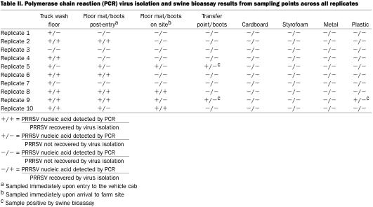
Polymerase chain reaction — Results of PCR testing for the 10 replicates are summarized in Table II. In 6 out of 10 replicates (replicates 2, 5, 6, 8, 9, and 10), PRRSV RNA was detected by PCR on the truck wash floor (sampling point 1) and inside the vehicle (sampling points 2 or 3). In 2 replicates (5 and 9), PRRSV RNA was detected on the floor of the anteroom (sampling point 4); however, in replicate 9 PRRSV RNA was detected on the ventral surface of a container (sampling point 5). The Fisher's exact test was used to assess the difference in the proportion of PCR positive results detected on containers in virus-positive replicates (1 out of 10) as compared to protocol control replicates (0 out of 10). This difference was not significant at P = 1.000. All samples collected from the truck wash floor following the sanitation protocol used between replicates, were PCR negative.
Table II.
Virus isolation — Infectious PRRSV was recovered from at least 1 sampling point in 4 out of 10 replicates (Table II). In these 4 replicates, PRRSV was either isolated from the truck wash floor, on the vehicle floor mats, or soles of boots either immediately following entry of the vehicle following washing (sampling point 2) or upon arrival at the farm premise (sampling point 3). All samples from sham-inoculated negative controls and protocol controls were VI negative on both PAM and MARC-145 cell lines. All samples collected from the truck wash floor following the sanitation protocol used between replicates were VI negative.
Swine bioassay — A total of 4 pigs were used, 2 inoculated with PCR-positive or VI-negative samples from the anteroom floor (replicates 5 and 9), 1 with a PCR-positive sample from the ventral surface of a plastic container (replicate 9), and 1 sham-inoculated negative control. All 3 inoculated pigs tested positive for PRRSV RNA by PCR on day 7 pi and for PRRSV-antibodies by ELISA on day 14 pi. The negative control pig remained PCR and ELISA negative throughout the testing period.
Nucleic acid sequencing — Three PRRSV isolates recovered from the bioassay pigs inoculated with samples from replicate 5 (sampling point 4) and replicate 9 (sampling points 4 and 5), were nucleic acid sequenced and found to be 100% homologous to the original study isolate.
Discussion
The objective of this study was to use a model to assess mechanical transmission of PRRSV under field conditions during warm weather and compare these findings with those reported during cold weather (7). Differences between the studies were the type of carriers used (soil versus snow) and the environmental conditions under which the studies were conducted (warm weather versus cold weather). As expected, these changes resulted in strikingly different outcomes and proved the initial hypothesis that during periods of warm weather, mechanical transmission of PRRSV is an infrequent event. In the warm weather study, viable PRRSV was detected on the anteroom floor on the simulated farm premise in only 2 out of the 10 replicates, and on the ventral surface of a single container in only 1 replicate. In 7 of the remaining 8 replicates, it appeared that PRRSV was present on the floor of the truck wash or within the vehicle, but it was not possible to transfer the virus into the farm anteroom. In contrast, successful transmission and detection of PRRSV RNA on containers was observed in 8 out of 10 cold-weather replicates. When using the Fisher's exact test to compare the number of replicates with detectable PRRSV RNA on containers across the 2 studies, the difference was significant at P = 0.006. This difference may be explained by a number of reasons. First, the carrier may not have been conducive for supporting long-term PRRSV viability. The soil samples used to manufacture carriers had a very low percentage of moisture (mean 1.56%, range 1.04 to 2.07%). Secondly, due to the warm environmental temperatures on the 5 sampling days (10°C to 16°C), soil samples appeared to dry rapidly over the course of the 2 to 2.5 h period required to complete each replicate. As validated by the positive controls, contact with soil promoted a dry environment, and PRRSV was recovered for only 1 to 2 h pi. In contrast, positive control samples of PRRSV that were covered with soil demonstrated enhanced viability, and live virus could be recovered for up to 4 h pi. Not only does drying have a negative impact on PRRSV viability, it could have reduced the adherence of the soil to the soles of boots or the ventral surface of the containers. Although it was not quantified, the amount of residual soil in the transfer area was visibly variable across all 10 replicates. This may have affected the amount of PRRSV introduced into the anteroom, transfer area, and the contamination of the containers.
However, it must be recognized that even during conditions unfavorable to PRRSV survival outside of the host viable, infectious PRRSV was still detected in swab samples collected from the anteroom floor in 2 out of 10 replicates, and from the ventral surface of a plastic container in 1 replicate. The value of this information is important for a number of reasons. The fact that the results vary significantly from the cold weather study validated the ability of the field model to authentically replicate PRRSV transmission in the field under a variety of environmental conditions. It demonstrated that during springtime conditions in Minnesota, mechanical transmission of viable PRRSV onto a farm site and into a facility can occur, although at a significantly lower rate than during the winter (P = 0.006). It substantiated previously identified risk factors to farm biosecurity, including the risk of traffic from an infected farm; such as, commercial truck washing facilities, contaminated boots and containers, the vehicle interior, and the farm anteroom (7). Finally, these results confirmed previously published laboratory data regarding the stability of the virus outside of the host (8,9).
Similar to the cold weather study, the strengths of the model were the use of field conditions and the testing of standard operating procedures employed by swine producers and practitioners. It used examples of surfaces and containers used in swine facilities, a field isolate of PRRSV, and a large number of replicates. It used multiple diagnostic methods to track the virus and to document the presence of infectious PRRSV in the simulated farm and on a plastic container. The study was well controlled, each replicate possessing a set of positive and negative controls, as well as a protocol control replicate. The purpose of the protocol control was to insure that accidental contamination of samples with the study isolate of PRRSV or unidentified field isolates of PRRSV did not occur between replicates or during replicates. As with all studies, there were known limitations prior to initiation of the work. It is unknown whether the carrier used in this study was realistic. Although vehicles frequently accumulate some amount of soil on the undercarriage area during the winter, no studies have attempted to isolate or quantify the amount of PRRSV present in these types of samples collected from actual livestock vehicles. It was not known whether the concentration of PRRSV used to inoculate the carriers in this study was representative of field conditions. Furthermore, during the process of washing, study personnel were aware of the presence of contaminated carriers and made direct efforts to come into contact with them, a situation that most likely would not occur in the field. Finally, regarding the assumption that contaminated fomites can introduce PRRSV to naïve populations, we only documented that infectious virus was present on the surface of a single plastic container, and we cannot definitively conclude that pigs exposed to this container would have become infected.
To conclude, one must acknowledge the fact that our study demonstrated that while mechanical transmission of PRRSV during warm weather is a relatively infrequent event, it might still occur. Therefore, strict biosecurity measures should be maintained throughout the year. Furthermore, it may also be prudent for swine producers to focus control measures on other proven vectors of PRRSV known to be present in high concentrations during warmer periods of the year, such as mosquitoes and houseflies.
Footnotes
Acknowledgments
The authors thank the Utah Pork Producers Association and Pig Improvement Company for their generous assistance and Genetiporc for the use of animals during this study.
Address all correspondence and reprint requests to Dr. S.A. Dee; 385c Animal Science/Veterinary Medicine Building, 1988 Fitch Avenue, St. Paul, Minnesota, USA 55108; telephone: (612) 625-4786; fax: (612) 625-1210; e-mail: deexx004@umn.edu
Received May 22, 2002. Acepted August 20, 2002.
References
- 1.Dee SA, Joo HS, Pijoan C. Controlling the spread of PRRS virus in the breeding herd through management of the gilt pool. Swine Health Prod 1994;3:64–69.
- 2.Christopher-Hennings J, Nelson EA, Hines RJ, et al. Persistence of porcine reproductive and respiratory syndrome virus in serum and semen of adult boars. J Vet Diagn Invest 1995;7:456–464. [DOI] [PubMed]
- 3.Otake S, Dee SA, Rossow KD, Joo HS, Deen J, Molitor TW. Transmission of porcine reproductive and respiratory syndrome virus by fomites. Swine Health Prod 2002;10:59–65. [PubMed]
- 4.Otake S, Dee SA, Rossow KD, Joo HS, Deen J, Molitor TW. Transmission of porcine reproductive and respiratory syndrome virus by needles. Vet Rec 2002;150:114–115. [PubMed]
- 5.Otake S, Dee SA, Rossow KD, Moon RD, Pijoan C. Mechanical transmission of porcine reproductive and respiratory syndrome virus by mosquitoes Aedes vexans. Can J Vet Res 2002;66:191–195. [PMC free article] [PubMed]
- 6.Otake S, Dee SA, Rossow KD, Moon RD, Pijoan C. Transmission of porcine reproductive and respiratory syndrome virus by houseflies. [DOI] [PubMed]
- 7.Dee SA, Deen J, Rossow KD, Weise C, Otake S, Joo HS, Pijoan C. Mechanical transmission of porcine reproductive and respiratory syndrome virus through a coordinated sequence of events during cold weather. Can J Vet Res 2002;66:232–239. [PMC free article] [PubMed]
- 8.Benfield D, Nelson E, Collins J, et al. Characterization of PRRS virus (isolate ATCC-VR 2332). J Vet Diagn Invest 1992;4:127–133. [DOI] [PubMed]
- 9.Pirtle EP, Beran GW. Stability of porcine reproductive and respiratory syndrome virus in the presence of fomites commonly found on farms. JAVMA 1996;208:390–392. [PubMed]
- 10.Miller SO, Kotuby-Armacher J, Rodriguez JB. Particle size analysis (hydrometer method). Western states laboratory-testing program: soil and plant analytical methods. Version 4.0 1997;96–99.
- 11.Watson ME, Brown JR. pH and lime requirement. Recommended chemical soil test procedures for the North Central region. Missouri Agricultural Experiment Station SB 1001 North Central Regional Research Publication No. 221 (Revised) January 1998.
- 12.Bierk MD, Dee SA, Rossow KD, Collins JE, Pijoan C, Guedes MI, Molitor TW. Diagnostic investigation of chronic PRRS virus infection in a breeding herd of pigs. Vet Rec 2001;148:687–690. [DOI] [PubMed]
- 13.Bautista EM, Goyal S, Yoon IJ, Joo HS, Collins J. Comparison of porcine alveolar macrophages and CL 2621 for the detection of porcine reproductive and respiratory syndrome virus (PRRSV) and anti-PRRSV antibody. J Vet Diagn Invest 1993;5:163–165. [DOI] [PubMed]
- 14.Molitor TW, Tune KA, Shin J, Collins J, Kapur V. Applications of TaqManTM PCR in the detection of porcine reproductive and respiratory syndrome virus. Proc AD Leman Swine Conf 1997;173–175.
- 15. Murtaugh MP, Elam MR, Kakach LT. Comparison of the structural protein coding sequences of the VR-2332 and Lelystad virus strains of the porcine reproductive and respiratory syndrome virus. Arch Virol 1995;40:1451–1460. [DOI] [PMC free article] [PubMed]
- 16.Swenson SL, Hill HT, Zimmerman JJ. Excretion of porcine reproductive and respiratory syndrome virus after experimentally induced infection in boars. JAVMA 1994;204:1943–1948. [PubMed]
- 17.Snyder ML, Mermer B, Anderson PR, Wensvoot G, Hill HT. Evaluation data for an immunological ELISA for porcine reproductive and respiratory syndrome. Proc 2nd Int Symp PRRS 1995;15.


