Abstract
Background
The quasistatic neutral zone is a surrogate for neutral region stiffness of spinal motion segments. No similar measure of dynamic stiffness has been validated. Because parameters related to stiffness are likely to be affected by loading rate and disc degeneration, we examined the effect of those factors on motion parameters derived from continuous motion data.
Methods
Fifteen human lumbar motion segments were tested with continuous flexion-extension pure moments at 0.5, 3.0 and 6.0 degrees/second. Range of motion, width of the hysteresis loop, transitional zone width, and slopes of the upper and lower arms of the hysteresis loop within the transitional zone were measured. Discs were then graded for degeneration.
Findings
As the loading rate increased from 0.5 to 6.0 degree/second there were significant increases in ROM, hysteresis area, hysteresis loop width, and the upper and lower transitional zone slopes. At the same time transitional zone width decreased significantly. Degeneration had a significant effect on all parameters except hysteresis loop width. The transition zone slopes appeared to best discriminate between normal and degenerative discs.
Interpretation
Loading rate had a significant effect on all parameters. As degeneration increased consistent effects were observed indicating decreasing stiffness from grade 1 to grade 3 then slightly increased stiffness in grade 4 specimens. The slopes of the transitional zone have potential to be a useful measure of neutral region stiffness during dynamic motion testing.
Keywords: neutral zone, lumbar vertebrae, intervertebral disc degeneration, methods, biomechanics
1. Introduction
Low back pain (LBP) is a common condition that results in great societal and monetary loss. The lifetime prevalence of LBP in North America has been estimated to be 79% to 84% and approximately 10% of persons with LBP were noted to be disabled by their pain during the previous 6 months (Cassidy et al., 1998; Walker et al., 2004). Disability from LBP is responsible for most of its costs (Hashemi et al., 1997; Williams et al., 1998). Severe, disabling LBP is often attributed to ‘clinical instability’ but defining instability has been problematic (O'Sullivan, 2000; Vo and MacMillan, 1994).
Several objective parameters have been used in an attempt to characterize clinical instability including anterior and posterior translation, (Kirkaldy-Willis, 1983) abnormal range of motion (ROM) (Nagel et al., 1981) and dispersion of the centers of rotation (Seligman et al., 1984). Clinical studies using x-ray have found excessive anterior vertebral translation during flexion to be associated with disc degeneration and facet arthrosis (Fujiwara et al., 2000) and pain (Iguchi et al., 2004).
A large gap exists between the laboratory characterization of segmental motion and clinical application. The understanding of abnormal lumbar segmental motion has been informed primarily by in vitro studies. The most common parameter used to characterize the degree of laxity in the neutral region is the neutral zone (NZ). The NZ has been defined as “that part of the range of physiological intervertebral motion, measured from the neutral position, within which spinal motion is produced with a minimal internal resistance” (Panjabi, 1988). The NZ is measured using a quasistatic technique and has been shown to correlate with injury and degenerative change (Mimura et al.; 1994, Oxland and Panjabi, 1992). Unfortunately, quasistatic methods cannot be used clinically. Further, they do not provide direct information about neutral region behavior during motion in vitro.
Continuous loading methods are considered more physiologic and provide continuous data rather than only discrete points at specific loads. Parameters derived from continuous loading curves have been proposed as the dynamic equivalent of the NZ. Wilke proposed the width of the hysteresis loop as a measure of the neutral laxity (Wilke et al., 1998b). Goertzen et al. compared the ROM and the width of the hysteresis loop between quasistatic and continuous loading methods. They found both ROM and hysteresis loop width to be less in the continuous motion protocol (Goertzen et al., 2004). Thompson et al proposed the amount of movement occurring while the slope of the moment-displacement curve is near 0 as a dynamic NZ. Using that definition, they found a NZ to be present only during flexion-extension motion in a sheep model (Thompson et al., 2003). Other measures of motion segment laxity have been proposed such as the lax zone, which extends beyond the NZ into the area where there is minimal ligamentous resistance (the transition between NZ and elastic zone). The lax zone was compared to the quasistatic NZ and found to be distinct from the NZ and less variable (Crawford et al., 1998).
We have previously compared biomechanical parameters from continuous motion load-displacement curves (loading rate 3 degrees/second) with the quasistatic NZ. We identified candidate measures of neutral region laxity during motion that included ROM, hysteresis (H width; width of the hysteresis loop on the X axis at 0 load), transitional zone (TZ; the motion in degrees between the intersections of the initial 0 load line and the tangents of the loading arm tails), and the TZ slopes (slopes of the upper and lower load-deformation curve in the TZ). Additionally, we studied the effect of degeneration on both quasistatic and dynamic parameters. The TZ slope had the strongest correlation with neutral zone and appeared to best detect differences between grades of degeneration (Gay et al., 2006). That report only considered a single loading rate (3 degrees/second). Continuous motion parameters that reflect neutral region stiffness are likely to be affected by loading rate, disc degeneration and other factors that influence the viscoelastic response of the motion segment. This study extends our previous work by using the same sample of specimens to examine the effect of loading rate and degenerative change on the candidate dynamic motion parameters.
2. Methods
2.1 Specimens
This study was approved by the institutional review board and is an extension of the previously reported results. Eight fresh, frozen (−20 C) cadaveric adult lumbar spines (L1-S1) were obtained from the institutional Anatomical Bequest Program. Specimens were screened for HIV/AIDS, Hepatitis B and C, tuberculosis, and Creutzfeldt-Jakob Disease. Radiographs were used to exclude specimens with post-traumatic deformity or significant anatomical anomaly.
The spines were thawed to a room temperature of 65 degrees F prior to use and kept moist with normal saline soaked toweling throughout preparation and testing. Non-ligamentous soft tissues were removed leaving the intact vertebral bodies and ligamentous structures including the anterior longitudinal ligament, posterior longitudinal ligament, interspinous ligaments, and facet joint capsules. Each specimen was divided into motion segments (functional spinal units) by separating them at the disc and facet joints. Motion segments were prepared and tested as previously described (Gay et al., 2006). Screws and K wires were passed through the vertebral body and facet joints and polymethylmethacrylate orthodontic resin was used to secure the upper and lower vertebral bodies of each motion segment in cylindrical acrylic fixtures.
2.2 Experimental set-up
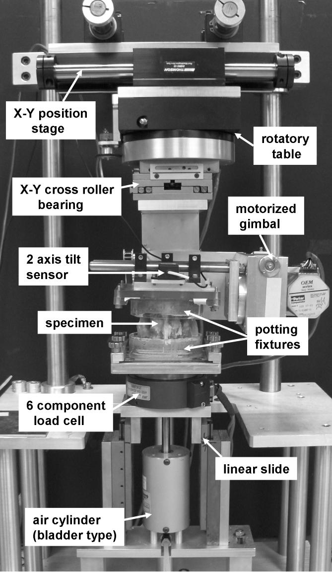
Specimens were first mounted in a custom testing device to measure quasistatic ROM and NZ in the sagittal plane. After quasistatic testing, the specimens were attached to a custom dynamic testing device with a 3 axis gimbal and stepper motors (Fig. 1). The device had an unconstrained XY stage between the rotatory table for internal-external rotation and the flexion-extension and lateral bending gimbal portion of the device. Forces and moments were recorded by a 6 component load cell (JR3, Woodland, CA, USA). The device effectively applied a pure bending moment to the specimen. This was verified by observing the force and moment outputs from the 6 component load cell. Angular changes were measured by miniature CXTA tilt sensors (Crossbow Technology, San Jose, CA, USA) with range of +/− 20 degrees, resolution of 0.05 degrees RMS, and accuracy less than 0.4 degrees for the +/−20 degree range. The device was controlled by a LabVIEW program (National Instruments Corp., Austin, TX, USA) that allowed adjustment of loading rates and limits.
Fig. 1.
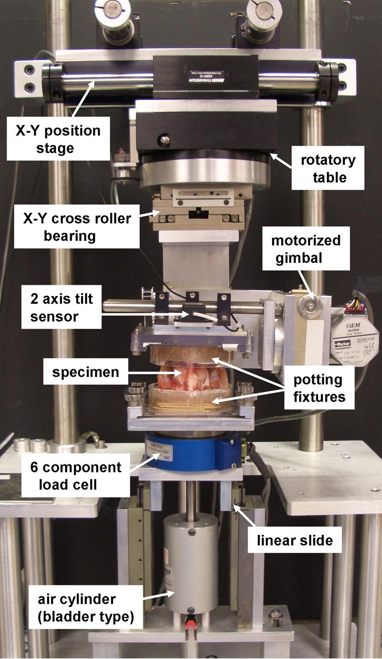
Single lumbar motion segment in the custom spine testing apparatus
2.3 Testing procedures
All specimens were tested in the same manner as follows:
a- Axial compression of 300 N was applied for 30 minutes to expel excess fluid and avoid disc hydration beyond the normal in vivo range (Adams et al., 1987, Edwards et al., 1987).
b- Quasistatic loading was performed and ROM and NZ measured (Panjabi, 1992).
c- A period of 30 minutes was allowed between quasistatic and dynamic testing for viscoelastic recovery.
d- Dynamic testing: Each motion segment was subjected to 8 cycles of dynamic flexion/extension motion. Continuous loads were applied in flexion to a limit of 5 Nm then the moment was reversed until an extension moment of 5 Nm was reached, then reversed again until reaching the starting position. Cycles 1 through 4 were preconditioning cycles at a rate of 1 degree/second. Cycles 5, 6 and 7 were completed with three loading rates in random order (0.5, 3.0 or 6.0 degrees/second). An 8th cycle was performed using the first randomized loading rate (same as cycle 5). The 8th cycle was compared to the 5th cycle to examine the effect of repetitive testing. Loading rates were chosen to reflect the range of loading rates that the lumbar motion segments are to exposed to during activities of daily living (Harada et al., 2000; Wilke et al., 1998a). Data (load, rotation, and force) were collected digitally and stored in a secure server.
After testing was completed motion segments were refrozen and then divided in axial and sagittal planes with a band saw and allowed to thaw at room temperature. The amount of degeneration was graded visually from 1 (normal) to 4 (severe degeneration) by 2 of the investigators using the scale of Adams et al (Adams et al., 1996). In the event of disagreement between the graders a third examiner's grade decided the final grade. Graders were blinded to test results of individual specimens.
2.4 Analysis
We have previously reported the quasistatic data and its association with 3 degree per second dynamic measures and degeneration (Gay et al., 2006). In this report we have analyzed only the dynamic parameters but at the 3 loading rates. The force-displacement curves were analyzed using custom written Matlab (The Mathworks, Novi, MI, USA) programs. The programs used linear regression to calculate the slope and intercept parameters of three independent lines through the data set. The optimal slope and intercept parameters for each line were those which minimized the difference (residual) between the calculated line and the acquired data. The TZ (laxity region) was calculated as the distance between the location of intersection of the toe and first linear region (first and second line) and the location of intersection of the toe and second linear region (second and third line) (Koff et al., 2006; Markolf et al., 1976).
The parameters examined (Fig. 2) included: 1) ROM: range of flexion-extension motion in degrees; 2) hysteresis width: width of the hysteresis loop in degrees (on the X axis at 0 load); 3) hysteresis area: area within the hysteresis loop indicating dissipated energy; 4) TZ width: width of the TZ in degrees; and 5) TZ slopes: slopes of the 2 lines within the TZ in Nm/degree.
Fig. 2.
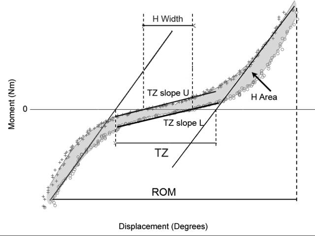
Parameters of dynamic motion derived from the force displacement curves
(TZ = transitional zone, laxity region between the intersection of the toe and first linear region (first and second line) and the intersection of the toe and second linear region (second and third line); TZ slope U = transitional zone slope-upper arm; TZ slope L = transitional zone slope-lower arm; ROM = range of motion; H Width = width of the hysteresis loop at 0 load; H Area = area within hysteresis loop)
The effect of repetitive testing was assessed by comparing test cycle 5 with cycle 8. The effect of loading rate and degeneration were assessed concurrently on each parameter by repeated measures analysis of variance (ANOVA). The model allowed us to consider the interaction of degenerative grade (independent effect) and loading rate (dependent effect). To estimate the overall differences between degenerative grades a one-way ANOVA and post hoc Tukey-Kramer tests were performed for the average of each parameter (mean of 3 loading rates for each parameter in each motion segment). All statistical tests were two-sided; p-values less than 0.05 were considered statistically significant. Statistical analysis was performed with JMP 5.1.2 (SAS Institute, Inc., Cary NC, USA).
3. Results
Mean (SD) age of specimens was 70.9 (+/− 9.95) with a range of 40 to 82 years. There were 7 males and 1 female. Motion segments were reasonably distributed by degenerative grade with the exception that there were only two grade 1 (normal) discs. The spines yielded 15 testable motion segments of varied spinal levels and a wide distribution of degenerative change based on the screening radiographs. The disc level distribution was L1-2 (3); L2-3 (4); L3-4 (2); L4-5 (5); and L5-S1 (1). The distribution of motion segments by grade was grade 1 (2), grade 2 (6), grade 3 (3), and grade 4 (4). We did not examine the effect of disc level because of the small number of specimens for each level which posed risk of error. However, we performed repeated-measures ANOVA on each parameter with level as the effect and found no significant differences among the disc levels for any parameter, suggesting that pooling of the levels was reasonable.
3.1 Effect of repetitive testing
The percent difference in parameter means from the 5th to the 8th cycle (same loading rate, all degenerative grades combined) have been previously reported and ranged from 1% to 7% (Gay et al., 2006). The upper and lower slopes within the TZ were not significantly different at 0.5 or 3.0 degree/second but a difference was noted at 6.0 degree/s (Wilcoxen sign-rank test, P = 0.048) Despite this difference, R2 was 0.93. Both the upper and lower slopes are reported below.
3.2 Effect of loading rate
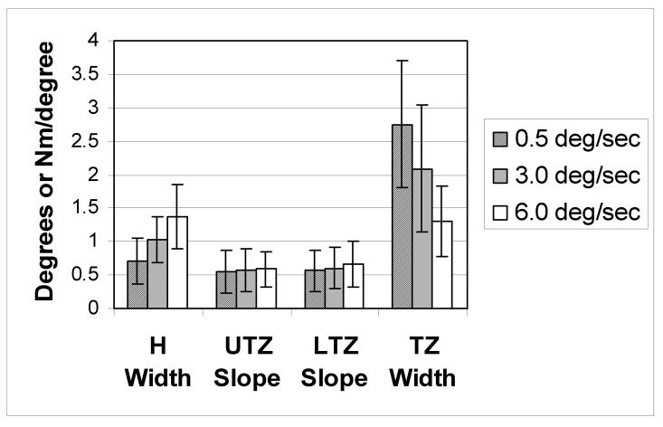
Repeated measures ANOVA showed that loading rate had a significant effect on all parameters. Fig. 3 shows the mean and standard deviations (SD) of the neutral region parameter values for each loading rate for all 15 specimens combined. As the rate increased from 0.5 to 6.0 degree/second ROM increased (P = 0.0006) as did the area within the hysteresis loop (P < 0.0001), the width of the hysteresis loop (P < 0.0001) and the upper and lower TZ slopes (P = 0.017 and 0.001 respectively). At the same time the width of the TZ decreased (P < 0.0001). The only parameter found to have a significant interaction between loading rate and grade was the upper TZ slope (P = 0.001).
Fig. 3.
Mean and Standard Deviation of Dynamic Motion Parameters (all specimens)
H width = hysteresis loop width; UTZ slope = transitional zone slope upper arm; LTZ slope = transitional zone slope lower arm; TZ Width = transitional zone width. Loading rate had a statistically significant effect on all parameters (repeated measures ANOVA).
3.3 Effect of degeneration
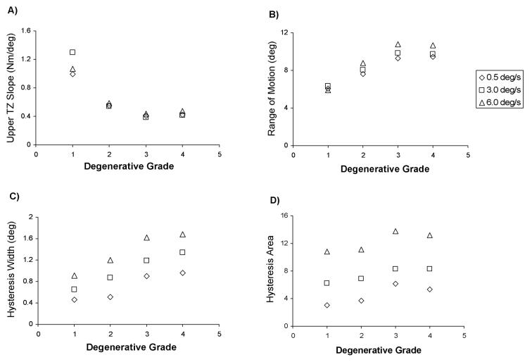
One way ANOVA of the averaged parameter values (mean of 3 loading rates) with grade as effect demonstrated associations between degenerative grade and several motion parameters (Table 1). Degeneration had a significant effect on the TZ, TZ slopes, hysteresis area, and ROM. Post-hoc Tukey-Kramer tests found differences primarily between Grade 1 discs and grade 3 or 4 discs (Table 1). The only parameter with no differences between grades was the hysteresis loop width. Fig. 4a-d illustrates the trends of parameter change for each of the 3 loading rates in each degenerative grade.
Table 1.
Effect of degenerative grade (1-4) on average motion parameters (mean of 3 loading rates)
| P value | differences | |
|---|---|---|
| TZ | 0.0123 | 1 and 3 or 4 |
| TZ slope U | 0.0007 | 1 and 2, 3 or 4 |
| TZ slope L | 0.0030 | 1 and 2, 3, or 4 |
| ROM | 0.0010 | 1 and 3 or 4 |
| 2 and 4 | ||
| H Width | 0.0494 | none* |
| H Area | 0.0040 | 1 and 3 or 4 |
| 2 and 3 or 4 |
TZ = transitional zone, TZ slope U = transitional zone slope upper arm, TZ slope L = transitional zone slope lower arm, ROM = range of motion, H Width = width of the hysteresis loop at 0 load, H Area = area within hysteresis loop.
P values from one way ANOVA of average parameter (specimen mean of value for the 3 loading rates, n = 15).
Pair-wise differences between grades based on post hoc Tukey-Kramer tests.
No differences between grade pairs on post hoc Tukey-Kramer tests despite P value <0.05
Fig. 4.
Effect of degeneration and loading rate on 4 continuous motion parameters
a) upper transitional zone slope
b) range of motion
a) hysteresis loop width
b) hysteresis loop area
3.4 Summary of results
Both the rate of loading and the presence of disc degeneration affected all the parameters examined. As loading rate increased, the ROM, hysteresis area, and width of the hysteresis loop increased while the TZ width decreased. As degeneration increased from grade 1 to grade 4 there were consistent effects on TZ width, TZ slope, ROM and hysteresis width and area. The observed changes indicated decreasing stiffness from grade 1 to grade 3 then slightly increased stiffness in grade 4 specimens.
4. Discussion
The NZ, as defined by Panjabi (Panjabi, 1992) is a widely used indicator of laxity around the neutral position of motion segments. Although it has been shown to change with degeneration and trauma, it cannot be measured during dynamic testing. With the increasing use of dynamic methods to measure spine function, parameters are needed to describe the characteristics of dynamic motion in the neutral region. Dynamic surrogates of the quasistatic NZ have been previously suggested. The width of the hysteresis loop as proposed by Wilke et al (Wilke et al., 1998b) has been reported to be smaller than the quasistatic NZ despite the terminal load being the same (Goertzen et al., 2004). This is likely due to the creep associated with quasistatic loading. Thompson et al. suggested the amount of movement occurring while the slope of the moment-displacement curve is near 0 as a dynamic equivalent of the NZ (Thompson et al., 2003). Using that definition, they found a dynamic NZ only in sagittal plane motion in a sheep model. To our knowledge these proposed measures have not been systematically examined to determine how they are affected by loading rate or degeneration, both of which can change the viscoelastic response to loading.
Identifying specific points on continuous load-displacement curves to define a parameter is not without problems. Crawford et al. (Crawford et al., 1998) explored this issue by comparing 2 methods. The first extrapolated a line fit to the elastic (stiff) zone to the zero-load line, the other method fitted a tangent to the elbow region that occurs between zero-load and the elastic region of the load-displacement curve, based on the work of Markolf et al. (Markolf et al., 1976) . The later technique had slightly less variability in flexion-extension curves. We therefore used a variation of that method in the current study.
We purposefully have not used the term “dynamic neutral zone” to describe the parameters examined in this study so as to avoid confusion with the quasistatic NZ. The concept of neutral region laxity might well be represented by both quasistatic and dynamic measures. We previously reported the relationship between the quasistatic NZ and dynamic motion parameters (3 degree/second) using data generated from the same specimens as used in the current study (see methods). We found that the NZ was moderately correlated with hysteresis loop width (r = 0.69) and strongly correlated with the TZ slope (r = −0.80). We found degenerative grade to have a significant effect on ROM and TZ size and slope. Differences were found in several parameters between grade 1 (normal) discs and higher grades but only the TZ slope was different between grades 1 and 2 (Gay et al., 2006).
The motivation of this study was to identify parameters of dynamic motion that best indicate stiffness in the neutral region during motion. Dynamic motion measures of spine stiffness (or laxity) that change in a predictable way with disc degeneration, injury or disease might be helpful in detecting critical levels of biomechanical failure if efficient and safe in vivo measurement methods are developed. Although current technology does not allow routine measurement of force during motion, methods of quantifying dynamic motion are improving (De Stefano et al.; 2004, Teyhen et al., 2005). It is plausible that clinical kinematic data (for example from videofluoroscopy studies) can be combined with calculated loads to generate in vivo load-displacement curves. Control of the loading rate during in vivo studies will be difficult. Therefore, measures used will need to be robust to changes in loading rate and yet sensitive to changes in stiffness. All of the dynamic parameters considered in the current study were affected by both loading rate and degeneration. The width of the hysteresis loop increased markedly with increasing loading rate. Likewise, the area within the hysteresis loop and the width of the TZ were greatly affected by loading rate and therefore would be less reproducible if measured in vivo. Among the parameters tested the TZ slopes and ROM were least affected by loading rate. Both were found to discriminate between discs with little or no degeneration and those with advanced degeneration but the limited number of specimens in this study limits any conclusion regarding which might be the most useful clinically. Also, we did not examine the effect of disc level, which may confound our results, particularly in regard to ROM.
In our earlier report we suggested that the TZ slope appeared to be the best candidate for a dynamic measure of neutral region stiffness based on its ability to discriminate between grades of degeneration. That enthusiasm is tempered by the current results showing TZ slope to be the only parameter to have an interaction between degeneration and loading rate. Larger studies are needed to clarify these relationships.
There are several limitations of this study that should be considered. The use of previously frozen cadaveric spines may not reflect in vivo material properties as closely as necessary. The limited number of specimens is also a significant shortcoming as it did not allow the examination of disc level effect. Additionally, only 2 of the 15 discs were graded as normal (grade 1) which might have resulted in error. We did not examine load displacement curves from coronal or axial plane motion; the sensitivity of the dynamic parameters to loading rate and degeneration may be different in those planes. We did not include a preload or follower load in our method of testing. This was done purposefully so that the NZ could be directly compared to dynamic parameters as reported in our previous paper (Gay et al., 2006). Future experiments should use a follower load, which will likely increase the overall stiffness of the motion segment (Crawford et al., 1998) and change the sensitivity of dynamic parameters to loading rate and degeneration. The fact that single motion segments were tested rather than multisegment spines may also affect the measured response. Finally, the anatomic grade was based on gross inspection and may not accurately reflect the mechanical or radiographic properties of the discs.
5. Conclusions
The slope of the force-displacement curve in the TZ has potential as a measure of neutral region stiffness in lumbar motion segments during continuous sagittal plane motion. It is sensitive to changes associated with disc degeneration and does not appear to be affected by loading rate as much as the comparative parameters. Further study is needed to determine the usefulness of the TZ slope as a biomarker of lumbar motion segment behavior.
Acknowledgments
The authors thank Lawrence Berglund, engineer, for numerous valuable contributions including design and construction of the spine testing apparatus. This study was supported in part by grant AT000972 from the National Center for Complementary and Alternative Medicine/National Institutes of Health.
Footnotes
Conflict of Interest Statement
The authors hold no commercial interests or personal relationships that constitute or infer conflict of interest.
Publisher's Disclaimer: This is a PDF file of an unedited manuscript that has been accepted for publication. As a service to our customers we are providing this early version of the manuscript. The manuscript will undergo copyediting, typesetting, and review of the resulting proof before it is published in its final citable form. Please note that during the production process errors may be discovered which could affect the content, and all legal disclaimers that apply to the journal pertain.
References
- Adams MA, Dolan P, Hutton WC. Diurnal variations in the stresses on the lumbar spine. Spine. 1987;12:130–7. doi: 10.1097/00007632-198703000-00008. [DOI] [PubMed] [Google Scholar]
- Adams MA, McNally DS, Dolan P. ’Stress’ distributions inside intervertebral discs. The effects of age and degeneration. J. Bone Joint Surg. Br. 1996;78:965–72. doi: 10.1302/0301-620x78b6.1287. [DOI] [PubMed] [Google Scholar]
- Cassidy JD, Carroll LJ, Cote P. The Saskatchewan health and back pain survey. The prevalence of low back pain and related disability in Saskatchewan adults. Spine. 1998;23:1860–6. doi: 10.1097/00007632-199809010-00012. [DOI] [PubMed] [Google Scholar]
- Crawford NR, Peles JD, Dickman CA. The spinal lax zone and neutral zone: measurement techniques and parameter comparisons. J. Spinal Disord. 1998;11:416–29. [PubMed] [Google Scholar]
- De Stefano A, Allen R, White PR. Noise reduction in spine videofluoroscopic images using the undecimated wavelet transform. Comput. Med. Imaging Graph. 2004;28:453–9. doi: 10.1016/j.compmedimag.2004.07.003. [DOI] [PubMed] [Google Scholar]
- Edwards WT, Hayes WC, Posner I, White AA, 3rd, Mann RW. Variation of lumbar spine stiffness with load. J. Biomech. Eng. 1987;109:35–42. doi: 10.1115/1.3138639. [DOI] [PubMed] [Google Scholar]
- Fujiwara A, Tamai K, An HS, Kurihashi T, Lim TH, Yoshida H, Saotome K. The relationship between disc degeneration, facet joint osteoarthritis, and stability of the degenerative lumbar spine. J. Spinal Disord. 2000;13:444–50. doi: 10.1097/00002517-200010000-00013. [DOI] [PubMed] [Google Scholar]
- Gay RE, Ilharreborde B, Zhao KD, Zhao C, An KN. Sagittal plane motion in the human lumbar spine: Comparison of the in vitro quasistatic neutral zone and dynamic motion parameters. Clin. Biomech. 2006;21:914–919. doi: 10.1016/j.clinbiomech.2006.04.009. [DOI] [PubMed] [Google Scholar]
- Goertzen DJ, Lane C, Oxland TR. Neutral zone and range of motion in the spine are greater with stepwise loading than with a continuous loading protocol. An in vitro porcine investigation. J. Biomech. 2004;37:257–61. doi: 10.1016/s0021-9290(03)00307-5. [DOI] [PubMed] [Google Scholar]
- Harada M, Abumi K, Ito M, Kaneda K. Cineradiographic motion analysis of normal lumbar spine during forward and backward flexion. Spine. 2000;25:1932–7. doi: 10.1097/00007632-200008010-00011. [DOI] [PubMed] [Google Scholar]
- Hashemi L, Webster BS, Clancy EA, Volinn E. Length of disability and cost of workers' compensation low back pain claims. J. Occup. Environ. Med. 1997;39:937–45. doi: 10.1097/00043764-199710000-00005. [DOI] [PubMed] [Google Scholar]
- Iguchi T, Kanemura A, Kasahara K, Sato K, Kurihara A, Yoshiya S, Nishida K, Miyamoto H, Doita M. Lumbar instability and clinical symptoms: which is the more critical factor for symptoms: sagittal translation or segment angulation? J. Spinal Disord. Tech. 2004;17:284–90. doi: 10.1097/01.bsd.0000102473.95064.9d. [DOI] [PubMed] [Google Scholar]
- Kirkaldy-Willis WH. The three phases of the spectrum of degenerative disease. In: Kirkaldy-Willis WH, Burton CV, editors. Managing Low Back Pain. Churchill Livingston; New York; 1983. p. 111. [Google Scholar]
- Koff MF, Shrivastava N, Gardner TR, Rosenwasser MP, Mow VC, Strauch RJ. An in vitro analysis of ligament reconstruction or extension osteotomy on trapeziometacarpal joint stability and contact area. J. Hand Surg. Am. 2006;31:429–39. doi: 10.1016/j.jhsa.2005.11.010. [DOI] [PubMed] [Google Scholar]
- Markolf KL, Mensch JS, Amstutz HC. Stiffness and laxity of the knee--the contributions of the supporting structures. A quantitative in vitro study. J. Bone Joint Surg. Am. 1976;58:583–94. [PubMed] [Google Scholar]
- Mimura M, Panjabi MM, Oxland TR, Crisco JJ, Yamamoto I, Vasavada A. Disc degeneration affects the multidirectional flexibility of the lumbar spine. Spine. 1994;19:1371–1380. doi: 10.1097/00007632-199406000-00011. [DOI] [PubMed] [Google Scholar]
- Nagel DA, Koogle TA, Piziali RL, Perkash I. Stability of the upper lumbar spine following progressive disruptions and the application of individual internal and external fixation devices. J. Bone Joint Surg. Am. 1981;63:62–70. [PubMed] [Google Scholar]
- O'Sullivan PB. Lumbar segmental ‘instability’: clinical presentation and specific stabilizing exercise management. Man. Ther. 2000;5:2–12. doi: 10.1054/math.1999.0213. [DOI] [PubMed] [Google Scholar]
- Oxland TR, Panjabi MM. The onset and progression of spinal injury: a demonstration of neutral zone sensitivity. J. Biomech. 1992;25:1165–72. doi: 10.1016/0021-9290(92)90072-9. [DOI] [PubMed] [Google Scholar]
- Panjabi MM. Biomechanical evaluation of spinal fixation devices: I. A conceptual framework. Spine. 1988;13:1129–34. doi: 10.1097/00007632-198810000-00013. [DOI] [PubMed] [Google Scholar]
- Panjabi MM. The stabilizing system of the spine. Part II. Neutral zone and instability hypothesis. J. Spinal Disord. 1992;5:390–6. doi: 10.1097/00002517-199212000-00002. [DOI] [PubMed] [Google Scholar]
- Seligman JV, Gertzbein SD, Tile M, Kapasouri A. Computer analysis of spinal segment motion in degenerative disc disease with and without axial loading. Spine. 1984;9:566–73. doi: 10.1097/00007632-198409000-00006. [DOI] [PubMed] [Google Scholar]
- Teyhen DS, Flynn TW, Bovik AC, Abraham LD. A new technique for digital fluoroscopic video assessment of sagittal plane lumbar spine motion. Spine. 2005;30:E406–13. doi: 10.1097/01.brs.0000170589.47555.c6. [DOI] [PubMed] [Google Scholar]
- Thompson RE, Barker TM, Pearcy MJ. Defining the Neutral Zone of sheep intervertebral joints during dynamic motions: an in vitro study. Clin. Biomech. 2003;18:89–98. doi: 10.1016/s0268-0033(02)00180-8. [DOI] [PubMed] [Google Scholar]
- Vo P, MacMillan M. The aging spine: clinical instability. South. Med. J. 1994;87:S26–35. [PubMed] [Google Scholar]
- Walker BF, Muller R, Grant WD. Low back pain in Australian adults: prevalence and associated disability. J. Manipulative Physiol. Ther. 2004;27:238–44. doi: 10.1016/j.jmpt.2004.02.002. [DOI] [PubMed] [Google Scholar]
- Wilke HJ, Jungkunz B, Wenger K, Claes LE. Spinal segment range of motion as a function of in vitro test conditions: effects of exposure period, accumulated cycles, angular-deformation rate, and moisture condition. Anat. Rec. 1998a;251:15–9. doi: 10.1002/(SICI)1097-0185(199805)251:1<15::AID-AR4>3.0.CO;2-D. [DOI] [PubMed] [Google Scholar]
- Wilke HJ, Wenger K, Claes L. Testing criteria for spinal implants: recommendations for the standardization of in vitro stability testing of spinal implants. Eur.Spine J. 1998b;7:148–54. doi: 10.1007/s005860050045. [DOI] [PMC free article] [PubMed] [Google Scholar]
- Williams DA, Feuerstein M, Durbin D, Pezzullo J. Health care and indemnity costs across the natural history of disability in occupational low back pain. Spine. 1998;23:2329–36. doi: 10.1097/00007632-199811010-00016. [DOI] [PubMed] [Google Scholar]


