Abstract
A 55-year-old man who is 178 cm tall and weighs 95 kg is hospitalized with community-acquired pneumonia and progressively severe dyspnea. His arterial oxygen saturation while breathing 100% oxygen through a face mask is 76%; a chest radiograph shows diffuse alveolar infiltrates with air bronchograms. He is intubated and receives mechanical ventilation; ventilator settings include a tidal volume of 1000 ml, a positive end-expiratory pressure (PEEP) of 5 cm of water, and a fraction of inspired oxygen (FiO2) of 0.8. With these settings, peak airway pressure is 50 to 60 cm of water, plateau airway pressure is 38 cm of water, partial pressure of arterial oxygen is 120 mm Hg, partial pressure of carbon dioxide is 37 mm Hg, and arterial blood pH is 7.47. The diagnosis of the acute respiratory distress syndrome (ARDS) is made. An intensive care specialist evaluates the patient and recommends changing the current ventilator settings and implementing a low-tidal-volume ventilation strategy.
THE CLINICAL PROBLEM
Acute lung injury is defined by the American–European Consensus Conference as the acute onset of impaired gas exchange (the ratio of the partial pressure of arterial oxygen in millimeters of mercury to the FiO2 of <300) and the presence of bilateral alveolar or interstitial infiltrates in the absence of congestive heart failure.1 Acute lung injury has an incidence of 86 cases per 100,000 person-years and a mortality rate of 39%. In the United States, there are an estimated 190,600 cases annually, leading to 74,500 deaths and 3.6 million hospital days.2 ARDS is a more severe form of lung injury, defined by a ratio of the partial pressure of arterial oxygen to FiO2 of less than 200.1 The incidence of ARDS is 64 cases per 100,000 person-years, and the mortality rate is 40 to 50%. Common causes of ARDS are sepsis (with or without a pulmonary source), trauma, aspiration, multiple blood transfusions, pancreatitis, inhalation injury, and certain types of drug toxicity.2,3
PATHOPHYSIOLOGICAL CHARACTERISTICS AND EFFECT OF THERAPY
Acute lung injury can be defined physiologically as acute respiratory failure due to pulmonary edema in the absence of an elevation in the hydrostatic pressure in the pulmonary veins. The syndrome is characterized by diffuse alveolar damage associated with increased permeability of the alveolar–capillary membrane. Edema fluid and plasma proteins leak from the vasculature into the alveolar spaces. Macrophages and neutrophils accumulate in the interstitium, and proinflammatory cytokines are released into the lungs. Hyaline membranes form in the alveoli. The chemical composition and functional activity of surfactant can be altered in patients with ARDS, resulting in an elevation in surface tension, which tends to promote regional alveolar collapse.4,5 The efficiency of gas exchange deteriorates precipitously.
Endotracheal intubation and mechanical ventilation are almost always necessary to manage the severe hypoxemia of ARDS. In the past, the primary goal of ventilation had been to increase arterial oxygenation to an acceptable range (principally, an arterial oxyhemoglobin saturation of 88 to 95%, but also normal partial pressure of carbon dioxide and pH). This objective was usually met with the use of a high FiO2 and a high minute ventilation. Tidal volumes were correspondingly high. Although the practice was variable, tidal volumes of 10 to 15 ml per kilogram of body weight (as compared with a normal tidal volume of 5 to 7 ml per kilogram for spontaneously breathing controls at rest) were commonly used.6,7 The concept of “recruitment” (i.e., the opening of previously collapsed alveoli) was thought to provide a justification for such high-volume ventilation.
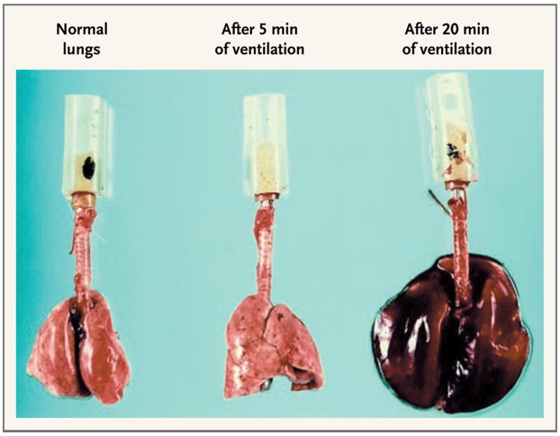
More recently, it has been recognized that mechanical ventilation, although potentially lifesaving, can contribute to the worsening of lung injury. This phenomenon is called ventilator-induced lung injury (Fig. 1). The volume of aerated lung in patients with ARDS is considerably reduced because of edema and atelectasis. As a result, ventilation with the use of high tidal volumes may cause hyperinflation of relatively normal regions of aerated lung. Since nonaerated lung tissue is stiffer than normal lung tissue, compliance is reduced and airway pressure is increased. Excessive volume and pressure, with correspondingly high transpulmonary pressure (the difference in pressure between the airway and the pleural space), contribute to ventilator-induced lung injury. In addition, the inflation of normal alveoli adjacent to noninflated, abnormal alveoli may create high shear forces that can contribute to injury of the lung parenchyma, even at modest applied pressures.9 The consequences of lung overdistention include direct physical damage, with disruption of the alveolar epithelium and capillary endothelium, as well as the induction of an inflammatory response, with the release of cytokines and other mediators.8,10–13 Some evidence suggests that the inflammatory response induced during ventilator-induced lung injury has systemic consequences, contributing to the pathogenesis of multisystem organ failure in patients with ARDS.14,15
Figure 1. Normal Rat Lungs and Rat Lungs after Receiving High-Pressure Mechanical Ventilation at a Peak Airway Pressure of 45 cm of Water.

After 5 minutes of ventilation, focal zones of atelectasis were evident, in particular at the left lung apex. After 20 minutes of ventilation, the lungs were markedly enlarged and congested; edema fluid filled the tracheal cannula. Adapted from Dreyfuss et al.8 with the permission of the publisher.
In 1993, a consensus conference of the American College of Chest Physicians recommended that applied tidal volume be decreased in patients with ARDS who had a plateau pressure of 35 cm of water or more, even though such a decrease can cause some degree of hypercapnia (sometimes referred to as permissive hypercapnia).16 This recommendation was based largely on data from studies of animals, since at that time there were few clinical studies of low-tidal-volume ventilation and no definitive data showing an outcome benefit with this approach. The use of PEEP was endorsed as a means of supporting oxygenation, but it was also noted that excessive PEEP may be associated with deleterious effects; the role and optimal use of PEEP in a low-tidal-volume ventilation strategy was not specified, owing to the lack of clinical-trial data addressing this issue.
CLINICAL EVIDENCE
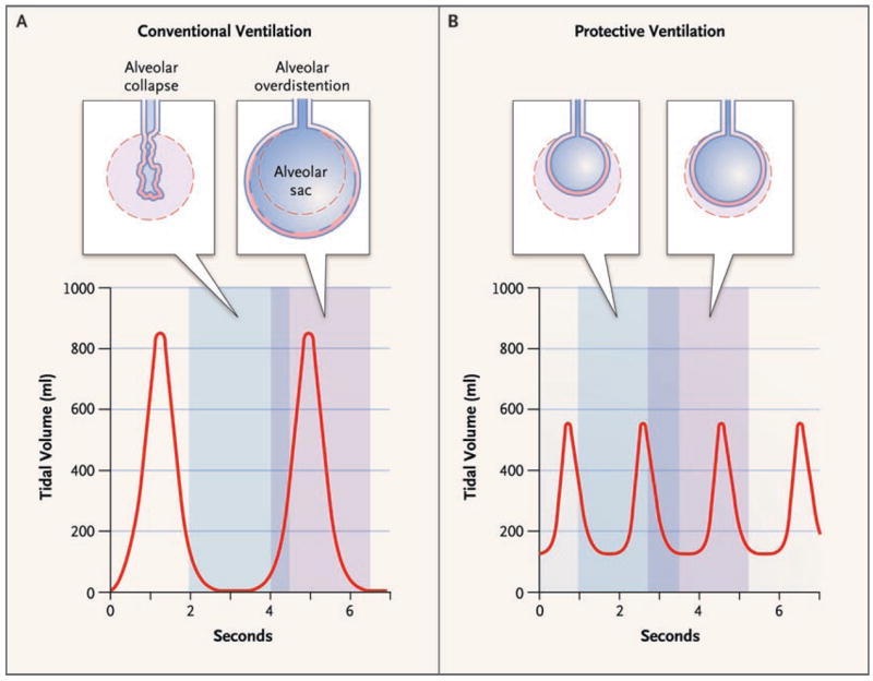
The first major randomized clinical trial to provide direct evidence of a potential benefit of low-tidal-volume ventilation in patients with ARDS was published in 1998.17 Amato et al. compared conventional ventilation with a low-tidal-volume, “protective ventilation” strategy in 53 patients (Fig. 2). Conventional ventilation involved a tidal volume of 12 ml per kilogram of body weight, a low PEEP, and a partial pressure of carbon dioxide of 35 to 38 mm Hg. Protective ventilation involved a tidal volume at or below 6 ml per kilogram, a high PEEP, and permissive hypercapnia. The mortality rate at 28 days was significantly lower with protective ventilation than with conventional ventilation (38% vs. 71%). There was also significantly less clinical barotrauma and a significantly higher rate of weaning from ventilation in the protective-ventilation group. Although some criticized this study for the high mortality rate in the conventional-ventilation group, the patients studied were extremely ill (with failure of a mean of 3.6 organs per patient).
Figure 2. Conventional Ventilation as Compared with Protective Ventilation.

This example of ventilation of a 70-kg patient with ARDS shows that conventional ventilation at a tidal volume of 12 ml per kilogram of body weight and an end-expiratory pressure of 0 cm of water (Panel A) can lead to alveolar overdistention (at peak inflation) and collapse (at the end of exhalation). Protective ventilation at a tidal volume of 6 ml per kilogram (Panel B) limits overinflation and end-expiratory collapse by providing a low tidal volume and an adequate positive end-expiratory pressure. Adapted from Tobin.18
In a subsequent, larger study by the Acute Respiratory Distress Syndrome Network (ARDSNet), 861 patients with acute lung injury or ARDS were randomly assigned to receive ventilatory support involving a tidal volume of either 12 or 6 ml per kilogram of predicted body weight.19 Although tidal volume was the manipulated variable, a major goal of the ventilatory strategy was to keep the plateau airway pressure below 30 cm of water; therefore, the group that underwent ventilation at 6 ml per kilogram of predicted body weight is often referred to as the low-stretch group. The low-stretch strategy was associated with a significantly lower mortality rate (31%, vs. 40% with ventilation at 12 ml per kilogram of predicted body weight). Therefore, the best available evidence is for a ventilation strategy using a tidal volume of 6 ml per kilogram of predicted body weight for patients with acute lung injury or ARDS.
Three other small, randomized trials, performed during the same period, failed to demonstrate a benefit of low-tidal-volume ventilation in patients with acute lung injury or ARDS.20–22 The reasons for this apparent inconsistency in study results are not clear, but they may have included differences in the airway pressures required for conventional ventilation in each trial. A significant survival benefit has been shown in trials in which conventional ventilation was associated with marked elevations in airway pressures.23 This finding suggests that the benefit of low-tidal-volume ventilation is a function of plateau pressure. However, the relationship between plateau pressure and risk of injury from ventilation may be continuous, since subsequent data have failed to confirm the concept of a threshold below which airway pressure is no longer injurious.24 Furthermore, evidence of hyperinflation may occur at a low volume or pressure, depending on the amounts of poorly aerated and nonaerated lung tissue.25 In addition, theoretically, as described above, high shear forces can create injury at junctions of normal and abnormal lung tissue, even when the applied pressures are below 30 cm of water.9
CLINICAL USE
Low-tidal-volume ventilation should be implemented in the context of a broader strategy of critical care management in a patient with acute lung injury or ARDS. An initial tidal volume of 6 ml per kilogram of predicted, not actual, body weight should be used, as in the ARDSNet trial.19 The predicted body weight (PBW) is calculated as follows: for men, PBW = 50.0 + 0.91 (height in centimeters – 152.4); and for women, PBW = 45.5 + 0.91 (height in centimeters – 152.4).
The concept underlying this approach is that it normalizes the tidal volume to lung size, since lung size has been shown to depend most strongly on height and sex. For example, a person who ideally weighs 70 kg and who then gains 35 kg has essentially the same lung size as he or she did when at a weight of 70 kg and should not receive ventilation with a higher tidal volume just because of the weight gain.
The initial respiratory rate should be set in the range of 18 to 22 breaths per minute. This is a somewhat higher rate than is used in other ventilatory schemes; it is intended to maintain a minute ventilation that is high enough to avoid marked hypercapnia. However, some degree of hypercapnia is to be expected with low-tidal-volume ventilation. Ideally, the partial pressure of carbon dioxide should rise gradually to prevent acute acidemia and to ensure hemodynamic stability. Specific target values of partial pressure of carbon dioxide and pH are debatable, although some clinicians would argue to keep the current guidelines of a partial pressure of carbon dioxide of less than 80 mm Hg and a pH of greater than 7.20. Although the administration of sodium bicarbonate has sometimes been advocated to maintain an acceptable pH, this is controversial in theory and rarely necessary in practice. In fact, mean partial pressures of carbon dioxide below 50 mm Hg were usually achieved in the ARDSNet study in the low-stretch group.
The response to low-tidal-volume ventilation should be assessed initially on the basis of plateau airway pressure. The goal should be to maintain a plateau airway pressure (i.e., the pressure during an end-inspiratory pause) of 30 cm of water or less; if this target is exceeded, the tidal volume should be further reduced to a minimum of 4 ml per kilogram of predicted body weight. An important caveat relates to patients who have stiff chest walls (for example, those with massive ascites). In such patients, it is reasonable to allow the plateau pressure to increase to values greater than 30 cm of water, since the pleural pressures are elevated and hence the transpulmonary pressures are not elevated (i.e., there is not necessarily alveolar over-distention). Whether the tidal volume should be increased in the patient with a plateau pressure substantially lower than 30 cm of water is less clear; given the lack of evidence of a safe threshold, some experts would argue that the lower the plateau pressure, the better, provided that the patient is comfortable and that gas-exchange goals are reached.
The optimal FiO2 also requires consideration in the context of low-stretch ventilation. Since severe hypoxemia is a characteristic feature of ARDS, efforts to improve oxygenation and to achieve a target arterial oxyhemoglobin saturation of about 90% may initially require high FiO2 levels. However, the prolonged use of high FiO2 levels can theoretically increase the risk of oxygen toxicity, which may actually increase injury to the lung parenchyma. Therefore, other adjustments may be necessary to improve oxygenation while reducing the FiO2. One approach is to use PEEP to increase oxygenation, although this should be done while plateau airway pressure is monitored. In the ARDSNet trial,19 combinations of FiO2 and PEEP values were specified for both study groups according to predefined settings (Table 1). However, the level of oxygenation is a poor predictor of outcome. In the ARDSNet trial, oxygenation was worse in the low-stretch group, despite a reduced mortality rate. Therefore, some experts recommend the application of PEEP based on lung mechanics rather than gas exchange (see below).
Table 1.
Settings for Positive End-Expiratory Pressure (PEEP), According to the Required Fraction of Inspired Oxygen (FiO2).*
| FiO2 | PEEP |
|---|---|
| 0.3 | 5 |
| 0.4 | 5–8 |
| 0.5 | 8–10 |
| 0.6 | 10 |
| 0.7 | 10–14 |
| 0.8 | 14 |
| 0.9 | 14–18 |
| 1.0 | 18–24 |
Settings are from the ARDSNet trial.19 The required FiO2 is the lowest value that maintains arterial oxyhemoglobin saturation above 90%. After the corresponding level of PEEP is selected, arterial oxyhemoglobin saturation and plateau airway pressure should be monitored in the patient.
Alternatives to low-tidal-volume ventilation either have been unsuccessful (e.g., partial liquid ventilation26) or are unproven (e.g., high-frequency oscillation27,28). However, many unproven strategies, such as open-lung protective ventilation or prone positioning, may be useful in combination with low-tidal-volume ventilation29,30 and thus should not be considered to be competing therapies.
ADVERSE EFFECTS
Low-tidal-volume ventilation can result in an increase in the partial pressure of carbon dioxide to above the normal range (permissive hypercapnia). As noted above, permissive hypercapnia results in respiratory acidosis, which can be mitigated to some degree by means of increasing the respiratory rate and gradual renal buffering. Potentially harmful consequences of permissive hypercapnia include pulmonary vasoconstriction and pulmonary hypertension, proarrhythmic effects of increased discharge of the sympathetic nervous system, and cerebral vasodilation yielding increased intracranial pressure. However, experimental data have suggested that permissive hypercapnia is not only safe but potentially beneficial.31 In most cases, hemodynamic characteristics actually improve owing to the release of catecholamines.32 Nonetheless, permissive hypercapnia should probably be used with caution in patients with heart disease and is relatively contraindicated in those with elevated intracranial pressure.
For at least some patients, low-tidal-volume ventilation is associated with a sensation of dyspnea that is uncomfortable and poorly tolerated.33 Such patients may require substantial sedation to maintain patient–ventilator synchrony, although sedation requirements were equivalent in patients receiving ventilation with a low tidal volume and those receiving ventilation with a high tidal volume in the ARDSNet trial.34 If discomfort is an issue, either minor elevations in delivered tidal volume can be made or sedation can be increased. Sedation can generally be managed with the use of short-acting agents like propofol and with daily interruptions to determine whether the requirement for sedation is ongoing.35,36
AREAS OF UNCERTAINTY
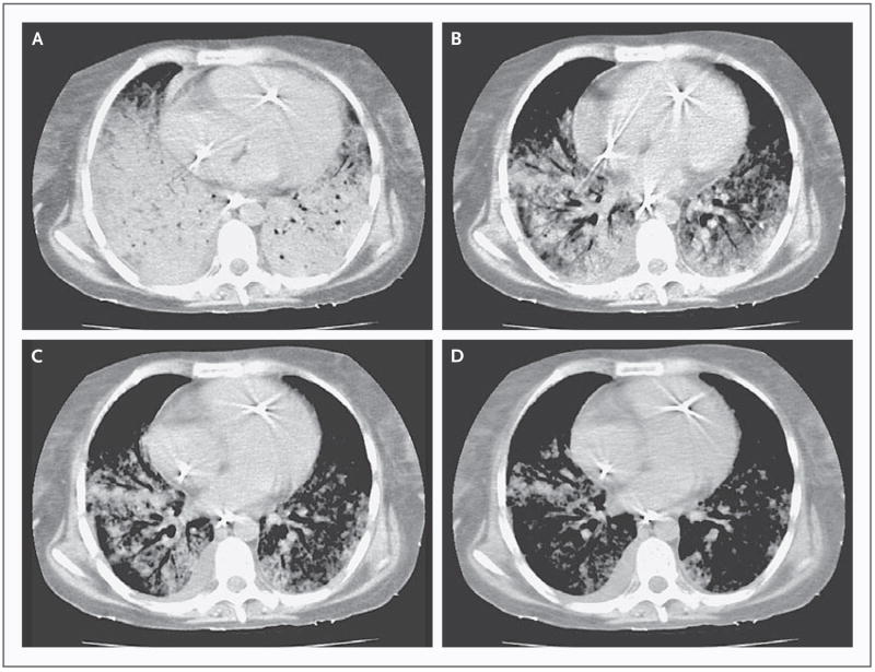
As noted above, PEEP is commonly adjusted in accordance with FiO2 in low-stretch ventilation, as was systematically defined in the ARDSNet trial. Whether high levels of PEEP may be beneficial in this setting has not been clearly established. PEEP can prevent the collapse of small airways and alveoli (referred to as derecruitment), further improving oxygenation and ventilation–perfusion matching. High PEEP values may also minimize a phenomenon called “atelectrauma,” which is the repetitive opening and closing of alveoli, with the propensity for collapse owing to either surfactant dysfunction resulting in high surface tension or elevated pleural pressures that promote regional lung collapse.37 Results of trials evaluating the role of a high PEEP have been inconsistent with regard to its potential benefit.17,29,38,39 At the bedside, the PEEP can often be adjusted on the basis of responses in individual patients. For example, recruitment can be inferred if the plateau pressure does not rise substantially after the PEEP is increased while the tidal volume remains fixed. In contrast, a rise in plateau pressure that is equal to or greater than the increase in PEEP would suggest that recruitment has not occurred and possibly that there is overdistention or regional hyperinflation. In the patient in whom there is recruitment, sustained high-pressure inflations (referred to as recruitment maneuvers), followed by the administration of a high PEEP and a low tidal volume, may reduce lung injury from shear forces by promoting homogeneity of inflation within the lung (Fig. 3). However, this approach, which has been designated “open-lung protective ventilation,” remains unproved in clinical trials.
Figure 3. Effects of Recruitment Maneuvers to Promote Homogeneity within the Lung.
Panels A through D show the progressive resolution of infiltrates after application of inflations of increasing pressure. Reprinted from Borges et al.40
Patients who do not have acute lung injury or ARDS may also benefit from the limiting of lung stretch. Some observational data provide support for the concept that inappropriate ventilator settings may contribute to the development of ARDS.41 That is, ARDS may be iatrogenic in some cases.42 In addition, because of the occasional failure to diagnose and appropriately manage acute lung injury or ARDS once it develops, some have made the argument to limit lung stretch in all patients undergoing mechanical ventilation, including during the perioperative period.43 However, there have been few randomized trials in this area.
GUIDELINES
As noted above, a consensus conference of the American College of Chest Physicians recommended in 1993 that low-tidal-volume ventilation be used in patients with ARDS.16 No subsequent formal guidelines dealing with low-tidal-volume ventilation have been developed by the American College of Chest Physicians, the American Thoracic Society, or the Society of Critical Care Medicine. However, all three organizations endorsed a set of industry-funded guidelines, called the Surviving Sepsis Campaign, published in 2004.44 The process by which these guidelines were developed has been criticized,45 although the recommendations with regard to mechanical ventilation are generally accepted.
The Surviving Sepsis guidelines endorse low-tidal-volume ventilation (6 ml per kilogram of predicted body weight), with a goal of maintaining end-inspiratory plateau pressures of less than 30 cm of water.44 Hypercapnia is deemed acceptable in this context, in the absence of increased intracranial pressure. The use of PEEP is recommended to prevent alveolar collapse at the end of expiration and to maintain adequate oxygenation.
RECOMMENDATIONS
The patient described in the vignette is an appropriate candidate for low-tidal-volume ventilation, given the diagnosis of ARDS and the high plateau airway pressure attained with the use of conventional mechanical ventilation. The tidal volume should be reduced to 6 ml per kilogram of predicted body weight, according to the ARDSNet formula (resulting in a tidal volume of 440 ml for this patient). I would then increase the ventilatory rate to 20 breaths per minute and observe the resulting plateau pressure and arterial blood gas levels, making subsequent adjustments as appropriate. For a patient with a plateau pressure above 30 cm of water, I would reduce the tidal volume further, by 1 ml per kilogram of predicted body weight, and I would then remeasure the plateau pressure. For a patient with an arterial oxyhemoglobin saturation below 88 to 90%, I would attempt to increase the PEEP to improve oxygenation without exceeding the target plateau pressure. Depending on the individual case, I will often attempt to perform a recruitment maneuver (sustained high-pressure inflation under heavy sedation and adequate fluid resuscitation) and then attempt to maintain recruitment by applying increased levels of PEEP and observing the resulting change in plateau pressure.
Acknowledgments
Supported by grants from the National Institutes of Health (P50 HL060292-09, AG024837, and RO1-HL73146).
I thank Dr. Arthur Slutsky of the University of Toronto for helpful comments and suggestions.
Footnotes
This Journal feature begins with a case vignette that includes a therapeutic recommendation. A discussion of the clinical problem and the mechanism of benefit of this form of therapy follows. Major clinical studies, the clinical use of this therapy, and potential adverse effects are reviewed. Relevant formal guidelines, if they exist, are presented. The article ends with the author’s clinical recommendations.
Dr. Malhotra reports receiving consulting fees from Respironics. No other potential conflict of interest relevant to this article was reported.
References
- 1.Bernard GR, Artigas A, Brigham KL, et al. Report of the American-European Consensus Conference on acute respiratory distress syndrome: definitions, mechanisms, relevant outcomes, and clinical trial coordination. J Crit Care. 1994;9:72–81. doi: 10.1016/0883-9441(94)90033-7. [DOI] [PubMed] [Google Scholar]
- 2.Rubenfeld GD, Caldwell E, Peabody E, et al. Incidence and outcomes of acute lung injury. N Engl J Med. 2005;353:1685–93. doi: 10.1056/NEJMoa050333. [DOI] [PubMed] [Google Scholar]
- 3.Piantadosi CA, Schwartz DA. The acute respiratory distress syndrome. Ann Intern Med. 2004;141:460–70. doi: 10.7326/0003-4819-141-6-200409210-00012. [DOI] [PubMed] [Google Scholar]
- 4.Faridy EE, Permutt S, Riley RL. Effect of ventilation on surface forces in excised dogs’ lungs. J Appl Physiol. 1966;21:1453–62. doi: 10.1152/jappl.1966.21.5.1453. [DOI] [PubMed] [Google Scholar]
- 5.Spragg RG, Lewis JF, Walmrath H-D, et al. Effect of recombinant surfactant protein C–based surfactant on the acute respiratory distress syndrome. N Engl J Med. 2004;351:884–92. doi: 10.1056/NEJMoa033181. [DOI] [PubMed] [Google Scholar]
- 6.Wheeler AP, Bernard GR. Acute lung injury and the acute respiratory distress syndrome: a clinical review. Lancet. 2007;369:1553–65. doi: 10.1016/S0140-6736(07)60604-7. [DOI] [PubMed] [Google Scholar]
- 7.MacIntyre NR. Current issues in mechanical ventilation for respiratory failure. Chest. 2005;128(Suppl 2):561S–567S. doi: 10.1378/chest.128.5_suppl_2.561S. [DOI] [PubMed] [Google Scholar]
- 8.Dreyfuss D, Saumon G. Ventilator- induced lung injury: lessons from experimental studies. Am J Respir Crit Care Med. 1998;157:294–323. doi: 10.1164/ajrccm.157.1.9604014. [DOI] [PubMed] [Google Scholar]
- 9.Mead J, Takishima T, Leith D. Stress distribution in lungs: a model of pulmonary elasticity. J Appl Physiol. 1970;28:596–608. doi: 10.1152/jappl.1970.28.5.596. [DOI] [PubMed] [Google Scholar]
- 10.Ranieri VM, Suter PM, Tortorella C, et al. Effect of mechanical ventilation on inflammatory mediators in patients with acute respiratory distress syndrome: a randomized controlled trial. JAMA. 1999;282:54–61. doi: 10.1001/jama.282.1.54. [DOI] [PubMed] [Google Scholar]
- 11.Matthay MA, Bhattacharya S, Gaver D, et al. Ventilator-induced lung injury: in vivo and in vitro mechanisms. Am J Physiol Lung Cell Mol Physiol. 2002;283:L678–L682. doi: 10.1152/ajplung.00154.2002. [DOI] [PubMed] [Google Scholar]
- 12.Imai Y, Parodo J, Kajikawa O, et al. Injurious mechanical ventilation and end-organ epithelial cell apoptosis and organ dysfunction in an experimental model of acute respiratory distress syndrome. JAMA. 2003;289:2104–12. doi: 10.1001/jama.289.16.2104. [DOI] [PubMed] [Google Scholar]
- 13.Tremblay L, Valenza F, Ribeiro SP, Li J, Slutsky AS. Injurious ventilatory strategies increase cytokines and c-fos mRNA expression in an isolated rat lung model. J Clin Invest. 1997;99:944–52. doi: 10.1172/JCI119259. [DOI] [PMC free article] [PubMed] [Google Scholar]
- 14.Slutsky AS, Tremblay LN. Multiple system organ failure: is mechanical ventilation a contributing factor? Am J Respir Crit Care Med. 1998;157:1721–5. doi: 10.1164/ajrccm.157.6.9709092. [DOI] [PubMed] [Google Scholar]
- 15.Dreyfuss D, Saumon G. From ventilator-induced lung injury to multiple organ dysfunction? Intensive Care Med. 1998;24:102–4. doi: 10.1007/s001340050529. [DOI] [PubMed] [Google Scholar]
- 16.Slutsky AS. Mechanical ventilation: American College of Chest Physicians’ Consensus Conference. Chest. 1993;104:1833–59. doi: 10.1378/chest.104.6.1833. Erratum, Chest 1994;106:656. [DOI] [PubMed] [Google Scholar]
- 17.Amato MBP, Barbas CSV, Medeiros DM, et al. Effect of a protective-ventilation strategy on mortality in the acute respiratory distress syndrome. N Engl J Med. 1998;338:347–54. doi: 10.1056/NEJM199802053380602. [DOI] [PubMed] [Google Scholar]
- 18.Tobin MJ. Advances in mechanical ventilation. N Engl J Med. 2001;344:1986–96. doi: 10.1056/NEJM200106283442606. [DOI] [PubMed] [Google Scholar]
- 19.The Acute Respiratory Distress Syndrome Network. Ventilation with lower tidal volumes as compared with traditional tidal volumes for acute lung injury and the acute respiratory distress syndrome. N Engl J Med. 2000;342:1301–8. doi: 10.1056/NEJM200005043421801. [DOI] [PubMed] [Google Scholar]
- 20.Stewart TE, Meade MO, Cook DJ, et al. Evaluation of a ventilation strategy to prevent barotrauma in patients at high risk for acute respiratory distress syndrome. N Engl J Med. 1998;338:355–61. doi: 10.1056/NEJM199802053380603. [DOI] [PubMed] [Google Scholar]
- 21.Brochard L, Roudot-Thoraval F, Roupie E, et al. Tidal volume reduction for prevention of ventilator-induced lung injury in acute respiratory distress syndrome. Am J Respir Crit Care Med. 1998;158:1831–8. doi: 10.1164/ajrccm.158.6.9801044. [DOI] [PubMed] [Google Scholar]
- 22.Brower RG, Shanholtz CB, Fessler HE, et al. Prospective, randomized, controlled trial comparing traditional versus reduced tidal volume ventilation in acute respiratory distress syndrome patients. Crit Care Med. 1999;27:1492–8. doi: 10.1097/00003246-199908000-00015. [DOI] [PubMed] [Google Scholar]
- 23.Tobin MJ. Culmination of an era in research on the acute respiratory distress syndrome. N Engl J Med. 2000;342:1360–1. doi: 10.1056/NEJM200005043421808. [DOI] [PubMed] [Google Scholar]
- 24.Hager DN, Krishnan JA, Hayden DL, Brower RG. Tidal volume reduction in patients with acute lung injury when plateau pressures are not high. Am J Respir Crit Care Med. 2005;172:1241–5. doi: 10.1164/rccm.200501-048CP. [DOI] [PMC free article] [PubMed] [Google Scholar]
- 25.Terragni PP, Rosboch G, Tealdi A, et al. Tidal hyperinflation during low tidal volume ventilation in acute respiratory distress syndrome. Am J Respir Crit Care Med. 2007;175:160–6. doi: 10.1164/rccm.200607-915OC. [DOI] [PubMed] [Google Scholar]
- 26.Kacmarek RM, Wiedemann HP, Lavin PT, Wedel MK, Tütüncü AS, Slutsky AS. Partial liquid ventilation in adult patients with acute respiratory distress syndrome. Am J Respir Crit Care Med. 2006;173:882–9. doi: 10.1164/rccm.200508-1196OC. [DOI] [PubMed] [Google Scholar]
- 27.Kacmarek RM, Malhotra A. High-frequency oscillatory ventilation: what large-animal studies have taught us! Crit Care Med. 2005;33(Suppl 3):S148–S154. doi: 10.1097/01.CCM.0000156786.43935.A0. [DOI] [PMC free article] [PubMed] [Google Scholar]
- 28.Derdak S, Mehta S, Stewart TE, et al. High-frequency oscillatory ventilation for acute respiratory distress syndrome in adults: a randomized, controlled trial. Am J Respir Crit Care Med. 2002;166:801–8. doi: 10.1164/rccm.2108052. [DOI] [PubMed] [Google Scholar]
- 29.Villar J, Kacmarek RM, Pérez-Méndez L, Aguirre-Jaime A. A high positive end-expiratory pressure, low tidal volume ventilatory strategy improves outcome in persistent acute respiratory distress syndrome: a randomized, controlled trial. Crit Care Med. 2006;34:1311–8. doi: 10.1097/01.CCM.0000215598.84885.01. [DOI] [PubMed] [Google Scholar]
- 30.Malhotra A, Ayas N, Kacmarek R. Prone positioning of patients with acute respiratory failure. N Engl J Med. 2002;346:296. [PubMed] [Google Scholar]
- 31.Kavanagh BP, Laffey JG. Hypercapnia: permissive and therapeutic. Minerva Anestesiol. 2006;72:567–76. [PubMed] [Google Scholar]
- 32.Carvalho CR, Barbas CS, Medeiros DM, et al. Temporal hemodynamic effects of permissive hypercapnia associated with ideal PEEP in ARDS. Am J Respir Crit Care Med. 1997;156:1458–66. doi: 10.1164/ajrccm.156.5.9604081. [DOI] [PubMed] [Google Scholar]
- 33.Kallet RH, Alonso JA, Diaz M, Camp-bell AR, Mackersie RC, Katz JA. The effects of tidal volume demand on work of breathing during simulated lung-protective ventilation. Respir Care. 2002;47:898–909. [PubMed] [Google Scholar]
- 34.Kahn JM, Andersson L, Karir V, Polissar NL, Neff MJ, Rubenfeld GD. Low tidal volume ventilation does not increase sedation use in patients with acute lung injury. Crit Care Med. 2005;33:766–71. doi: 10.1097/01.ccm.0000157786.41506.24. [DOI] [PubMed] [Google Scholar]
- 35.Carson SS, Kress JP, Rodgers JE, et al. A randomized trial of intermittent lorazepam versus propofol with daily interruption in mechanically ventilated patients. Crit Care Med. 2006;34:1326–32. doi: 10.1097/01.CCM.0000215513.63207.7F. [DOI] [PubMed] [Google Scholar]
- 36.Kress JP, Pohlman AS, O’Connor MF, Hall JB. Daily interruption of sedative infusions in critically ill patients undergoing mechanical ventilation. N Engl J Med. 2000;342:1471–7. doi: 10.1056/NEJM200005183422002. [DOI] [PubMed] [Google Scholar]
- 37.Slutsky AS. Lung injury caused by mechanical ventilation. Chest. 1999;116(Suppl 1):9S–15S. doi: 10.1378/chest.116.suppl_1.9s-a. [DOI] [PubMed] [Google Scholar]
- 38.Meade MO, Cook DJ, Arabi Y, et al. A multinational randomized controlled trial of a lung open ventilation strategy in ALI/ARDS — preliminary results. Am J Respir Crit Care Med. 2007;175:A506. abstract. [Google Scholar]
- 39.Mercat A, Richard JC, Brochard L, et al. Comparison of two strategies for setting PEEP in ALI/ARDS (ExPress study) Am J Respir Crit Care Med. 2007;175:A507. abstract. [Google Scholar]
- 40.Borges JB, Carvalho CRR, Amato MBP. Lung recruitment in patients with ARDS. N Engl J Med. 2006;355:319–20. doi: 10.1056/NEJMc061434. [DOI] [PubMed] [Google Scholar]
- 41.Gajic O, Frutos-Vivar F, Esteban A, Hubmayr RD, Anzueto A. Ventilator settings as a risk factor for acute respiratory distress syndrome in mechanically ventilated patients. Intensive Care Med. 2005;31:922–6. doi: 10.1007/s00134-005-2625-1. [DOI] [PubMed] [Google Scholar]
- 42.Villar J. Ventilator or physician-induced lung injury? Minerva Anestesiol. 2005;71:255–8. [PubMed] [Google Scholar]
- 43.Cho MH, Malhotra A, Donahue DM, et al. Mechanical ventilation and air leaks after lung biopsy for acute respiratory distress syndrome. Ann Thorac Surg. 2006;82:261–6. doi: 10.1016/j.athoracsur.2006.02.022. [DOI] [PMC free article] [PubMed] [Google Scholar]
- 44.Dellinger RP, Carlet JM, Masur H, et al. Surviving Sepsis Campaign guidelines for management of severe sepsis and septic shock. Crit Care Med. 2004;32:858–73. doi: 10.1097/01.ccm.0000117317.18092.e4. Errata, Crit Care Med 2004;32:1448, 2169–70. [DOI] [PubMed] [Google Scholar]
- 45.Eichacker PQ, Natanson C, Danner RL. Surviving sepsis — practice guidelines, marketing campaigns, and Eli Lilly. N Engl J Med. 2006;355:1640–2. doi: 10.1056/NEJMp068197. [DOI] [PubMed] [Google Scholar]


