
Figure 3. Effect of anti-Lex mAbs on adhesion of MCF-7 cells and PMN to activated and non-activated HUVEC.
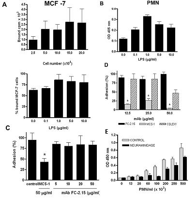
a) Adhesion assay to HUVEC was performed with different numbers of [3H] labeled MCF-7 cells (5×10⁴ loaded cells corresponded to 20,020 cpm) (upper panel). In the lower panel, HUVEC were activated with 0, 0.1, 1, 5, and 10 μg/ml of LPS, before incubation with [3H] labeled MCF-7 cells (5×10⁴ loaded cells corresponded to 22,619 cpm). Data shown are mean of bound cpm ± SD of triplicate determinations in three independent experiments. b) A PMN adhesion assay was performed by the myeloperoxidase technique as described under Methods. HUVEC were previously activated with 0, 0.1, 1, 5, and 10 μg/ml LPS. Paired Student's t test, P<0.05. Data shown are mean ± SD of triplicate determinations in three independent experiments. c) Adhesion assay was performed with [3H] labeled MCF-7. Left panel: Incubations were performed in the presence of PBS (control), MCS-1 (50 μg/ml), and FC-2.15 (5, 10, 20, and 50 μg/ml) (5×10⁴ loaded cells corresponded to 58,114 cpm). Paired Student's t test, P<0.05. Data shown are mean + SD of triplicate determinations in three independent experiments. Right panel: Adhesion assay was performed with [3H] labeled MCF-7 digested with neuraminidase (NANAse) or not (control) (5×10⁴ loaded cells corresponded to 11,210 cpm). d) PMN adhesion to HUVEC was assayed in the presence of the following mAbs: FC-2.15, MCS-1, and CSLEX1 at 12.5, 25, 50 μg/ml. Paired Student's t test, P<0.05. e) PMN adhesion to activated HUVEC was assayed after PMN treatment with 5 U/ml of neuraminidase from Clostridium perfringens or with PBS (control). X-axis represents the number of PMN/ml. Paired Student's t test, P<0.05. Data shown are mean ± SD of triplicate determinations in three independent .