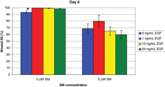
Figure 1.
NHEK cells exposed to 5 μM of SM and treated with EGF. Cells were treated daily with 0, 1, 10, or 20 ng/mL of EGF and stained with 0.1% crystal violet 6 days after wounding. For 0 μM of SM, a significant difference* (P ≤ .05) in wound fill was observed between the cells treated with 0 ng/mL of EGF and the 1, 10, and 20 ng/mL of EGF groups. For 5 μM of SM, cells treated with 1 ng/mL of EGF had the greatest percentage of wound fill, but no statistically significant differences were observed between the EGF treatment groups. Data points represent mean values ± SEM of 3 determinations. A 1-factor ANOVA was used to compare treatment groups at each SM concentration.

