

Abstract
A rapidly inactivating K+ current (A-type current; IA) present in murine colonic myocytes is important in maintaining physiological patterns of slow wave electrical activity. The kinetic profile of colonic IA resembles that of Kv4-derived currents. We examined the contribution of Kv4 α-subunits to IA in the murine colon using pharmacological, molecular and immunohistochemical approaches. The divalent cation Cd2+ decreased peak IA and shifted the voltage dependence of activation and inactivation to more depolarized potentials. Similar results were observed with La3+. Colonic IA was sensitive to low micromolar concentrations of flecainide (IC50 = 11 μM). Quantitative PCR indicated that in colonic and jejunal tissue, Kv4.3 transcripts demonstrate greater relative abundance than transcripts encoding Kv4.1 or Kv4.2. Antibodies revealed greater Kv4.3-like immunoreactivity than Kv4.2-like immunoreactivity in colonic myocytes. Kv4-like immunoreactivity was less evident in jejunal myocytes. To address this finding, we examined the expression of K+ channel-interacting proteins (KChIPs), which act as positive modulators of Kv4-mediated currents. Qualitative PCR identified transcripts encoding the four known members of the KChIP family in isolated colonic and jejunal myocytes. However, the relative abundance of KChIP transcript was 2.6-fold greater in colon tissue than in jejunum, as assessed by quantitative PCR, with KChIP1 showing predominance. This observation is in accordance with the amplitude of the A-type current present in these two tissues, where colonic myocytes possess densities twice that of jejunal myocytes. From this we conclude that Kv4.3, in association with KChIP1, is the major molecular determinant of IA in murine colonic myocytes.
Potassium (K+) currents are important physiological regulators of membrane potential in excitable tissues, including gastrointestinal smooth muscle (see Nelson & Quayle, 1995; Koh et al. 1999b). K+ currents present in the colonic smooth muscle syncytium modulate responses from pacemaker and neural inputs (see Horowitz et al. 1999). Thus, these important currents participate in shaping and defining colonic electrical and mechanical responses. In a previous report, we characterized a rapidly inactivating 4-aminopyridine (4-AP)-sensitive K+ current (A-type current; IA) in murine colonic myocytes (Koh et al. 1999b). The macroscopic A-type current was later shown to be primarily due to 19 pS channels (Amberg et al. 2001). In cells that display repetitive firing, A-type currents participate in regulation of the interspike period (Connor & Stevens, 1971; McCormick & Huguenard, 1992). Application of 4-AP to intact colon muscles results in continuous spiking with a loss of physiological patterns of slow wave activity (Koh et al. 1999b). The inactivation kinetics of the A-type current in colonic muscle cells are dynamically regulated by calcium-calmodulin-dependent protein kinase II (Koh et al. 1999a) and calcineurin (Amberg et al. 2001).
The kinetic profile of native colonic IA resembles macroscopic currents formed by the Kv4 (Shal) family of K+ channel α-subunits (Serodio et al. 1994; Koh et al. 1999b). This observation was supported using the polymerase chain reaction, which demonstrated smooth muscle-specific expression of transcripts encoding Kv4 isoforms but not other Kv family members known to give rise to A-type currents (e.g. Kv1.4). However, the molecular identity of colonic IA presently remains unresolved. In studies of other cell types, several tests have been performed to determine the participation of Kv4 channels in A-type currents (e.g. Watkins & Mathie, 1994; Yeola & Snyders, 1997; Faivre et al. 1999; Wickenden et al. 1999). These include functional tests, such as the shifting of the voltage dependence of activation and inactivation by di- and trivalent cations and block by flecainide. Together with information about specific expression, these tests can lend support to the hypothesis that Kv4 contributes to whole-cell A-type currents.
We examined the contribution of the three known Kv4 isoforms (Kv4.1, Kv4.2 and Kv4.3) to the A-type current in murine colonic and jejunal myocytes. We also determined the relationship between the KChIP (K+ channel-interacting protein; An et al. 2000) family of Kv4 modulatory subunits and IA in the gastrointestinal tract. Using pharmacological, molecular and immunohistochemical techniques we have attempted to clarify the molecular nature of the A-type current in murine colonic and jejunal myocytes.
METHODS
Preparation and collection of isolated myocytes
Smooth muscle cells were prepared from the tunica muscularis of proximal colons and jejunums removed from BALB/c mice. Mice were anaesthetized with isoflurane (Aerane, Baxter Healthcare Corp., Deerfield, IL, USA) and killed by cervical dislocation. The Institutional Animal Care and Use Committee approved the housing and protocols for the killing of animals. Bowel segments were removed and opened along the longitudinal axis. The resulting sheets were pinned out in a Sylgard-lined dish and washed with Ca2+-free, phosphate-buffered saline (PBS) containing (mm): 125 NaCl, 5.36 KCl, 15.5 NaOH, 0.336 Na2HPO4, 0.44 KH2PO4, 10 glucose, 2.9 sucrose and 11 Hepes; pH adjusted to 7.4 with NaOH. Mucosa and submucosa were removed with fine-tipped forceps.
Pieces of muscle were incubated in Ca2+-free PBS supplemented with 4 mg ml−1 fatty acid-free bovine serum albumin (BSA; Sigma, St Louis, MO, USA), 20 U ml−1 papain (Sigma), 270 U ml−1 collagenase (Worthington Biochemical, Lakewood, NJ, USA) and 1 mm dithiothreitol (Sigma); tissue was incubated at 37 °C in this enzyme solution for 8-12 min and then washed with Ca2+-free PBS. Tissue pieces were agitated gently to create a cell dispersion. Cells were stored at 4 °C in Ca2+-free solution supplemented with minimum essential medium for suspension culture (S-MEM; Sigma) and 0.5 mm CaCl2, 0.5 mm MgCl2, 4.17 mm NaHCO3 and 10 mm Hepes; pH adjusted to 7.4 with Tris.
Dispersed colonic and jejunal smooth muscle cells were collected (Epperson et al. 1999) for RNA isolation (see below). Cells were allowed to settle in a glass-bottomed chamber located on an inverted microscope. Individual myocytes were selected by the same criteria used during electrophysiological experiments (elongated, spindle-shaped cells, 100-500 μm long, 5-10 μm in diameter) and aspirated into large-bore pipettes (tip diameters > 10 μm). After 60 smooth muscle cells were collected, the contents of the pipette were expelled into RNase-free tubes, frozen in liquid nitrogen and stored at −70 °C.
Voltage-clamp methods
All experiments were performed at room temperature (25 °C) within 6 h of dispersing cells using a perfused recording chamber mounted on an inverted microscope. Currents were amplified with an Axopatch 200B amplifier and digitized with a DigiData 1200 A/D converter (Axon Instruments, Union City, CA, USA). Data were digitized at 4 kHz, filtered at 1 kHz and recorded using pCLAMP 6 software (Axon Instruments). Fire-polished glass pipettes with resistances of 1-4 MΩ were used. For determination of whole-cell current densities (pA pF−1), cell membrane capacitance was calculated from the time constant of a capacitance current elicited by a 5 mV depolarization from −60 mV. Series resistance (2-5 MΩ) was compensated to at least 70 %. The myocytes were bathed in a nominally Ca2+-free solution containing (mm): 5 KCl, 135 NaCl, 2 MnCl2, 10 glucose, 1.2 MgCl2 and 10 Hepes; pH adjusted to 7.4 with Tris. The pipette solution contained (mm): 130 KCl, 5 MgCl2, 2.7 K2ATP, 0.1 Na2GTP, 2.5 creatine phosphate disodium, 5 Hepes and 10 BAPTA; pH adjusted to 7.2 with Tris. Cadmium (CdCl2; Sigma), lanthanum (LaCl3; Sigma), flecainide (acetate salt; Sigma) and tetraethylammonium (TEA, chloride salt; Sigma) were dissolved in deionized water. Desired concentrations were obtained by further dilution in the extracellular solution. These agents were applied after completion of control recordings by exchanging the external solution in a continuous fashion.
Data are reported as the mean ± s.e.m.; n refers to the number of cells (from at least 3 animals) from which recordings were made. Statistical significance was evaluated by Student's paired t test or two-way analysis of variance, where appropriate. P values less than 0.05 were considered significant. Methods of curve fitting were performed using pCLAMP 6 (Axon Instruments) or GraphPad Prism (GraphPad Software Inc., San Diego, CA, USA).
Total RNA isolation and quantitative PCR
Total RNA was isolated from mouse proximal colon and jejunum tissue (mucosa and submucosa removed) and isolated cells using the SNAP Total RNA isolation kit (Invitrogen, Carlsbad, CA, USA), according to the manufacturer's protocol. Total RNA was also isolated from whole mouse brain and ventricle. Briefly, the animals were anaesthetized by inhalation of isoflurane and killed by decapitation. Tissues were obtained by gross dissection. First-strand cDNA was prepared from the total RNA using the Superscript Reverse Transcriptase kit (Gibco, Gaithersburg, MD, USA). One microgram of total RNA was reverse transcribed with 200 units reverse transcriptase in a 20 μl reaction mixture containing 25 ng oligo-dT primer, 500 μM each dNTP, 75 mm KCl, 3 mm MgCl2, 10 mm dithiothreitol and 50 mm Tris-HCl (pH 8.3). As a control, PCR primers specific for β-actin (GenBank accession no. V01217) nucleotides 2383-2402 and 3071-3091 were used to establish that the cDNA prepared above was non-genomic. The β-actin-specific primers amplified only the intron-less amplification product from all cDNA samples, indicating that these preparations were free of genomic DNA contamination (data not shown).
The cDNA reverse transcription products were amplified with Kv4.1, Kv4.2, Kv4.3, KChIP1, KChIP2, KChIP3, KChIP4 and β-actin-specific primers by reverse transcriptase (RT)-PCR using AmpliTaq Gold reagents (PE Applied Biosystems, Foster City, CA, USA). The primer pairs used are listed in Table 1. The amplification protocol for these primer pairs was as follows: 95 °C for 10 min to activate the AmpliTaq polymerase, followed by 40 cycles of 95 °C for 15 s and 60 °C for 1 min. Primers specific for KChAP (K+ channel-associated protein; GenBank accession no. NM031784) nucleotides 729-752 and 842-865 were amplified using the above protocol modified to 35 cycles of 95 °C for 15 s and 60 °C for 1 min. Aliquots of the PCR reactions were analysed by 2 % agarose gel electrophoresis and visualized by ethidium bromide fluorescence. PCR amplification products from each primer pair were extracted and identities confirmed by DNA sequencing.
Table 1.
Real-time PCR primer pairs
| mRNA (accession no.) | Position | Primer sequence (5′ to 3′) |
|---|---|---|
| Kv4.1 (M64226) | 1538–1559 | GCCGCAGTACCTCAGTATCATC |
| 1632–1653 | GACAGAGGCAGTAGAGTTGGCA | |
| Kv4.2 (AF107780) | 1529–1549 | ATCGCCCATCAAGTCACAGTC |
| 1619–1639 | CCGACACATTGGCATTAGGAA | |
| Kv4.3L (AF107781) | 1398–1418 | CAAGACCACCTCACTCATCGA |
| 1553–1573 | TCGAGCTCTCCATGCAGTTCT | |
| KChIP1 (AB075041) | 126–145 | ACCGGCCTGAGGGACTGGAG |
| 270–289 | GCTGGCATCTCCGTGAGGGA | |
| KChIP2 (AB044570) | 494–513 | TTGTGGCTGGCTTGTCGGTG |
| 664–683 | TGTTCCCTTGGGGCCTCCTC | |
| KChIP3 (AF287733) | 176–195 | GGGCGCATACCACTGAGCAA |
| 324–343 | CTGATGGCGCACCGTGGATA | |
| KChIP4 (AF305071) | 229–250 | GAGGCCCAGAGCAAATTCACCA |
| 394–414 | TCCATTGTGGTCCGTGTCGAA | |
| β-Actin (V01217) | 2206–2223 | GCTGTGTTGTCCCTGTAT |
| 2385–2402 | GTGGTGGTGAAGCTGTAG |
Included are the transcript of interest and corresponding GenBank sequence accession number used for primer design, position of the primers in the sequence and the primer sequence (5′ to 3′).
Real-time RT-PCR was used to quantify the relative expression levels of Kv4 and KChIP isoforms using SYBR Green I as the fluorescent probe on an ABI 5700 sequence detector (PE Applied Biosystems). Real-time PCR was performed in triplicate using the same amplification protocol described above. Reaction mixtures lacking cDNA (no-template controls) were included during each session to assess contamination and non-specific amplification. To examine Kv4 and KChIP primer efficiencies, standard curves were generated for each primer pair by regression analysis of PCR amplifications on log10 serial dilutions of cDNA. For each Kv4 and KChIP isoform, the β-actin standard curve was used to determine the relative abundance of each transcript, which was then normalized to the amount of β-actin transcript present within the same sample (Walker et al. 2001).
Data are reported as the mean ± s.e.m.; n refers to the number of animals from which tissues were collected. Statistical significance was evaluated by one-way analysis of variance with Tukey's multiple comparison test. P values less than 0.05 were considered significant.
Immunohistochemistry
Mouse proximal colon and jejunum were collected and flushed with PBS pH 7.4. The tissues were fixed with paraformaldehyde (4 %) in PBS for 20 min. The fixed sections were cryoprotected in increasing gradients of sucrose in PBS (5-20 %). Tissues were embedded in Tissue Tek (Miles Scientific, Naperville, IL, USA) and rapidly frozen in isopentane pre-cooled in liquid nitrogen. Cryosections were cut at 8 μm (Leica CM 3050); endogenous peroxide was quenched by incubating in 0.03 % hydrogen peroxide in PBS for 20 min. The sections were blocked in 1 % BSA containing 0.1 % Triton-X for 1 h at room temperature. Endogenous biotin was blocked with a biotin-blocking kit (Molecular Probes, Eugene, OR, USA) according to the manufacturer's instructions. Excess blocking serum was removed and sections were incubated with 1:100 primary antibody (Kv4.2 or Kv4.3) overnight at room temperature. The anti-Kv4.2 and −Kv4.3 antibodies were obtained commercially (Alomone Labs, Jerusalem, Israel) and have been used previously (e.g. Anderson et al. 2000; Zhang, T. T. et al. 2001). The Kv4.3 antibody recognizes long and short forms of Kv4.3. Biotinylated goat anti-rabbit immunoglobulin and horseradish peroxidase-conjugated antibiotin antibody were applied to the sections for 30 min at room temperature. Peroxidase activity was visualized by applying 3,3′-diaminobenzidine containing 0.05 % hydrogen peroxide for 5 min at room temperature, and a Haematoxylin counterstain was applied. The sections were rinsed in tap water, dehydrated, cleared and mounted with coverslips. Negative control sections were tested with each antibody and were processed as above except that primary antibodies were substituted with: (1) PBS; and (2) pre-absorbed antibody (2 h at room temperature with antigens supplied by Alomone Labs). Photomicrographs were made with a Nikon eclipse E600 microscope incorporating Nomarski optics.
RESULTS
Divalent and trivalent inorganic cations modulate murine colonic myocyte A-type currents
The kinetic profile of IA in the murine colon resembles that of currents formed by α-subunits of the Kv4 family of voltage-gated potassium channels (Serodio et al. 1994; Koh et al. 1999b). We examined the effect of inorganic di- and trivalent cations on IA in murine colonic myocytes because these agents have been used previously in an attempt to correlate the behaviour of currents due to Kv4 channels and native A-type currents in ventricular myocytes (e.g. Fiset et al. 1997; Faivre et al. 1999; Wickenden et al. 1999). A-type currents were recorded from colonic myocytes with the conventional whole-cell patch-clamp technique. To minimize contamination from Ca2+-activated currents (i.e. large conductance Ca2+-activated K+ currents), we used an external solution containing Mn2+ (2 mm) and included BAPTA (10 mm) in the pipette solution.
From a holding potential of −80 mV, step depolarizations evoked inactivating A-type currents with an additional sustained component (Fig. 1A). Addition of Cd2+ (500 μM) reduced peak current at voltages positive to −20 mV (P < 0.05; n = 7; Fig. 1B). Digitally subtracted difference currents (Fig. 1C), which represent the Cd2+-sensitive component, indicated that the current affected by Cd2+ is principally a rapidly inactivating, A-type current. However, at +20 mV, Cd2+ also decreased the peak and sustained components of the voltage-dependent outward current (P < 0.05; n = 7; Fig. 1D). The sustained component of the voltage-dependent outward current in colonic myocytes resembled a classical delayed rectifier (Koh et al. 1999b). It is therefore possible that a portion of the observed effect of Cd2+ may have resulted from an effect on the delayed rectifier current instead of IA. To examine this, we applied a 250 ms prepulse to −30 mV to inactivate IA and retested the effects of Cd2+. This technique effectively isolated the delayed rectifier component of outward current (data not shown). Using this protocol, application of Cd2+ (500 μM) increased the peak current while the sustained current decreased, as above (data not shown; P < 0.05; n = 6). The effects of Cd2+ on outward currents are consistent with a shift in the voltage dependence of activation and inactivation of IA (e.g. Agus et al. 1991). In comparison to control conditions, 500 μM Cd2+ shifted the voltage of half-activation by 12.08 ± 1.9 mV in the positive direction (P < 0.05; n = 6; Fig. 1E), while the voltage of half-inactivation was shifted 14.23 ± 1.4 mV in the positive direction (P < 0.05; n = 6; Fig. 1F).
Figure 1. Cadmium decreases peak colonic A-type current and shifts the voltage dependence of activation and inactivation to more depolarized potentials.
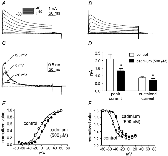
A and B, whole-cell A-type currents recorded from a colonic myocyte before (A) and after (B) Cd2+ (500 μM). The membrane potential was stepped for 500 ms from −80 mV to potentials between −80 and +40 mV. C, difference currents obtained by digitally subtracting records in B from those in A. D, summarized data quantifying the effect of Cd2+ (500 μM) on peak and sustained current at a test potential of +20 mV. * Significant reduction in peak and sustained current amplitude after Cd2+ compared to control (P < 0.05; n = 7). E, voltage dependence of activation of A-type current K+ permeabilities. Peak K+ currents (at test potentials between −80 and +40 mV; not shown) were converted into permeabilities using the Goldman-Hodgkin-Katz current equation. Permeabilities were then normalized, plotted as a function of test potential and fitted with a Boltzmann function. F, voltage dependence of inactivation of A-type current. Normalized peak currents at +20 mV (I/Imax; not shown) are plotted as a function of the conditioning potential ranging from −80 to +20 mV for 3 s and fitted with a Boltzmann function.
La3+ has been shown to inhibit cerebellar granule neuronal A-type currents, which are reported to be due to Kv4 channels (Watkins & Mathie, 1994; Shibata et al. 1999, 2000). In murine colonic myocytes, La3+ (100 μM) decreased IA (Fig. 2A and B). The La3+-sensitive current (Fig. 2C) was limited to IA. For example, at +20 mV, La3+ decreased only the peak current (Fig. 2D; P < 0.05; n = 4), while the sustained current was not significantly affected (Fig. 2D; P > 0.05; n = 4). As with Cd2+, La3+ shifted the voltage dependence of activation and inactivation to more positive potentials (i.e. half-activation shifted by +20.93 ± 1.69 mV and half-inactivation shifted by +18.25 ± 1.07 mV; P < 0.05; n = 4; Fig. 2E and F).
Figure 2. Lanthanum decreases peak colonic A-type current and shifts the voltage dependence of activation and inactivation to more depolarized potentials.
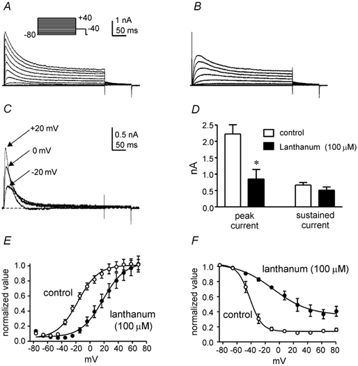
A and B, whole-cell A-type currents recorded from a colonic myocyte before (A) and after (B) La3+ (100 μM). The membrane potential was stepped for 500 ms from −80 mV to potentials between −80 and +40 mV. C, difference currents obtained by digitally subtracting records in B from those in A. D, summarized data quantifying the effect of La3+ (100 μM) on peak and sustained current at a test potential of +20 mV. * Significant reduction in peak current amplitude after La3+ compared to control (P < 0.05; n = 4). E, voltage dependence of activation of A-type current K+ permeabilities. Peak K+ currents (at test potentials between −80 and +40 mV; not shown) were converted into permeabilities using the Goldman-Hodgkin-Katz current equation. Permeabilities were then normalized, plotted as a function of test potential and fitted with a Boltzmann function. F, voltage dependence of inactivation of A-type current. Normalized peak currents at +20 mV (I/Imax; not shown) are plotted as a function of the conditioning potential (ranging from −80 to +20 mV for 3 s) and fitted with a Boltzmann function.
Sensitivity of murine colonic myocyte A-type currents to flecainide
We also tested the effects of flecainide on IA of colonic myocytes. Previous studies have shown that flecainide blocks voltage-dependent K+ channels. Kv4 K+ channels have been shown to have a higher sensitivity to flecainide than Kv1 channels (Grissmer et al. 1994; Yamagishi et al. 1995; Yeola & Snyders, 1997). Exposure of colonic myocytes to flecainide (10 μM) resulted in a decrease in peak IA (P < 0.05; n = 5; Fig. 3A and B). These effects of flecainide were dose dependent with an IC50 of 11 ± 1 μM (on peak current with step depolarizations to 0 mV; Fig. 3C and D; n = 5). Similar IC50 values have been reported for heterologously expressed Kv4 isoforms (Yeola & Snyders, 1997).
Figure 3. Inhibition of colonic A-type current by flecainide.
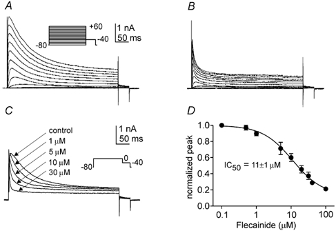
A and B, whole-cell A-type currents recorded from a colonic myocyte before (A) and after (B) flecainide (10 μM). The membrane potential was stepped for 500 ms from −80 mV to potentials between −80 and +40 mV. C, whole-cell A-type currents recorded from a colonic myocyte before and after different concentrations of flecainide (concentrations indicated in figure). The membrane potential was stepped for 500 ms from −80 to 0 mV. D, dose-dependent inhibition of peak A-type current by flecainide. Normalized peak currents at 0 mV (I/Imax; not shown) were plotted as a function of flecainide concentration (ranging from 0.1 to 100 μM) and fitted with a variable slope logistic equation, from which an IC50 of 11 ± 1 μM was determined.
Expression of Kv4 isoforms in murine colon and jejunum
Previously, we demonstrated expression of Kv4.1, Kv4.2 and Kv4.3 transcripts in isolated murine colonic myocytes (Koh et al. 1999b). In the present study we performed quantitative analyses to determine which isoform is predominantly expressed in murine colonic smooth muscles. We also tested expression in jejunal muscle for comparison. Relative expression levels of transcripts encoding each Kv4 isoform were determined by real-time PCR. Qualitative RT-PCR was used initially to test Kv4-specific primers suitable for real-time PCR. Consistent with our previous findings, transcripts for each of the three Kv4 isoforms were found in colonic cDNA (Fig. 4A). Each Kv4 isoform was also detected in jejunal cDNA (Fig. 4B). For each primer pair, only a single product of the correct size was visualized. Amplicon identity was confirmed by DNA sequence analysis of gel-extracted products (data not shown). The primer pair for Kv4.3 flanked the alternatively spliced region of Kv4.3 (e.g. Ohya et al. 1997; Takimoto et al. 1997). We found only the long isoform of Kv4.3 in colonic and jejunal muscles.
Figure 4. Quantification of Kv4 transcripts in colon and jejunum.
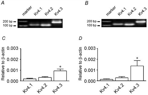
A and B, RT-PCR analysis of primer pairs used for real-time PCR in colon (A) and jejunum (B). From left to right: 100 bp marker; Kv4.1 (amplicon = 116 bp); Kv4.2 (amplicon = 111 bp); Kv4.3, long isoform (amplicon = 176 bp). Amplicon identity confirmed by DNA sequencing; see Table 1 for primer sequences. C and D, Kv4.1, Kv4.2 and Kv4.3 gene expression relative to β-actin in colon (C) and jejunum (D) as determined by real-time PCR. * Significantly greater expression of Kv4.3 transcripts relative to Kv4.1 or Kv4.2 within the same tissue (P < 0.05; n = 5).
Following RT-PCR analysis, to assess primer efficiency, standard curves (threshold cycle vs. log10 [amplicon]) were generated and slopes determined for each primer pair. The slopes obtained for the Kv4.1, Kv4.2 and Kv4.3 primer pairs were similar (3.4, 3.7 and 3.5, respectively) and were within the range of the calculated standard deviations for each pair (P > 0.05; n = 3). The efficiencies of each primer pair were thus considered equal, allowing for relative quantification of Kv4 transcripts.
The primer pairs were used to perform quantitative real-time PCR on murine colonic and jejunal cDNA (mucosa and submucosa removed as described above). Amplification in no-template controls was never observed. Relative quantifications were normalized between samples and PCR sessions using endogenous β-actin as a standard. As illustrated in Fig. 4C and D, in murine colonic and jejunal smooth muscle, transcripts encoding Kv4.3 were present in greater relative abundance than those encoding Kv4.1 and Kv4.2 (P < 0.05; n = 5 by one-way analysis of variance with Tukey's multiple comparison test). For each Kv4 isoform, the relative expression between colon and jejunum was not significantly different (P > 0.05; n = 5). As a control, each Kv4 primer pair was tested on cDNA isolated from whole murine brain and ventricle. Consistent with previous reports (e.g. Dixon & McKinnon, 1994; Serodio et al. 1996), the rank order of transcript abundance was Kv4.2 > Kv4.3 ≫ 4.1 with a ratio of 1.0:0.47:0.27 in brain and 1.0:0.28:0.05 in ventricle.
Antibodies raised against specific epitopes of Kv4.2 and Kv4.3 channels were used to assess the expression of channel proteins in the murine proximal colon and jejunum. Antibodies for Kv4.1 were not available. In the colon, intense Kv4.3-like immunoreactivity was observed in smooth muscle cells of the longitudinal and circular muscle layers (Fig. 5B). Substantially weaker Kv4.2-like immunoreactivity was resolved in colonic smooth muscle cells (Fig. 5A). In addition, Kv4.2- and Kv4.3-like immunoreactivity was also detected in other cell types (e.g. myenteric neurons) in the colon. Considerably weaker Kv4-like immunoreactivity was observed in jejunal myocytes of the longitudinal and circular muscle layers (Fig. 5C and D). Two independent negative control experiments were performed to assess non-specific binding of Kv4 antibodies. Immunoreactivity was not observed in control sections in which primary antibodies were omitted, and immunoreactivity was not detected when primary antibodies were pre-absorbed (data not shown).
Figure 5. Kv4.2- and Kv4.3-like immunoreactivity in the tunica muscularis of murine colon and jejunum.
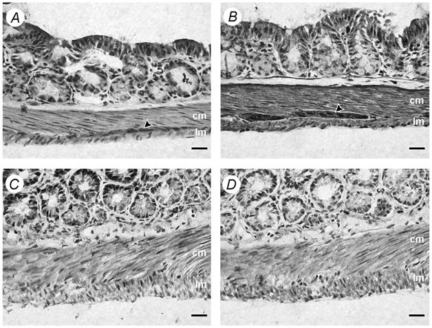
Haematoxylin counterstain. A and B, Kv4.2-like (A) and Kv4.3-like (B) immunoreactivity (in brown) throughout the circular (cm) and longitudinal (lm) muscle layers of the tunica muscularis in murine colon. Arrowheads indicate Kv4-like immunoreactivity found within myenteric ganglia. C and D, Kv4.2-like (C) and Kv4.3-like (D) immunoreactivity (in brown) throughout the circular (cm) and longitudinal (lm) layers of the tunica muscularis in murine jejunum. Scale bars, 20 μm.
A-type current in murine colonic and jejunal myocytes
To assess the functional relevance of the contrasting observations in colon and jejunum, we compared the density of A-type current (pA pF−1) in dispersed colonic and jejunal myocytes. Measured membrane capacitances ranged between 30 and 40 pF with no significant difference between cells of the two tissues (P > 0.05; n = 10). Current densities were determined in the presence of external TEA (10 mm) to minimize contamination from the sustained component of the voltage-dependent outward current in these cells (see Koh et al. 1999b). From a holding potential of −80 mV, typical responses of colonic and jejunal myocytes to 500 ms step depolarizations (potentials between −70 and +20 mV) are shown in Fig. 6A and B, respectively. When compared to jejunal myocytes, A-type current densities were significantly greater in colonic myocytes at step potentials positive to −40 mV (P < 0.05; n = 5; Fig. 6C). At +20 mV, the density of colonic myocyte A-type current was 36.6 ± 3.1 pA pF−1; in jejunal myocytes the density was 18.4 ± 2.9 pA pF−1. As with colonic myocytes, A-type current of jejunal myocytes displayed dose-dependent sensitivity to flecainide with observable effects at low micromolar concentrations (IC50 = 24 ± 2 μM; n = 3; see Fig. 6D). Jejunal A-type currents also displayed rapid recovery from inactivation, typical of Kv4 conductances, with a time constant for recovery of 72 ms at −80 mV following a 1 s prepulse to 0 mV (n = 6; Fig. 6B inset).
Figure 6. Comparison of colonic and jejunal A-type currents.
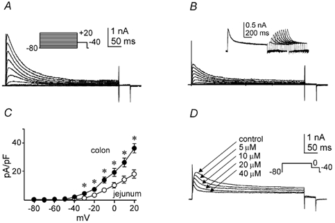
A and B, whole-cell A-type currents recorded from a colonic (A) and a jejunal (B) myocyte in the presence of TEA (10 mm). The membrane potential was stepped for 500 ms from −80 mV to potentials between −70 and +20 mV. Inset in B ahows representative traces demonstrating jejunal IA recovery from inactivation. The membrane potential was stepped for 1 s from −80 to 0 mV followed by a repolarization to −80 mV. Recovery from inactivation was then determined by stepping the membrane potential back to 0 mV after incrementally (50 ms) increasing periods of time. C, peak current density (pA pF−1) as a function of voltage in colonic and jejunal myocytes. * Significantly greater current density in colonic myocytes relative to jejunal myocytes (P < 0.05; n = 5). D, whole-cell A-type currents recorded from a jejunal myocyte before and after different concentrations of flecainide (concentrations indicated in figure). The membrane potential was stepped from −80 to 0 mV for 500 ms.
Expression of KChIP isoforms in murine colon and jejunum
Transcriptional expression of Kv4 channels was equivalent in colonic and jejunal myocytes, but qualitative analysis of protein expression by immunohistochemical analysis and measurements of current densities differed between these cell types. Further studies were performed to attempt to determine the reason why colonic and jejunal cells differed in A-type current density. Recent studies have shown that functional expression of Kv4 currents depends upon parallel expression of chaperone proteins, such as KChIP, that appear to facilitate trafficking of translated protein to the plasma membrane (An et al. 2000; Bahring et al. 2001). Real-time PCR was used to determine the relative expression of each KChIP isoform in murine colonic and jejunal smooth muscles. As with the Kv4 primer pairs, qualitative RT-PCR was used initially to test KChIP gene-specific primers suitable for real-time PCR. Transcripts encoding each of the four KChIP isoforms were present in cDNA prepared from isolated colonic and jejunal myocytes (Fig. 7A and B). For each primer pair, only a single product of the correct size was visualized and amplicon identity was confirmed by DNA sequence analysis of gel-extracted products. Where appropriate, the KChIP primer pairs were designed to amplify all known KChIP splice variants, with no attempt at assessing the relative contribution of individual splice variants.
Figure 7. Quantification of KChIP transcripts in colon and jejunum.
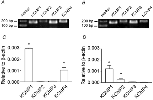
A and B, detection of KChIP transcripts in isolated colonic (A) and jejunal (B) myocytes and RT-PCR analysis of primer pairs used for real-time PCR. From left to right: 100 bp marker; KChIP1 (amplicon = 164 bp); KChIP2 (amplicon = 190 bp); KChIP3 (amplicon = 168 bp); and KChIP4 (amplicon = 186 bp). Amplicon identity confirmed by DNA sequencing; see Table 1 for primer sequences. C, KChIP1, KChIP2, KChIP3 and KChIP4 gene expression relative to β-actin in colon as determined by real-time PCR. * Significantly greater expression of KChIP1 transcripts relative to KChIP2, KChIP3 or KChIP4 (P < 0.05; n = 5); † significantly greater expression of KChIP4 transcripts relative to KChIP2 or KChIP3 (P < 0.05; n = 5). D, KChIP1, KChIP2, KChIP3 and KChIP4 gene expression relative to β-actin in jejunum as determined by real-time PCR. * Significantly greater expression of KChIP1 transcripts relative to KChIP2, KChIP3 or KChIP4 (P < 0.05; n = 5); † significantly greater expression of KChIP2 transcripts relative to KChIP3 or KChIP4 (P < 0.05; n = 5).
The slopes obtained for the KChIP1, KChIP2, KChIP3 and KChIP4 primer pairs were similar (3.0, 2.8, 2.9 and 3.1, respectively) and were within the range of the calculated standard deviations for each pair (P > 0.05; n = 3). The primer pairs were therefore considered to have equal efficiency. Employing the same control strategies as for Kv4 quantification, these primers were used for relative quantification of KChIP expression in murine colonic and jejunal smooth muscle. In colon and jejunum, transcripts encoding KChIP1 predominated (P < 0.05; n = 5; Fig. 7C and D). In colon, the relative abundance of total KChIP transcript was 2.6-fold greater than in jejunum (P < 0.05; n = 5). As a control, each KChIP primer pair was tested on cDNA isolated from whole murine brain and ventricle. Consistent with previous reports, the rank orders of transcript abundance were KChIP3 > KChIP4 ≃ KChIP1 ≫ KChIP2 with a ratio of 1.0:0.60:0.53:0.08 in brain (e.g. An et al. 2000; Liss et al. 2001) and KChIP2 ≫ KChIP1 ≃ KChIP3 ≃ KChIP4 with a ratio of 1.0:0.003:0.002:0.001 in ventricle (e.g. Ohya et al. 2001; Rosati et al. 2001).
We also designed primers for an unrelated K+ channel-associated protein, KChAP, the co-expression of which is also known to increase Kv4 current density (Kuryshev et al. 2000, 2001). After 35 amplification cycles, RT-PCR detected KChAP transcripts in cDNA from mouse ventricle and brain, but did not detect KChAP transcripts in colonic or jejunal cDNA (n = 3; data not shown).
DISCUSSION
Previously, we characterized an A-type current (IA) in murine colonic myocytes that dampens excitability and may participate in maintaining the phasic pattern of electrical activity observed in intact colon tissue preparations (Koh et al. 1999b). Subsequent investigation identified 19 pS channels in colonic myocytes with voltage-dependent and regulatory properties consistent with macroscopic A-type currents (Amberg et al. 2001). Kinetic and molecular analysis of colonic IA suggested that Kv4 α-subunits, as opposed to other Kv family members (e.g. Kv1.4), may encode IA (Koh et al. 1999b). In the present study we sought to determine the relative contribution of Kv4 isoforms to A-type currents in the murine colonic cells. Using a variety of techniques we conclude that the A-type currents are likely to be due to Kv4 expression, and analyses of transcription and protein expression suggest that Kv4.3 is the predominant isoform. Our data also suggest that expression of KChIP1 in gastrointestinal myocytes may regulate the current density of A-type currents.
We used quantitative real-time PCR to establish the relative expression levels of transcripts encoding each Kv4 isoform in mouse proximal colon. For comparative purposes, we also determined relative expression of Kv4 isoforms in jejunal smooth muscles. We have previously demonstrated smooth muscle cell-specific expression of Kv4 transcripts using qualitative RT-PCR on isolated colonic myocytes (Koh et al. 1999b). In this study we showed that transcripts encoding Kv4.3 were 3-fold more abundant than Kv4.1 transcripts and 2-fold more abundant than Kv4.2 transcripts in colonic and jejunal smooth muscle. Kv4.3 appears to be alternatively spliced in some tissues (e.g. Ohya et al. 2001); we only detected the long form in colonic and jejunal muscles. This observation is consistent with a previous report describing tissue-specific expression of Kv4.3 splice variants (Ohya et al. 1997). There were no significant differences in the levels of Kv4 transcripts in colon and jejunum. A caveat to this conclusion is that RNA from colonic and jejunal muscles with mucosa and submucosa removed was used for the quantitative analysis of Kv4 expression. Cell types other than myocytes, including interstitial cells of Cajal and enteric neurons, are present in these muscles and contain transcripts that could influence absolute quantification.
To provide further support for the measurements of transcriptional expression and to address the issue of contamination from non-muscle cells, we investigated myocyte-specific expression of Kv4.2 and Kv4.3 channels with immunohistochemistry. Strong Kv4.3-like immunoreactivity was observed in colonic myocytes, whereas Kv4.2-like immunoreactivity was relatively weaker. Kv4.2- and Kv4.3-like immunoreactivities were also substantially weaker in jejunal myocytes. To further test these observations we also characterized the current density of IA in dispersed colonic and jejunal myocytes. The stronger Kv4.3-like immunoreactivity in the colon correlated with 2-fold greater current density than in jejunal myocytes.
There was a discrepancy between the levels of Kv4 transcript expression and the levels of Kv4 protein and IA density in colonic and jejunal myocytes. We considered the possibility that this discrepancy may be due to differential expression of KChIP proteins in these cells. KChIPs, which belong to the neuronal calcium sensor (NCS) family of proteins, are positive modulators of native and heterologously expressed Kv4-derived currents (An et al. 2000; Decher et al. 2001; Liss et al. 2001). These auxiliary proteins enhance Kv4 current density by increasing expression of the channels in the plasma membrane (An et al. 2000; Bahring et al. 2001). KChIPs also modify the kinetic behaviour of Kv4 channels (Beck et al. 2002). Kv4 channels underlie the A-type current (ITO) in ventricular myocytes (Xu et al. 1999; see Nerbonne, 2000), and the pattern of KChIP2 expression has recently been shown to mirror the transmural gradient of ITO in canine and human ventricles (Rosati et al. 2001). In transgenic mice harbouring a targeted null-KChIP2 allele, heterozygotes displayed ventricular ITO that was reduced by approximately half of the current in wild-type myocytes (Kuo et al. 2001). Homozygote null-KChIP2 mice did not express functional ITO. By analogy with cardiac muscle, we suggest that similar regulation of functional Kv4 channels by KChIPs may occur in gastrointestinal smooth muscles and explain the disparity between transcriptional expression of Kv4 isoforms and current density in colonic and jejunal muscles. We detected transcripts encoding KChIPs in colonic and jejunal myocytes and, in agreement with our hypothesis, total KChIP transcripts were 2.6-fold greater in colon than in jejunum. In these tissues KChIP1 was the dominant isoform.
Our data suggest that in gastrointestinal smooth muscles, functional expression of Kv4 may be regulated by the pattern of KChIP expression. Another member of the NCS protein family, frequenin (NCS-1), has been shown to act as a positive modulator of Kv4 currents (Nakamura et al. 2001b). Although examination of other NCS family members in gastrointestinal smooth muscle is warranted, differences, namely recovery from inactivation and increased current density, between heterologously expressed Kv4 channels and native colonic IA are more consistent with the actions of KChIP than those of frequenin (An et al. 2000; Nakamura et al. 2001a,b). Similarly, expression of other modulatory subunits such as minK-related peptide 1(MiRP1; Zhang, M. et al. 2001) and Kvβ (Yang et al. 2001) should be examined, although the importance of these proteins may be tentatively discounted for similar reasons to frequenin. Expression of another positive effector of Kv4 channels, KChAP (Kuryshev et al. 2000, 2001), was not evident in colonic and jejunal muscles.
The pharmacological characterization of colonic IA presented in this study provides additional supportive evidence linking Kv4 channels to this current. We examined the sensitivity of IA to the antiarrhythmic flecainide. A-type currents formed by Kv4 channels are more sensitive to inhibition by flecainide (IC50 ≤ 20 μM) than those formed by Kv1 channels (IC50 ≥ 50 μM; Grissmer et al. 1994; Yamagishi et al. 1995; Yeola & Snyders, 1997; Rolf et al. 2000). Colonic and jejunal IA were sensitive to low micromolar concentrations of flecainide, with IC50 values of 11 and 24 μM, respectively. These concentrations of flecainide are well below levels reported to inhibit Kv1 channels and are comparable with levels reported to inhibit expressed Kv4 channels.
With the exception of current density, jejunal IA is kinetically and pharmacologically similar to that found in colon (Koh et al. 1999b; present paper). Jejunal A-type currents recovered from inactivation rapidly (τrecovery of 72 ms) and were sensitive to micromolar levels of flecainide (IC50 of 24 μM), suggesting that they were also formed by Kv4 α-subunits. The minor differences in τrecovery and flecainide sensitivity may reflect the difficulty in isolating IA in jejunal myocytes, where IA is smaller in amplitude, and classical delayed rectifier-like currents are more dominant in macroscopic current recordings.
Several investigators have tested the effects of inorganic cations, such as Cd2+ and La3+, on native A-type currents (e.g. Mayer & Sugiyama, 1988; Imaizumi et al. 1990; Agus et al. 1991; Watkins & Mathie, 1994; Wickenden et al. 1999). As noted above, Kv4 channels underlie ITO in ventricular myocytes (see Nerbonne, 2000). The effects of Cd2+ on native ITO and heterologously expressed Kv4 currents are similar (Fiset et al. 1997; Faivre et al. 1999; Wickenden et al. 1999). These effects have been shown to depend on negatively charged sialic acid residues of Kv4 proteins (Ufret-Vincenty et al. 2001). In an analogous fashion, Cd2+ decreased the peak current of colonic myocyte IA and shifted the voltage dependences of activation and inactivation to more depolarized potentials. Qualitatively, the effect of Cd2+ on colonic IA resembles the effects observed on expressed Kv4 channels. However, the depolarizing shift of voltage dependence induced by Cd2+ in this study was less dramatic than those reported previously (e.g. Fiset et al. 1997). This is likely to be due to the presence of Mn2+ (2 mm) in the external solution we used to minimize Ca2+-activated currents. Procedures to minimize Ca2+-activated K+ currents (BK current) were necessary in our experiments because depolarizations positive to 0 mV were strongly contaminated with BK currents. For the A-type current of rat sensory neurons, Mn2+ (2.5 mm) shifted the voltage dependence of activation and inactivation by +7 and +14 mV, respectively (Mayer & Sugiyama, 1988). Taking these values into account, the depolarizing shift of voltage dependences observed with Cd2+ in this study is comparable to those seen in previous studies characterizing Kv4-derived A-type currents. We also examined the modulatory properties of La3+ on the A-type current of murine colonic myocytes. La3+ inhibited IA and shifted voltage dependence of activation and inactivation to more depolarized potentials. These results are consistent with a previous report on the effects of La3+ on the A-type current of cerebellar granule neurons (Watkins & Mathie, 1994), which appears to be formed by Kv4-type channels (Shibata et al. 1999, 2000).
In conclusion, Kv4 channels appear to play an important role in regulating the electrical activity of gastrointestinal smooth muscles. This conclusion is supported by previous functional characterization of colonic IA and by the molecular and pharmacological experiments in this study. Kv4.3 is the predominant molecular species expressed and is likely to be responsible for IA in murine colonic and intestinal myocytes. The degree to which Kv4 expression results in functional channels appears to depend upon parallel expression of KChIP. Future development of conditional knockout animals will be necessary to provide more definitive evidence regarding specific roles of Kv4.3 and KChIP1 in murine gastrointestinal IA as well as their importance in mediating gastrointestinal muscle responses.
Acknowledgments
This study was supported by a program project grant from National Institute of Diabetes and Digestive and Kidney Diseases (DK41315).
REFERENCES
- Agus ZS, Dukes ID, Morad M. Divalent cations modulate the transient outward current in rat ventricular myocytes. American Journal of Physiology. 1991;261:C310–318. doi: 10.1152/ajpcell.1991.261.2.C310. [DOI] [PubMed] [Google Scholar]
- Amberg GC, Koh SD, Perrino BA, Hatton WJ, Sanders KM. Regulation of A-type potassium channels in murine colonic myocytes by phosphatase activity. American Journal of Physiology – Cell Physiology. 2001;281:C2020–2028. doi: 10.1152/ajpcell.2001.281.6.C2020. [DOI] [PubMed] [Google Scholar]
- An WF, Bowlby MR, Betty M, Cao J, Ling HP, Mendoza G, Hinson JW, Mattsson KI, Strassle BW, Trimmer JS, Rhodes KJ. Modulation of A-type potassium channels by a family of calcium sensors. Nature. 2000;403:553–556. doi: 10.1038/35000592. [DOI] [PubMed] [Google Scholar]
- Anderson AE, Adams JP, Qian Y, Cook RG, Pfaffinger PJ, Sweatt JD. Kv4. 2 phosphorylation by cyclic AMP-dependent protein kinase. Journal of Biological Chemistry. 2000;275:5337–5346. doi: 10.1074/jbc.275.8.5337. [DOI] [PubMed] [Google Scholar]
- Bahring R, Dannenberg J, Peters HC, Leicher T, Pongs O, Isbrandt D. Conserved Kv4 N-terminal domain critical for effects of Kv channel-interacting protein 2. 2 on channel expression and gating. Journal of Biological Chemistry. 2001;276:23888–23894. doi: 10.1074/jbc.M101320200. [DOI] [PubMed] [Google Scholar]
- Beck EJ, Bowlby M, An WF, Rhodes KJ, Covarrubias M. Remodelling inactivation gating of Kv4 channels by KChIP1, a small-molecular-weight calcium-binding protein. Journal of Physiology. 2002;538:691–706. doi: 10.1113/jphysiol.2001.013127. [DOI] [PMC free article] [PubMed] [Google Scholar]
- Connor JA, Stevens CF. Voltage clamp studies of a transient outward membrane current in gastropod neural somata. Journal of Physiology. 1971;213:21–30. doi: 10.1113/jphysiol.1971.sp009365. [DOI] [PMC free article] [PubMed] [Google Scholar]
- Decher N, Uyguner O, Scherer CR, Karaman B, Yuksel-Apak M, Busch AE, Steinmeyer K, Wollnik B. hKChIP2 is a functional modifier of hKv4. 3 potassium channels: cloning and expression of a short hKChIP2 splice variant. Cardiovascular Research. 2001;52:255–264. doi: 10.1016/s0008-6363(01)00374-1. [DOI] [PubMed] [Google Scholar]
- Dixon JE, McKinnon D. Quantitative analysis of potassium channel mRNA expression in atrial and ventricular muscle of rats. Circulation Research. 1994;75:252–260. doi: 10.1161/01.res.75.2.252. [DOI] [PubMed] [Google Scholar]
- Epperson A, Bonner HP, Ward SM, Hatton WJ, Bradley KK, Bradley ME, Trimmer JS, Horowitz B. Molecular diversity of KVα- and β-subunit expression in canine gastrointestinal smooth muscles. American Journal of Physiology. 1999;277:G127–136. doi: 10.1152/ajpgi.1999.277.1.G127. [DOI] [PubMed] [Google Scholar]
- Faivre JF, Calmels TP, Rouanet S, Javre JL, Cheval B, Bril A. Characterisation of Kv4. 3 in HEK293 cells: comparison with the rat ventricular transient outward potassium current. Cardiovascular Research. 1999;41:188–199. doi: 10.1016/s0008-6363(98)00215-6. [DOI] [PubMed] [Google Scholar]
- Fiset C, Clark RB, Larsen TS, Giles WR. A rapidly activating sustained K+ current modulates repolarization and excitation-contraction coupling in adult mouse ventricle. Journal of Physiology. 1997;504:557–563. doi: 10.1111/j.1469-7793.1997.557bd.x. [DOI] [PMC free article] [PubMed] [Google Scholar]
- Grissmer S, Nguyen AN, Aiyar J, Hanson DC, Mather RJ, Gutman GA, Karmilowicz MJ, Auperin DD, Chandy KG. Pharmacological characterization of five cloned voltage-gated K+ channels, types Kv1. 1, 1.2, 1.3, 1.5, and 3.1, stably expressed in mammalian cell lines. Molecular Pharmacology. 1994;45:1227–1234. [PubMed] [Google Scholar]
- Horowitz B, Ward SM, Sanders KM. Cellular and molecular basis for electrical rhythmicity in gastrointestinal muscles. Annual Review of Physiology. 1999;61:19–43. doi: 10.1146/annurev.physiol.61.1.19. [DOI] [PubMed] [Google Scholar]
- Imaizumi Y, Muraki K, Watanabe M. Characteristics of transient outward currents in single smooth muscle cells from the ureter of the guinea-pig. Journal of Physiology. 1990;427:301–324. doi: 10.1113/jphysiol.1990.sp018173. [DOI] [PMC free article] [PubMed] [Google Scholar]
- Koh SD, Perrino BA, Hatton WJ, Kenyon JL, Sanders KM. Novel regulation of the A-type K+ current in murine proximal colon by calcium-calmodulin-dependent protein kinase II. Journal of Physiology. 1999a;517:75–84. doi: 10.1111/j.1469-7793.1999.0075z.x. [DOI] [PMC free article] [PubMed] [Google Scholar]
- Koh SD, Ward SM, Dick GM, Epperson A, Bonner HP, Sanders KM, Horowitz B, Kenyon JL. Contribution of delayed rectifier potassium currents to the electrical activity of murine colonic smooth muscle. Journal of Physiology. 1999b;515:475–487. doi: 10.1111/j.1469-7793.1999.475ac.x. [DOI] [PMC free article] [PubMed] [Google Scholar]
- Kuo HC, Cheng CF, Clark RB, Lin JJ, Lin JL, Hoshijima M, Nguyen-Tran VT, Gu Y, Ikeda Y, Chu PH, Ross J, Giles WR, Chien KR. A defect in the Kv channel-interacting protein 2 (KChIP2) gene leads to a complete loss of ITO and confers susceptibility to ventricular tachycardia. Cell. 2001;107:801–813. doi: 10.1016/s0092-8674(01)00588-8. [DOI] [PubMed] [Google Scholar]
- Kuryshev YA, Gudz TI, Brown AM, Wible BA. KChAP as a chaperone for specific K+ channels. American Journal of Physiology – Cell Physiology. 2000;278:C931–941. doi: 10.1152/ajpcell.2000.278.5.C931. [DOI] [PubMed] [Google Scholar]
- Kuryshev YA, Wible BA, Gudz TI, Ramirez AN, Brown AM. KChAP/Kvβ1. 2 interactions and their effects on cardiac Kv channel expression. American Journal of Physiology – Cell Physiology. 2001;281:C290–299. doi: 10.1152/ajpcell.2001.281.1.C290. [DOI] [PubMed] [Google Scholar]
- Liss B, Franz O, Sewing S, Bruns R, Neuhoff H, Roeper J. Tuning pacemaker frequency of individual dopaminergic neurons by Kv4. 3L and KChip3.1 transcription. EMBO Journal. 2001;20:5715–5724. doi: 10.1093/emboj/20.20.5715. [DOI] [PMC free article] [PubMed] [Google Scholar]
- McCormick DA, Huguenard JR. A model of the electrophysiological properties of thalamocortical relay neurons. Journal of Neurophysiology. 1992;68:1384–1400. doi: 10.1152/jn.1992.68.4.1384. [DOI] [PubMed] [Google Scholar]
- Mayer ML, Sugiyama K. A modulatory action of divalent cations on transient outward current in cultured rat sensory neurones. Journal of Physiology. 1988;396:417–433. doi: 10.1113/jphysiol.1988.sp016970. [DOI] [PMC free article] [PubMed] [Google Scholar]
- Nakamura TY, Nandi S, Pountney DJ, Artman M, Rudy B, Coetzee WA. Different effects of the Ca2+-binding protein, KChIP1, on two Kv4 subfamily members, Kv4. 1 and Kv4.2. FEBS Letters. 2001a;499:205–209. doi: 10.1016/s0014-5793(01)02560-1. [DOI] [PubMed] [Google Scholar]
- Nakamura TY, Pountney DJ, Ozaita A, Nandi S, Ueda S, Rudy B, Coetzee WA. A role for frequenin, a Ca2+-binding protein, as a regulator of Kv4 K+-currents. Proceedings of the National Academy of Sciences of the USA. 2001b;98:12808–12813. doi: 10.1073/pnas.221168498. [DOI] [PMC free article] [PubMed] [Google Scholar]
- Nelson MT, Quayle JM. Physiological roles and properties of potassium channels in arterial smooth muscle. American Journal of Physiology. 1995;268:C799–822. doi: 10.1152/ajpcell.1995.268.4.C799. [DOI] [PubMed] [Google Scholar]
- Nerbonne JM. Molecular basis of functional voltage-gated K+ channel diversity in the mammalian myocardium. Journal of Physiology. 2000;525:285–298. doi: 10.1111/j.1469-7793.2000.t01-1-00285.x. [DOI] [PMC free article] [PubMed] [Google Scholar]
- Ohya S, Tanaka M, Oku T, Asai Y, Watanabe M, Giles WR, Imaizumi Y. Molecular cloning and tissue distribution of an alternatively spliced variant of an A-type K+ channel α-subunit, Kv4. 3 in the rat. FEBS Letters. 1997;420:47–53. doi: 10.1016/s0014-5793(97)01483-x. [DOI] [PubMed] [Google Scholar]
- Ohya S, Tanaka M, Oku T, Furuyama T, Mori N, Giles WR, Watanabe M, Imaizumi Y. Regional expression of the splice variants of Kv4.3 in rat brain and effects of C-terminus deletion on expressed K+ currents. Life Sciences. 2001;68:1703–1716. doi: 10.1016/s0024-3205(01)00958-4. [DOI] [PubMed] [Google Scholar]
- Rolf S, Haverkamp W, Borggrefe M, Musshoff U, Eckardt L, Mergenthaler J, Snyders DJ, Pongs O, Speckmann EJ, Breithardt G, Madeja M. Effects of antiarrhythmic drugs on cloned cardiac voltage-gated potassium channels expressed in Xenopus oocytes. Naunyn-Schmiedeberg's Archives of Pharmacology. 2000;362:22–31. doi: 10.1007/s002100000257. [DOI] [PubMed] [Google Scholar]
- Rosati B, Pan Z, Lypen S, Wang H-S, Cohen I, Dixon JE, McKinnon D. Regulation of KChIP2 potassium channel β subunit gene expression underlies the gradient of transient outward current in canine and human ventricle. Journal of Physiology. 2001;533:119–125. doi: 10.1111/j.1469-7793.2001.0119b.x. [DOI] [PMC free article] [PubMed] [Google Scholar]
- Serodio P, Kentros C, Rudy B. Identification of molecular components of A-type channels activating at subthreshold potentials. Journal of Neurophysiology. 1994;72:1516–1529. doi: 10.1152/jn.1994.72.4.1516. [DOI] [PubMed] [Google Scholar]
- Serodio P, Vega-Saenz DM, Rudy B. Cloning of a novel component of A-type K+ channels operating at subthreshold potentials with unique expression in heart and brain. Journal of Neurophysiology. 1996;75:2174–2179. doi: 10.1152/jn.1996.75.5.2174. [DOI] [PubMed] [Google Scholar]
- Shibata R, Nakahira K, Shibasaki K, Wakazono Y, Imoto K, Ikenaka K. A-type K+ current mediated by the Kv4 channel regulates the generation of action potential in developing cerebellar granule cells. Journal of Neuroscience. 2000;20:4145–4155. doi: 10.1523/JNEUROSCI.20-11-04145.2000. [DOI] [PMC free article] [PubMed] [Google Scholar]
- Shibata R, Wakazono Y, Nakahira K, Trimmer JS, Ikenaka K. Expression of Kv3. 1 and Kv4.2 genes in developing cerebellar granule cells. Developmental Neuroscience. 1999;21:87–93. doi: 10.1159/000017370. [DOI] [PubMed] [Google Scholar]
- Takimoto K, Li D, Hershman KM, Li P, Jackson EK, Levitan ES. Decreased expression of Kv4.2 and novel Kv4.3 K+ channel subunit mRNAs in ventricles of renovascular hypertensive rats. Circulation Research. 1997;81:533–539. doi: 10.1161/01.res.81.4.533. [DOI] [PubMed] [Google Scholar]
- Ufret-Vincenty CA, Baro DJ, Santana LF. Differential contribution of sialic acid to the function of repolarizing K+ currents in ventricular myocytes. American Journal of Physiology – Cell Physiology. 2001;281:C464–474. doi: 10.1152/ajpcell.2001.281.2.C464. [DOI] [PubMed] [Google Scholar]
- Walker RL, Hume JR, Horowitz B. Differential expression and alternative splicing of TRP channel genes in smooth muscles. American Journal of Physiology – Cell Physiology. 2001;280:C1184–1192. doi: 10.1152/ajpcell.2001.280.5.C1184. [DOI] [PubMed] [Google Scholar]
- Watkins CS, Mathie A. Modulation of the gating of the transient outward potassium current of rat isolated cerebellar granule neurons by lanthanum. Pflügers Archiv. 1994;428:209–216. doi: 10.1007/BF00724499. [DOI] [PubMed] [Google Scholar]
- Wickenden AD, Tsushima RG, Losito VA, Kaprielian R, Backx PH. Effect of Cd2+ on Kv4. 2 and Kv1.4 expressed in Xenopus oocytes and on the transient outward currents in rat and rabbit ventricular myocytes. Cellular Physiology and Biochemistry. 1999;9:11–28. doi: 10.1159/000016299. [DOI] [PubMed] [Google Scholar]
- Xu H, Li H, Nerbonne JM. Elimination of the transient outward current and action potential prolongation in mouse atrial myocytes expressing a dominant negative Kv4 α subunit. Journal of Physiology. 1999;519:11–21. doi: 10.1111/j.1469-7793.1999.0011o.x. [DOI] [PMC free article] [PubMed] [Google Scholar]
- Yamagishi T, Ishii K, Taira N. Antiarrhythmic and bradycardic drugs inhibit currents of cloned K+ channels, Kv1. 2 and Kv1.4. European Journal of Pharmacology. 1995;281:151–159. doi: 10.1016/0014-2999(95)00240-l. [DOI] [PubMed] [Google Scholar]
- Yang EK, Alvira MR, Levitan ES, Takimoto K. Kvβ subunits increase expression of Kv4. 3 channels by interacting with their C termini. Journal of Biological Chemistry. 2001;276:4839–4844. doi: 10.1074/jbc.M004768200. [DOI] [PubMed] [Google Scholar]
- Yeola SW, Snyders DJ. Electrophysiological and pharmacological correspondence between Kv4. 2 current and rat cardiac transient outward current. Cardiovasular Research. 1997;33:540–547. doi: 10.1016/s0008-6363(96)00221-0. [DOI] [PubMed] [Google Scholar]
- Zhang M, Jiang M, Tseng GN. minK-related peptide 1 associates with Kv4.2 and modulates its gating function: potential role as β subunit of cardiac transient outward channel? Circulation Research. 2001;88:1012–1019. doi: 10.1161/hh1001.090839. [DOI] [PubMed] [Google Scholar]
- Zhang TT, Takimoto K, Stewart AF, Zhu C, Levitan ES. Independent regulation of cardiac Kv4. 3 potassium channel expression by angiotensin II and phenylephrine. Circulation Research. 2001;88:476–482. doi: 10.1161/01.res.88.5.476. [DOI] [PubMed] [Google Scholar]