Abstract
The real extent of damage in high-pressure injection injuries (grease gun injuries, paint gun injuries, pressure gun in juries) is hidden behind a small and frequently painless punctiform skin lesion on the finger or the hand. These kinds of injuries require prompt surgical intervention with surgical debridement of all ischemic tissue. Possibility of a general intoxication by the fluid must always be ruled out. Postoperative intensive physiotherapy is essential for the final hand function. The initial benign aspect is frequently causing a delay for an adequate treatment while in the mean time the possibility for subcutaneous damage continuously increases. Because of this delay the chance of permanent reduced functionality in the hand or finger amputation raises. Not only the latency time to adequate treatment but also the injected fluid’s nature, the pressure, the volume and the location of injection, has influence on the seriousness and extensiveness of subcutaneous damage. All these factors influence the functional outcome of the patient.
Keywords: Injection injuries, Hand, High-pressure injuries, Management, Function
Introduction
In spite of the multiple industrial usages of high-pressure guns, injection injuries of the hand only seldom occur. On average 1/600 hand traumatisms include an injection injury under high-pressure. Large surgical hand centres have on average 1–4 injection injury treatments every year [1]. These lesions are generally underestimated because of the initial minimal complaints of the patient and the clinical aspect of being a small-sized skin lesion [2, 3]. It generally concerns males and the accident happens mostly during working circumstances. The average age of patients with an injection injury is around 36 years. It mostly concerns the non-dominant hand [4] and the circumstances of the accident, are generally during cleaning the apparatus or during a leakage in one of the pipes. In the usual case of high compression injection of foreign material into the hand develops severe and sometimes catastrophic consequences related to the tamponade effect occurring from the compression force, the quantity of material injected, and the subsequent outpouring edema fluid occaisond by the chemical irritation of these substances within the tissues [5]. The amputation rate of these injuries is up to 30–48% [2] without adequate treatment. The importance of a fast and adequate treatment of such injury is discussed in this article by means of a case report. Also the results are being compared to data from the literature [2, 3, 6–8].
Case report
A 33-year-old, right-handed industrial painter injected an amount of oil-based paint, with his paint gun, in his left index finger by accident. He was immediately referred to a specialized hand centre.
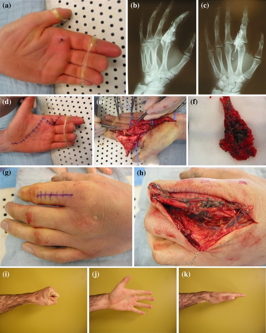
The composition of the paint was notified to value the risk of a systemic intoxication. Also a tetanus prophylaxis was given on the emergency department. Clinical examination showed only a small entry port at the palmar MP level of the index finger (MCP II) (Fig. 1a). A decreased capillary refill and hypersensibility of the hand was observed. On the X-ray of the left hand a large amount of radio dense material on the dorsal side from the MCP II joint to the DIP II joint and from the entry port until the carpal tunnel level, was present (Fig. 1b, c). Surgical exploration under tourniquet and general anaesthesia was decided to carry out. A palmar incision was made from the PIP joint, along the skin fold of the thenar muscle. Subsequently, the paint was removed and a debridement of all the ischemic tissue was performed, followed by a complete synovectomy and microsurgical neurolysis and arteriolysis of the second finger and open carpal tunnel release (Fig. 1d–f). By means of a second straight dorsal approach starting from P1 and going up to the MP level, the painting around the extensor tendons of digit II was removed (Fig. 1g, h). A suction drain was placed before closing the wound primarily.
Fig. 1.
High pressure injection injury in a 33-year-old industrial painter. a Clinical aspect at admission: small punctiform palmar skin lesion at left MCP II level. b X-ray of the left hand with radio dense fluid on MCP II and on the hand palm oblique view. c X-ray of the left hand with radio dense fluid on MCP II and on the hand palm dorso-palmar view. d Clinical aspect intraoperatively: planning of Incision. e Clinical aspect intraoperatively: exploration and debridement of the paint and necrotic tissue on the palmar side. f Clinical aspect intraoperatively: debrided tissue. g Clinical aspect intraoperatively: planning of dorsal Incision. h Clinical aspect intraoperatively: exploration of the dorsal side. i Clinical aspect 3 years post-operatively: complete finger flexion. j Clinical aspect 3 years post-operatively: complete finger extension (plantar view). k Clinical aspect 3 years post-operatively: complete finger extension (lateral view)
Postoperative the hand was placed on a palmar splint in intrinsic plus position. The patient received antibiotics intravenously for a duration of 5 days. There was a good primary wound healing. Immediately the patient had to start with passive physiotherapy of the hand and three weeks postoperative he could switch to an intensive physiotherapeutic training for about 6–12 months.
A year after the injury the patient was re-evaluated with special attention to the vascularisation, sensibility, active and passive range of motion and social reintegration. Vascularisation of the hand showed no changes at rest and at work compared to normal conditions, however paling of the skin as well as hypersensibility and dysfunction occurred by cold exposure. The static two-point discrimination amounted 4 mm for N3 and N4. There was a complete active and passive range of motion of the finger and hand. There was a soft scar on the palmar side and a slight hypertrophic scar on the dorsal side of the hand. Three years postoperatively the patient was re-evaluated once again. Hypersensibility by cold exposure was still present as well as the Raynaud complaints. Meanwhile the patient has changed his profession because of complaints. He also stopped competitive mountain-biking, because of pain caused by the repetitive bump movements. The hypertrophic scar on the dorsal side of the finger had disappeared. Both the maximum grip strength and the pinch strength of the left hand had diminished slightly. There was no change concerning static and dynamic two-point discrimination at the index level (Fig. 1i–k).
Discussion
High-pressure injection injuries mainly occur with industrial labourers. In the majority of the cases the injection place is the hand. Generally it concerns the non-dominant hand [4, 7–9], although in the study of Wieder et al. [10] 13 of 25 injections took place at the dominant hand. More than 50% of the injections occur in the index finger. The second most touched region is the thumb and only 10% of the injections occur in the hand palm or elsewhere [10].
The consequences for the hand function must not be underestimated. Therefore not only an adequate treatment, but also sufficient attention to the prevention of such hand traumatisms must be given. Prevention means a good education concerning the safe use of the high-pressure guns, regular functional and component controls, wearing protection clothes and giving information concerning the seriousness of a hand traumatism under high-pressure [2, 3, 6, 11, 12].
Pathophysiology
There are several mechanisms responsible for the irreversible damage of the tissues:
Firstly, the pressure plays an important role. In the literature it varies from 40 to 800 bar [10, 16]. A pressure of 7 bar is already sufficient to penetrate the skin. At higher pressures, direct contact with the skin is not necessary to infiltrate the subcutaneous tissues [13]. The injected fluid spreads along the neurovascular bundles through places with the lowest resistance [17]. This causes a traumatic dissection of the finger and compression of the neurovascular bundles with vascular spasms, tissue ischemia and thrombosis as a consequence. If the distension of the tissues, caused by the fluid itself and by swelling and oedema, creates a pressure build-up exceeding hydrostatic pressure, tissue perfusion will be limited similar to that of compartment syndrome.
Secondly, there is the chemical damage by the fluid himself. Some fluids have cytolytic properties and can cause tissue destruction, necrosis and intense inflammatory responses. Fibrosis arises around the tissues and can result in a strong restriction of the hand function [5, 7, 10, 16].
A final factor which plays a role in the vast destruction of tissues is infection. This can occur primarily during the injection, but more often it is a secondary infection that occurs. Ischemia and necrosis facilitate this secondary infection [7, 17]. The use of antibiotics which should cover both gram-positive and gram-negative organism is indicated [4]. The application of corticosteroids has no effect on the presence of infection and does not affect the incidence of amputation [2].
Symptoms
Initially there are only minimal complaints. Mostly there is only a small punctiform skin lesion. After some hours swelling, pain, functio laesa and sensibility impairments appear. Finally, a dysfunction of the perfusion occurs. The initially mild symptoms lead to a delay of treatment and so subcutaneous damage can spread out, increasing the chance on permanent complications and amputation. On average patients are seeing a doctor only after 9 h. The fluid can damage the soft tissues and can spread to neighbouring structures. When the injection takes place at the pink or small finger, the fluid can spread along the synovial sleeves like in a V-phlegmona [9, 13].
In literature some rare cases are described. There was a patient who developed a pneumomediastinum after injection of air in the hypothenar [13, 14]. Some rare perversions of granulomes, a sequel after a high-pressure injection injury by intense inflammatory response, in squameus carcinomas is also given [13, 15].
Prognostic influencing factors
The factors influencing the prognosis of the final hand function are mainly stipulated by the circumstances of the accident:
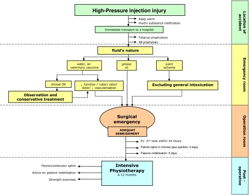
A first factor is the nature of the injected fluid. Injections with water, air or small quantities of veterinary vaccine only cause little damage and frequently have a good outcome, even without surgical intervention [2, 12, 10, 19]. Paints and solvents are more irritating substances and have larger cytolytic properties than water, some oils or greases. That is the reason why they also have a worse outcome than other fluids [17, 19, 20]. Solvents have a lower viscosity compared to paints and as a consequence a faster distribution along the tissues is apparent [21]. A further distinction can be made based on the paint type. Paints based on white-spirit cause damage by disintegration of cell membranes, oil-based paints cause intense inflammatory responses and latex paints based on water have been known to be less destructive (Fig. 2).
A second important factor determining the patients outcome is the pressure of the gun ejection. An injection under low pressure causes less damage than an injection under high pressure. That is why injections with veterinary vaccines in general give less damage than other injections.
A third factor which is determinative for the seriousness of the injury is the volume. A larger quantity of the injected fluid causes a higher pressure in the tissues and therefore a larger risk on compression of the neurovascular bundles and tissue ischemia.
A fourth important factor is the site of injection, especially concerning large volumes. The hand palm has a lager expansion capacity than a finger top. Therefore an injection with the same quantity of fluid at both sites, results into faster development of a compartment syndrome in the finger top compared to the hand palm [16, 20]. The internal spreading of the injected material depends on the different strengths of the encountered tissues and can continue to enter until resistant structures are reached. The site of injection ascertains whether the fluid can penetrate in the tendon sheath itself or not. The flexor sheath is not uniform in consistency. The C-pulleys, overlying the interphalangeal joints, are flexible and thin. They allow penetration of the tendon sheath and surroundings by the injected material with lower chance of functional outcome. The A-pulleys on the other hand are rigid and fibrous structures, overlying the centres of the phalanx, and inducing deflection and lateral spreading of the injected material in the superficial tissues encircling the digit. Only cutaneous necrosis will be enhanced [21].
A fifth, and the only factor where a doctor or a patient can anticipate on, is the latency time between the accident and the establishing of an adequate treatment. Several authors consider this the most important prognostic influencing factor [2, 3, 6, 12]. Among others, the risk on amputation increases with time latency. Some studies report a time limit of 10 h on which amputation risk is strongly raised. Other studies showed no significant difference in prognosis if the patient is treated within the first 24 h [13,20]. Stark et al. [22] concluded that patients who underwent a decompression within the first 10 h had a better outcome. Pinto et al. showed also that the longer the latency time to adequate treatment was, the larger the risk on amputation. They had only been obliged to perform an amputation when the patient came to the emergency department after more than 72 h [8]. According to Christodoulou et al. this time factor is not always the most important variable. They state that the eventual prognosis is influenced by different factors and the prognosis of patients with an injection under very high pressure and with very toxic material is as bad as the prognosis of those people that are treated only after 10 h with a less detrimental injection [19].
Fig. 2.
Algorithm for the treatment of high-pressure inject injuries on the base of nature of the fluid
Treatment
Firstly, information about the fluid’s nature is to be gathered to exclude a general intoxication. If needed, contact with an anti-poison-centre can give information about an anti-dotum. Vital parameters must be followed up. The general systemic responses which can occur among others are renal failure, intoxication with lead, allergic responses and haemolysis. There is a big danger for intoxication in case of an injection with white-spirit or terebentine [16].
Most of the authors agree that only a fast and wide exploration under general anaesthesia or plexus block is the suitable treatment for a high-pressure injection injury [2, 3]. Pushing the fluid to the outside or making relieving incisions for decompression is insufficient to prevent additional subcutaneous damage. Ring block of the finger should be avoided because of the possibility of further vascular compression and vasospasm by the extra injected volume [9, 13]. All injected material and necrotic substances must be removed, followed by a saline irrigation. The use of a solvent to remove the fluid is no solution, because most of the solvents themselves have cytolytic properties and can cause additional damage to the weak tissues. The procedure occurs under tourniquet but without using the Esmarch bandage for exsanguination of the arm to avoid further spreading of the injection material along the tendon sheaths and neurovascular bundles [13, 15]. There must be an optimalisation of the vascularisation of the injected hand. Therefore application of ice to reduce swelling is dissuaded (Table 1). Regional anesthesia of the stellate ganglion and brachial plexus produces analgesia and vasodilatation of peripheral arteries by inhibition of the sympathic tone [23]. If there is already a loss of sensibility and a poor vascularisation at arrival in the emergency department, immediate amputation must be discussed with the patient [16]. Frequently there is a need for several debridements or a reconstruction by means of skin grafts, local or free flaps [8, 24]. Sometimes there is a preference to open wound technique with regular salvage of the wound [8]. With this technique Pinto et al. had only an amputation risk of 16%, which lies much lower than the amputation risk that is described in other articles [2, 3, 12]. They applied the same wide exploration and debridement with leaving the wound open and regularly salvage in combination with early intensive physiotherapy treatment in all cases [8].
Table 1.
Do nots in high pressure injection injuries
| • Exploration under ring block of the finger |
| • Using of the Esmarch bandage |
| • Removing the material with a solvent |
| • Pushing the fluid to the outside or making relieving incisions for decompression |
| • Application of ice to reduce swelling |
In the study of Wong et al. the injection injuries were divided in mild, moderate and serious cases, based on the nature of the fluid, the latency time to adequate treatment and the clinical neurovascular status at arrival. Mild injuries can be treated conservatively with broad spectrum antibiotics, tetanus prophylaxis and observation of the neurovascular situation of the fingers. Patients with moderate or serious injuries underwent immediate surgical exploration and decompression with wide debridement in combination with antibiotics and tetanus prophylaxis. Six of seven mild injuries could be well treated with conservative therapy. One nevertheless still needed a surgical exploration. Sixteen patients with a moderate injury had good results. At three of the five serious high-pressure injection injuries an amputation could not be avoided. The other two had good results [24].
Preoperative X-rays can show the quantity and distribution of radio-opaque fluids. The distribution of radiolucent substances can sometimes be shown on X-rays by subcutaneous emphysema [7]. At arrival in the hospital a tetanus prophylaxis and antibiotic prophylaxis, under the form of 3e a generation cephalosporine must be administered. In literature a controverse concerning the use of corticosteroids for high-pressure injection injuries exists. There is a theoretical evidence for the use of corticosteroids in the case of intense inflammatory reactions and at late presentations with diffuse oedema and erythema. Corticosteroids can avoid an acute response to the strange fluid and functional sequels [15]. In the study of Lewis et al. all patients received 100 mg hydrocortisone/6 h intravenously and later 25 mg prednisolone/24 h orally while diminishing the concentration in order to stop within 3–5 days. [16] Other authors dissuade the use of corticosteroids because of the possible disadvantages. Corticosteroids oppress the leukocyte response and raise the infection risk. The chance on infection increases more within necrotic tissues and diminished vascularisation [24, 25]. A recent review of the literature [2] however shows, that the application of corticosteroids has no effect on the presence of infection and does not affect the incidence of amputation [2].
Postoperative the patient receives a palmar splint. It is very important to start immediately with physiotherapy to build up the hand function as well as possible. In the first three weeks patients only receive active and passive mobilisation of the fingers. After three weeks they can start with an intensive physiotherapeutic scheme for 6 up to 12 months.
Outcome
The outcome after a high-pressure injection injury is frequently disappointing, even after immediate adequate treatment. The patient has to be informed previously concerning the possible restrictions in hand function and the chance on finger amputation. The amputation risk is valued on 16–55%. With solvents it goes up to 50–80% [12, 13, 16]. When there are already impairments of the vascularisation during the first medical examination or when the pressure was more than 490 bar, amputation risk reaches the 100% [13]. Permanent complaints of the patient among others are hyperesthesia, continuous pain, cold intolerance, contracture, and reduced sensitivity. Amputation and aesthetic problems are two other complications. Only a small percentage of the patients can resume its original work [10].
Conclusion
High-pressure injection injuries to the hand are characterised by a small and punctiform skin lesion but with severe subcutaneous damage of the tissues. The initial clinical presentation can be misleading as a result of which an adequate treatment is frequently postponed. In the first place an intoxication caused by the fluid must be excluded. The surgical treatment must happen under complete anaesthesia or plexus block. An immediate wide microsurgical exploration must be carried out with complete debridement of the foreign material and necrotic tissue. If there is no immediate intervention, additional damage occurs, with a decrease of the functionality of the hand. Frequently an amputation of the finger can no longer be avoided. A long-term and intensive physiotherapy afterwards will influence the outcome of the hand function in a positive way. Therefore it is very important to inform users of high-pressure guns about the seriousness of such injuries and to take preventive measures.
References
- 1.Neal NC, Burke FD (1991) High-pressure injection injuries. Injury 22(6):467–470 [DOI] [PubMed]
- 2.Hogan CJ, Ruland RT (2006) High-pressure injection injuries to the upper extremity: a review of the literature. J Orthop Trauma 20(7):503–511 [DOI] [PubMed]
- 3.Gonzalez R, Kasdan ML (2006) High pressure injection injuries of the hand. Clin Occup Environ Med 5(2):407–411 [DOI] [PubMed]
- 4.Sirio CA, Smith JS Jr, Graham WP 3rd (1989) High-pressure injection injuries of the hand. A review. Am Surg 55(12):714–718 [PubMed]
- 5.Lewis RC Jr (1985) High-compression injection injuries to the hand. Emerg Med Clin North Am 3(2):373–381 [PubMed]
- 6.Fialkov JA, Freiberg A (1991) High pressure injection injuries: an overview. J Emerg Med 9(5):367–371 [DOI] [PubMed]
- 7.Moutet F, Chaussard C, Guinard D, Corcella D (1998) Injections à haute pression au niveau de la main. p 83–91. Monographie de la société française de chirurgie de la main no. 25, Infections de la main
- 8.Pinto M, Turkula-Pinto L, Cooney W, Wood M;B, Dobyns J (1993) High-pressure injection injuries of the hand. Review of 25 patients managed by open wound technique. J Hand Surg [Am] 18(1):125–130 [DOI] [PubMed]
- 9.Stoffelen D., De Smet L., Broos P (1994) Delayed diagnosis of high-pressure injection injuries to the finger. A case report and review of the literature. Acta Orhop Belg 60:332–333 [PubMed]
- 10.Wieder A, Lapid O, Plakht Y, Sagi A (2006) A. Long term follow-up of high-pressure injection injuries to the hand. Plast Reconstr Surg 117(1):186–189 [DOI] [PubMed]
- 11.Hart R, Smith D, Haq A (2006) Prevention of high-pressure injection injuries to the hand. Am J Emerg Med 24(1):73–76 [DOI] [PubMed]
- 12.Schnall SB, Mirzayan R (1999) High-pressure injection injuries to the hand. Hand Clin 15(2):245–248 [PubMed]
- 13.Tempelman T, Borg D, Kon M (2004) Verwonding van de hand door een hogedrukspuit: vaak grote onderhuidse schade. Ned Tijdschr Geneeskd 148(47):2334–2338 [PubMed]
- 14.Temple C, Richards RS, Dawson WB (2000) Pneumomediastinum after injection injury to the hand. Ann Plast Surg 45:64–66 [DOI] [PubMed]
- 15.Mizani M, Weber B (2000) High-pressure injection injury of the hand. The potential for disastrous results. Postgrad Med 108(1):183–185–189–190 [DOI] [PubMed]
- 16.Lewis H, Clarke P, Kneafsey B, Brennen M (1998) A 10–year review of high-pressure injection injuries to the hand. J Hand Surg [Br] 23B(4):479–481 [DOI] [PubMed]
- 17.Valentino M, Rapisarda V, Fenga C (2003) Hand injuries due to high-pressure injection devices for painting in shipyards: circumstances, management and outcome in twelve patients. Am J Ind Med 43(5):539–542 [DOI] [PubMed]
- 18.Mirzayan R, Schnall SB, Chon JH, Holtom PD, Patzakis MJ, Stevanovic MV (2001) Culture results and amputation rates in high-pressure paint gun injuries of the hand. Orthopedics 24(6):587–589 [DOI] [PubMed]
- 19.Christtodoulou L, Melikyan E, Woodbridge S, Burke F (2001) Functional outcome of high-pressure injection injuries of the hand. J Trauma 50(4):717–720 [DOI] [PubMed]
- 20.Obert L, Lepage D, Jeunet D, Gereard P, Garbuio P, Tropet Y (2002) Traumatisme de la main par injection sous pression: Spécificité lésionnelle de l’huile industrielle. Chir Main 21(6):43–49 [DOI] [PubMed]
- 21.Vasilevski D, Noorbergen M, Depierreux M, Lafontaine M (2000) High-pressure injection injuries to the hand. Am J Emerg Med 18(7):820–824 [DOI] [PubMed]
- 22.Stark HH, Asworth CR, Boyes JH (1967) Paint gun injuries of the hand. J Bone Joint Surg Am 49:637–647 [PubMed]
- 23.Beguin JM, Poilvache G, Van Meerbeeck J, de Coninck A (1985) Hand injuries caused by high pressure injection. Contribution of loco-regional anaesthesia. Ann Chir Main 4(1):37–42 [DOI] [PubMed]
- 24.Wong T, Ip F, Wu W (2005) High-pressure injection injuries of the hand in a Chinese population. J Hand Surg [Br] 30(6):588–592 [DOI] [PubMed]
- 25.Gutowski K, Chu J, Choi M, Friedman D (2003) High-pressure hand injection injuries caused by dry cleaning solvents: case reports, review of the literature, and treatment guidelines. Plast Reconstr Surg 111:174–177 [DOI] [PubMed]


