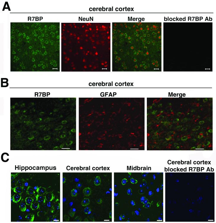
Figure 7.
R7BP is expressed in neurons but not astrocytes. R7BP protein expression in neurons or astrocytes was analyzed by performing fluorescence confocal microscopy of sections co-stained with chicken anti-R7BP antibody (green) and NeuN antibody (red; panel A), which specifically marks neuronal cell bodies, or GFAP antibody (red; panel B), which s astro Adjacent cortical sections stained with blocked R7BP antibodies are shown. C) High magnification images of R7BP immunoreactivity detected in hippocampal, midbrain, and cerebellar sections stained with chicken anti-R7BP antibodies (green), and the DNA dye DAPI (blue). Scale bars =20 μm (A and B), 10 μm (C). Results shown are representative of 2-5 independent experiments in which sections of brains from 3 animals were used.

