Abstract
The virulence determinants for highly pathogenic avian influenza viruses (AIVs) are considered multigenic, although the best characterized virulence factor is the hemagglutinin (HA) cleavage site. The capability of influenza viruses to reassort gene segments is one potential way for new viruses to emerge with different virulence characteristics. To evaluate the role of other gene segments in virulence, we used reverse genetics to generate two H5N1 recombinant viruses with differing pathogenicity in chickens. Single-gene reassortants were used to determine which viral genes contribute to the altered virulence. Exchange of the PB1, PB2, and NP genes impacted replication of the reassortant viruses while also affecting the expression of specific host genes. Disruption of the parental virus' functional polymerase complexes by exchanging PB1 or PB2 genes decreased viral replication in tissues and consequently the pathogenicity of the viruses. In contrast, exchanging the NP gene greatly increased viral replication and expanded tissue tropism, thus resulting in decreased mean death times. Infection with the NP reassortant virus also resulted in the upregulation of gamma interferon and inducible nitric oxide synthase gene expression. In addition to the impact of PB1, PB2, and NP on viral replication, the HA, NS, and M genes also contributed to the pathogenesis of the reassortant viruses. While the pathogenesis of AIVs in chickens is clearly dependent on the interaction of multiple gene products, we have shown that single-gene reassortment events are sufficient to alter the virulence of AIVs in chickens.
The eight-segment negative-sense single-strand RNA genome of influenza A viruses provides for the possibility of genetic reassortment between different influenza viruses that contributes to the natural evolution of these viruses. Wild aquatic birds are known to be natural reservoirs for avian influenza viruses (AIVs) (58), and coinfection of these avian hosts occurs commonly (49). Live poultry markets, where many birds of different species share close quarters, can also contribute to the reassortment between different influenza viruses because of the potential for dual infection.
Reassortment of one or more viral gene segments can lead to attenuation and plays a major role for influenza viruses when crossing host barriers (13, 33, 51, 56). Just as reassortment can lead to attenuation, the converse is also true, and the emergence of viruses with increased pathogenicity or increased host range was an important occurrence in past outbreaks and is also likely in future outbreaks. Of particular interest is the role of reassortment in past human influenza outbreaks in which avian gene segments reassorted with human viruses, resulting in the pandemic outbreaks of 1957 and the pandemic outbreak of 1968, in which avian hemagglutinin (HA), NA, and PB1 genes and HA and PB1 avian genes, respectively, underwent reassortment with circulating human viruses (29, 46).
The host tropism and pathogenicity of influenza viruses are considered to be multigenic, and the primary virulence factor is the presence of multiple basic amino acids or the insertion of amino acids at the HA cleavage site (8, 9, 25, 47). Reverse genetic techniques and classical reassortment studies have elucidated several genes and specific mutations as being important contributors in host range determination and the pathogenicity of influenza viruses in mammals. In addition to the HA cleavage site, a lysine residue at amino acid 627 in PB2 has been found in most influenza viruses pathogenic to humans and also influences virulence in mice (22, 36, 50, 54, 55). The alternate splice product of the PB1 gene, PB1-F2, has also been linked to the increased virulence of the 1918 virus in mice (14). PA and NS1 have also been shown to contribute to pathogenicity for mice, and NS1 also influences the pathogenicity of influenza viruses in pigs (16, 32, 45, 48, 52).
Despite what is known about the virulence of influenza viruses in mammals, the role of the genes important for host tropism and the virulence of AIVs in avian species remains largely undetermined. Currently, pathogenesis studies of AIVs in chickens clearly indicate that the proteolytic cleavage site of HA (22, 25, 27) and changes in the NS1 protein (11, 33) are important determinants of virulence. Recently it was reported that PA and PB1 gene mutations were responsible for differences between nonpathogenic and highly pathogenic viral clones in ducks (26), and mixtures of polymerase components from different viruses have been found to lead to the attenuation of AIVs in chickens (43). However, the K627 mutation of PB2, important for virulence in mammals, does not seem to influence AIV virulence in avian cell lines (31).
In this study, we used reverse genetics to generate two H5N1 AI recombinant viruses with different pathogenicity levels in chickens. Single-gene reassortant viruses were generated in order to better understand the role of gene reassortment events as a way of generating variant AIVs and to study the role of each gene segment in viral pathogenicity in chickens. Our results indicate that the HA, NS, NP, and M2 proteins are important contributing factors in the increased virulence of our reassortant viruses in chickens. Three genes, PB1, PB2, and NP, impacted replication of the reassortant viruses, emphasizing the importance of these proteins in the replication process. In addition, infection with the reassortant viruses affected host gene expression levels of alpha interferon (IFN-α), the orthomyxovirus resistance gene 1 (Mx1), IFN-γ, and/or inducible nitric oxide synthase (iNOS), suggesting that the regulation of host genes may contribute to the differences in replication observed in chickens.
MATERIALS AND METHODS
Generation of infectious reassortant viruses.
Two high-pathogenicity recombinant H5N1 AIVs were derived from A/Egret/Hong Kong/757.2/02 and A/Chicken/Indonesia/7/03 as follows and are referred to as rEgret and rIndo, respectively. RNA was extracted from virus stocks of the wild-type viruses, after propagation in embryonating chicken eggs (ECEs), using Trizol LS (Invitrogen, Inc., Carlsbad, CA) according to the manufacturer's instructions. Transcription and expression plasmids were constructed as previously described (38). 293T cells were transfected with 1 μg of each of the eight transcription and four protein expression plasmids and 11 μl of Lipofectamine 2000 (Invitrogen) in a 2-ml volume of OptiMEM I (Invitrogen). Cells were washed after 4 h at 37°C, and medium was replaced with Dulbecco's modified Eagle medium I supplemented with 10% fetal bovine serum (Invitrogen) for 72 h. ECEs were inoculated with 100 μl of the cell supernatant. Virus was harvested from the allantoic fluid of eggs 36 to 48 h after inoculation and titrated in ECEs. Titration end points were calculated by the method of Reed and Muench (42). In addition to the reconstitution of the parental strains by reverse genetics, six single-gene reassortants containing one of the rEgret genes were produced in the rIndo background. All experiments using HPAI H5N1 viruses were conducted using biosafety level 3 Ag (BSL-3 Ag) containment at SEPRL, USDA, in Athens, GA (5).
Sequencing of influenza virus genes.
Genes from the rescued viruses were sequenced to confirm the reassortment. Viral RNA was extracted from infectious allantoic fluid from ECEs using Trizol LS reagent (Invitrogen). Gene sequences were obtained by reverse transcription (RT)-PCR by use of a Qiagen One-Step RT-PCR kit (Qiagen, Valencia, CA) using specific primers for each influenza virus gene. The primer sequences and RT-PCR conditions used are available upon request. PCR products were extracted from agarose gels, using a GenScript QuickClean gel extraction kit (GenScript, Piscataway, NJ). An ABI BigDye Terminator version 1.1 sequencing kit (Applied Biosystems, Foster City, CA) run on a 3730xl DNA analyzer (Applied Biosystems) was used for sequencing PCR products. The MegAlign program (Lasergene 7.1; DNAStar, Madison, WI) was used to compare nucleotide sequences, using the Clustal W alignment algorithm.
In vivo characterization of reassortant viruses.
In order to determine the pathogenic phenotypes of the reassortant viruses in chickens, 2-week-old specific-pathogen-free White Leghorn chickens (Gallus gallus domesticus) (from SEPRL in-house flocks) were inoculated intranasally (IN) with the reassortant viruses and evaluated for clinical signs for 10 days. The birds were housed in self-contained isolation cabinets that were ventilated under negative pressure with HEPA-filtered air and maintained under continuous lighting. The birds had ad libitum access to feed and water. Each group, containing 11 birds, was inoculated IN with 0.1 ml of an inoculum containing 106 50% egg infective doses (EID50)/ml of one of the viruses. Three birds from each group were euthanized and necropsied at 2 days postinoculation (dpi). Gross lesions were recorded, and tissue samples (lung, spleen, and brain) were collected separately from two birds for virus isolation. Lung, bursa, kidney, adrenal gland, gonad, thymus, thyroid, brain, liver, heart, ventriculus, pancreas, intestine, spleen, trachea, and thigh muscle specimens were collected in 10% neutral buffered formalin solution from the same birds. Sample birds, moribund birds, and all birds remaining at the end of a 10-day period were humanely euthanized. Mean death time (MDT) was calculated by determining the sum of the day of death for the chickens and dividing it by the total number of dead chickens.
Virus isolation and titrations.
Portions of the spleens, lungs, and brains from two birds per group were collected at 2 dpi in brain heart infusion medium (BHI) (BD Bioscience, Sparks, MD) and stored frozen at −70°C. Titers of infectious virus were determined as follows. Tissues were homogenized (10% [wt/vol]) and diluted in BHI. One hundred microliters of the homogenate dilutions was inoculated into the allantoic cavity of ECEs. Titration end points were calculated by the method of Reed and Muench (42) (n = 2). The threshold of detection was 102.4 EID50/g from tissues.
Histopathology and immunohistochemistry.
Collected tissues fixed by submersion in 10% neutral buffered formalin were routinely processed and embedded in paraffin. Sections were made at 5 μm and were stained with hematoxylin and eosin. A duplicate 4-μm section was immunohistochemically stained by first microwaving the sections in antigen retrieval citra solution (BioGenex, San Ramon, CA) for antigen exposure. A 1:2,000 dilution of a mouse-derived monoclonal antibody (P13C11) specific for a type A influenza virus nucleoprotein (developed at Southeast Poultry Research Laboratory, USDA) was applied and allowed to incubate for 2 h at 37°C. The primary antibody was then detected by the application of biotinylated goat anti-mouse immunoglobulin G secondary antibody using a biotin-streptavidin detection system (supersensitive multilink immunodetection system; BioGenex). Fast Red TR (BioGenex) served as the substrate chromogen, and hematoxylin was used as a counterstain. All tissues were systematically screened for microscopic lesions.
Total cellular RNA isolation.
Total cellular RNA for the evaluation of gene expression was prepared from spleen or lung tissues of chickens collected at 2 dpi. Tissue samples were homogenized in 3 ml minimal essential medium, Alpha 1× (Invitrogen), by passing the tissues through 100-μm cell strainers (BD Bioscience, Sparks, MD), added to 6 ml Trizol, inverted, and stored at −80°C. Chloroform (1.2 ml) was added and spun, and the aqueous phase was added to an equal volume of 70% ethanol. A Qiagen RNA midiprep kit (Qiagen) was used to isolate the RNA from the aqueous phase-ethanol solution.
Semiquantitative analysis of mRNA gene expression.
RT-PCR was carried out using a Qiagen OneStep PCR kit (Qiagen) according to the manufacturer's instructions, using 100 ng of pooled cellular RNA per group (33.3 ng of total RNA from three chickens per group) for each infected group 2 dpi in a 25-μl reaction volume (n = 1). The primers for IFN-α and β-actin were previously described (60). The IFN-γ, Mx1, and iNOS primers were designed using nucleotide sequences from NCBI GenBank and are available upon request. β-actin served as an amplification and loading control. DNA was visualized by ethidium bromide gel electrophoresis using an EDAS 290 imaging station (Kodak, New Haven, CT). Band intensities were analyzed using Kodak 1D version 3.6.1 imaging software (Kodak). Band intensity values for each tissue were normalized to the β-actin values, and the values for control birds were arbitrarily set at 1.
Statistical analyses.
Data were analyzed using Prism v.5.01 software (GraphPad Software, Inc., San Diego, CA), and values are expressed as the mean ± standard error of the mean. The Kaplan-Meier survival rate data were analyzed using the log rank test, and one-way analysis of variance with the Tukey-Kramer posttest was used to analyze MDTs. Statistical significance was set at a P value of <0.05.
RESULTS
Pathogenicity of the H5N1 recombinant viruses.
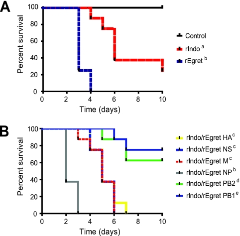
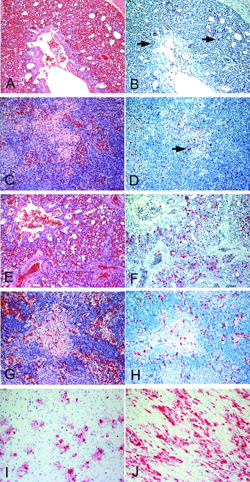
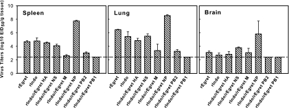
The two-parent recombinant H5N1 viruses used in this study were derived from A/Chicken/Indonesia/7/03 and A/Egret/Hong Kong/757.2/02 using reverse genetics. Table 1 shows the comparison of the amino acid sequences of the resulting recombinant viruses, rIndo and rEgret. The recombinant viruses that we generated have a PA gene with the same amino acid sequence, but the remaining viral genes have amino acid sequence differences (see Table S1 in the supplemental material). When administered to chickens IN, the rIndo virus resulted in a mortality of 6 of 8 and an MDT of 6.1 days, while the rEgret virus resulted in a higher mortality (8 of 8) and a shorter MDT (3.25 days) (Table 2). MDTs (Table 2) and survival rates were significantly different (P < 0.05) upon infection with rIndo and rEgret compared to the control group and also to each other (Fig. 1A). Chickens inoculated with rEgret presented histological lesions in tissues, which included diffuse interstitial pneumonia in the lung, moderate tracheitis, multifocal splenic necrosis, moderate cardiac degeneration, and multifocal nonsupurative encephalitis among others (Fig. 2; Table 3). AIV antigen was present in blood vessel endothelial cells in most tissues, tissue macrophages, cardiac and skeletal muscle myocytes, neurons and glial cells in the brain, pancreatic acinar cells, respiratory epithelia of the trachea, and adrenal corticotrophic cells. In contrast, chickens inoculated with rIndo presented lesions only in the trachea and lungs, and viral antigen was primarily detected in the cells of these tissues and infrequently in the liver, spleen, and brain (Table 3). Virus titers in the lung tissue show that the rEgret virus had slightly higher titers than the rIndo virus, although there was variation in the rIndo virus titers (Fig. 3).
TABLE 1.
Amino acid sequence comparisons of recombinant A/Egret/Hong Kong/757.2/02 and recombinant A/Chicken/Indonesia/7/03 viruses
| Gene product | Amino acid sequence similarity (%) | No. of different amino acids | Differing amino acid positions |
|---|---|---|---|
| PB2 | 98.7 | 10 | 105, 132, 221, 251, 288, 292, 309, 339, 389, 394 |
| PB1 | 99.3 | 5 | 110, 158, 215, 372, 400 |
| PB1-F2 | 98.8 | 1 | 73 |
| PA | 100 | 0 | |
| HA | 98.1 | 11 | 8, 110, 111, 136, 140, 145, 194, 231, 498, 504, 549 |
| NP | 99.2 | 5 | 22, 184, 400, 406, 423 |
| NA | 91.0 | 22 + 20-amino-acid deletiona | 8, 19, 29, 42, 48, 49-68, 74, 95, 100, 105, 224, 257, 258, 267, 270, 313, 338, 341, 346, 355, 378, 386, 454 |
| M1 | 100 | 0 | |
| M2 | 96.9 | 3 | 10, 31, 64 |
| NS1 | 96.0 | 9 | 7, 21, 48, 73, 112, 148, 200, 202, 209 |
| NS2 | 99.2 | 1 | 7 |
rIndo virus NA contains a 20-amino-acid stalk deletion as well as 22 other amino acid differences.
TABLE 2.
Morbidity, mortality, and MDT resulting from IN inoculation of 2-week-old chickens with 0.1 ml of inoculum containing 106 EID50/ml of the reassortant viruses
| Recombinant virus | No. of sick/dead/total | MDTa |
|---|---|---|
| rEgret | 8/8/8 | 3.25b |
| rIndo | 7/6/8d | 6.1c |
| rIndo/rEgret HA | 8/8/8 | 5.25c |
| rIndo/rEgret NA | Not rescued | Not rescued |
| rIndo/rEgret NS | 8/8/8 | 5.12c |
| rIndo/rEgret M | 8/8/8 | 5.0 |
| rIndo/rEgret NP | 8/8/8 | 2.3b |
| rIndo/rEgret PB2 | 8/3/8e | 6.3c |
| rIndo/rEgret PB1 | 8/2/8e | 6.5c |
The MDTs of all groups were significantly different from that of the control group.
MDTs were significantly different from the rIndo group.
MDTs were significantly different from the rEgret group.
Two birds survived. One presented no clinical signs; the other one presented only mild hemorrhages in the shanks and combs.
The surviving birds presented only mild hemorrhages in the shanks and combs.
FIG. 1.
Survival after IN inoculation with the reassortant viruses. Two-week-old chickens were inoculated with 0.1 ml of an inoculum containing 106 EID50/ml of the reassortant viruses, and mortality was monitored for 10 days. Parent virus and control survival analysis (A) and reassortant virus survival analysis (B). All groups are statistically different from rEgret and rIndo/rEgret NP (P < 0.05) as determined by the log rank test. Explanation of footnotes in figure key: a, survival is significantly different from the control group and also from the rEgret group; b, survival is significantly different from all groups and the control group; c, survival is significantly different from the control group but not the rIndo group; d, survival is not significantly different from the control group or the rIndo group; e, survival is significantly different from the rIndo group but not the control group.
FIG. 2.
Experimental studies of chickens that were IN inoculated with AIV H5N1 recombinant viruses; representative microscopic lesions and immunohistologic findings. Photomicrographs (×200) of tissue sections stained with hematoxylin and eosin (A, C, E, and G) or by immunohistochemistry to demonstrate AIV (B, D, F, H, I, and J). (A) Histiocytic interstitial pneumonia in a 2-week-old chicken inoculated with rEgret, 2 dpi, with congestion and fibrous exudates in the airways. (B) AI viral antigen (red, arrows) in macrophages and air capillary and blood vessel endothelium in lung tissue of the same chicken. (C) Spleen tissue of a 2-week-old chicken inoculated with rEgret, 2 dpi; mild focal splenitis. (D) AI viral antigen (red, arrow) in macrophages and vascular endothelial cells in spleen tissue of the same chicken. (E) Severe interstitial pneumonia in a 2-week-old chicken inoculated with rIndo/Egret NP, 2 dpi. There is congestion, mononuclear infiltrates, and serofibrinous exudates filling air capillaries. (F) Diffuse staining (red) for AI viral antigen in the endothelium and infiltrating macrophages in lung tissue of the same chicken. (G) Splenic necrosis in a 2-week-old chicken inoculated with rIndo/Egret NP, 2 dpi. (H) AI viral antigen (red) in macrophages in spleen tissue of the same chicken. (I) AI viral antigen (red) in brain tissue of a 2-week-old chicken inoculated with rIndo/Egret NP, 2 dpi. Staining present in neurons and glial cells of brain tissue. (J) AI viral antigen (red) in the myocardial cells of the heart of a 2-week-old chicken inoculated with rIndo/Egret NP, 2 dpi.
TABLE 3.
Severity of histological lesions and distribution of viral antigen in tissues from chickens IN inoculated with recombinant AIVsa
| Tissue | Histological lesions/viral antigen stainingb
|
|||||||
|---|---|---|---|---|---|---|---|---|
| rEgret | rIndo | rIndo/Egret HA | rIndo/Egret NS | rIndo/Egret M | rIndo/Egret NP | rIndo/Egret PB2 | rIndo/Egret-PB1 | |
| Trachea | ++/+ | ++/+ | ++/+ | ++/+ | ++/+ | +++/++ | +/− | +/− |
| Lung | ++/++ | ++/++ | ++/++ | ++/++ | ++/+ | +++/+++ | +/+ | +/+ |
| Heart | +/++ | −/− | +/+ | +/+ | +/++ | ++/+++ | −/− | −/− |
| Brain | +/++ | −/+ | +/+ | ++ | −/− | ++/+++ | −/− | −/− |
| Pancreas | +/+ | −/− | +/+ | −/− | −/+ | ++/++ | −/− | −/− |
| Adrenal | +/++ | −/− | −/− | −/− | −/− | ++/++ | −/− | −/− |
| Intestine | −/+ | −/− | −/− | −/− | −/− | +/+ | −/− | −/− |
| Liver | −/++ | −/+ | −/+ | −/+ | −/− | +/+ | −/− | −/− |
| Kidney | +/+ | −/− | +/+ | +/+ | −/+ | +/++ | −/− | −/− |
| Spleen | +/++ | −/++ | +/++ | ++/++ | +/+ | +++/+++ | −/− | −/− |
| Bursa | ++/+ | −/− | −/− | −/− | −/− | ++/+ | −/− | −/− |
| Thymus | +/+ | −/− | −/+ | +/+ | −/+ | +/++ | −/− | −/− |
| Muscle | −/+ | −/− | −/− | −/− | −/− | +/++ | −/− | −/− |
| Gizzard | −/+ | −/− | −/− | −/− | −/− | +/++ | −/− | −/− |
Tissues were taken 2 dpi and were immunohistochemically stained with antibodies to AIV nucleoprotein to visualize the viral antigen.
Lesions were scored as follows: −, no lesions; +, mild; ++, moderate; +++, severe lesions. The intensity of viral antigen staining in each section was scored as follows: −, no antigen staining; +, infrequent; ++, common; +++, widespread staining.
FIG. 3.
Virus titers in spleen, lung, and brain tissues. Tissues were taken 2 dpi and homogenized to a 10% (wt/vol) final concentration in BHI medium. A portion (100 μl) of 10-fold dilutions of the homogenates was inoculated into 10-day-old ECEs, and log10 EID50/g was calculated. Values are the mean ± standard error (n = 2) with the exception of the rIndo/rEgret PB1 lung titer (n = 1). The threshold of detection is 2.4 log10 EID50/gram of tissue.
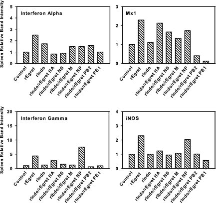
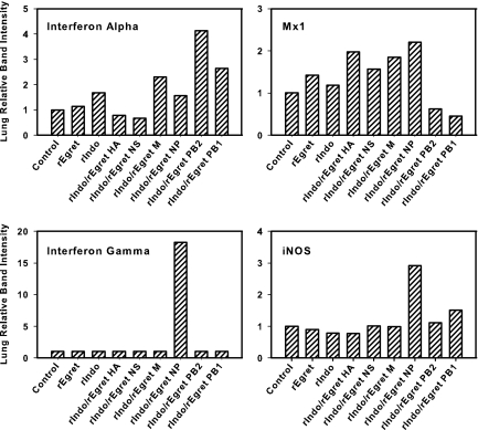
The expression levels of four different immune-related genes in spleen and lung tissues were compared 2 days after challenge with the different recombinant viruses (Fig. 4 and 5). Infection with the rEgret virus resulted in increased expression of IFN-α, IFN-γ, iNOS, and Mx1 in the infected chicken spleen tissues, but in the lung tissues, Mx1 was only slightly upregulated compared to the controls. Only IFN-α gene expression, in both tissues, was upregulated upon infection with rIndo compared to the control. These data suggest that the recombinants induce different host responses and/or are able to differentially suppress host gene response.
FIG. 4.
Semiquantitative analysis of differential mRNA gene expression in the spleen tissue of chickens infected with AIV recombinants. Total cellular RNA was extracted from spleen tissue collected 2 dpi from three chickens. Equal amounts of RNA from the three chickens per group were pooled prior to RT-PCR analysis. Analysis of the pooled RNA was carried out using different primer sets with β-actin as an amplification and loading control (n = 1). Bands were quantified, and intensities shown were normalized to the β-actin control. Gene expression of control birds was arbitrarily set to 1.
FIG. 5.
Semiquantitative analysis of differential mRNA gene expression in lung tissue from chickens infected with AIV recombinants. Total cellular RNA was extracted from spleen and lung tissues from three chickens 2 dpi. Equal amounts of RNA from the three chickens per group were pooled prior to RT-PCR analysis. Analysis was carried out using different primer sets with β-actin as an amplification and loading control. Bands were quantified, and intensities shown were normalized to the β-actin control. Gene expression of control birds was arbitrarily set to 1.
Six single-gene reassortants containing one of the rEgret HA, PB2, PB1, NS, NP, or M genes were generated in the rIndo background. We were unable to generate the rIndo/rEgret NA reassortant due to the limited replication of this reassortant virus in 293T cells or ECEs. The rest of the reassortant viruses grew to high titers in ECEs (≥106.6 EID50/ml). Inoculation with the single-gene reassortant viruses resulted in variable morbidity, mortality, and MDTs (Table 2; Fig. 1). The effect of the single-gene reassortments on pathogenicity of the viruses in chickens is discussed below.
Exchange of the HA gene resulted in increased mortality, expanded tissue range, and differential host gene expression levels.
The HA genes of both our H5N1 recombinant parent viruses contain identical multiple basic amino acid motifs adjacent to the HA0 cleavage site. However, there were 11 other amino acid differences outside of the cleavage site of the HA gene (Table 1). In order to evaluate the contribution of the HA gene in the pathogenesis of these viruses, we exchanged the HA gene of the rIndo virus with the rEgret virus HA gene. The resulting virus, referred to as rIndo/rEgret HA, resulted in increased mortality (8 of 8) over the rIndo parent virus (6 of 8). Exchange of the HA gene resulted in MDTs and survival rates that were not significantly different from those of the rIndo group (Fig. 1B). However, viral antigen staining of tissues from infected birds showed the presence of the virus in many organs, with an expanded tissue range with immunohistochemical staining for AIV in the heart, thymus, kidney, and pancreas (Table 3). Virus titers in the spleen, lung, and brain tissues, which had immunohistochemistry results comparable to those of rIndo, showed that rIndo/rEgret HA had titers similar to those of the rIndo parent virus in those tissues (Fig. 3). mRNA expression levels of the immune-related genes showed that rIndo/rEgret HA virus infection induced gene expression of IFN-γ and Mx1 in the spleen at levels similar to those of the rEgret parent virus (Fig. 4). Expression levels of IFN-α and iNOS in spleen tissue were more similar to the expression levels of the controls. Gene expression in lung tissue showed only increased expression levels of Mx1 over those of the rIndo and rEgret parent viruses upon infection with rIndo/rEgret HA (Fig. 5).
Exchange of the NS gene increases mortality and the tissue range of the reassortant virus.
Our recombinant viruses, rIndo and rEgret, have 9 amino acid sequence differences in NS1 and 1 difference in the NS2 protein (Table 1). Exchange of the NS gene resulted in increased mortality (8 of 8) compared to the rIndo parent (6 of 8) (Table 2). Infection with rIndo/rEgret NS did not result in a significant difference in MDT (Table 2) or survival rate (Fig. 1) compared to the rIndo parent. Viral antigen distribution in tissues was expanded and differed from the distribution of the parent rIndo virus among tissues and was more similar to the rIndo/rEgret HA virus distribution (Table 3). Exchange of the NS gene did not result in a replication advantage in spleen and lung tissues, as the virus titers remained at the rIndo parent levels (Fig. 3). Titers of rIndo/rEgret NS in brain tissue were slightly elevated over those of rIndo and rEgret. Expression of IFN-α was less than that of the rIndo virus in both spleen and lung tissues, while iNOS expression levels were similar to the control levels in both tissues (Fig. 4 and 5). IFN-γ could not be detected in lung tissue, and levels in spleen tissue were similar to the rIndo parent levels. Only the Mx1 gene expression levels were increased over those of rIndo in both spleen and lung tissues.
The M2 protein contributes to the expanded tissue range of the reassortant virus.
The rIndo and rEgret viruses share identical M1 protein sequences but differ in three amino acids in the M2 spliced gene product (Table 1). Therefore, the changes in pathogenicity that we saw when the M gene was exchanged are due to the differences in the M2 protein alone. Exchanging the M gene in rIndo led to increased mortality (8 of 8) compared to the rIndo parent virus (6 of 8) (Table 2) and expanded distribution of viral antigen in tissues (Table 3). Virus titers of the rIndo/rEgret M virus in spleen tissue remained just above the detection limit. Titers in lung tissue were lower than the rIndo titers, and titers in brain tissue were at the level of rEgret and rIndo despite the lack of viral antigen staining in the brain tissue of rIndo/rEgret M-infected chickens (Fig. 3). The MDT of rIndo/rEgret M-infected chickens was not significantly different from either rIndo or rEgret infection (Table 2), and the survival rates were also not significantly different from infection with the rIndo parent (Fig. 1B). Despite the altered pathogenesis observed with rIndo/rEgret M infection, mRNA gene expression in the spleen tissue of infected chickens 2 dpi did not differ greatly from the expression in chickens infected with the rIndo parent virus. Gene expression of Mx1 and IFN-α in lung tissue showed slight upregulation over levels in chickens with rIndo infection (Fig. 5).
The nucleoprotein is important for viral replication and pathogenicity of the reassortant virus.
There are five amino acid differences in the NP protein between the rIndo and rEgret viruses (Table 1). The exchange of the NP gene resulted in an increase in mortality (8 of 8) compared to the rIndo parent (Table 2). A significant decrease in MDT (2.3 days) over the rIndo parent infection (6.1 days) was seen; however, there was no significant difference in MDT compared to the rEgret parent (Table 2). Survival rates of birds infected with rIndo/rEgret NP were significantly different than the survival rates of all other groups, including rEgret (Fig. 1). Infection with the rIndo/rEgret NP virus also greatly increased viral antigen staining in nearly all tissues tested at 2 dpi (Table 3; Fig. 2). Titers from rIndo/rEgret NP-infected tissues demonstrate that the replication advantage conferred by the exchange of the NP gene resulted in increased viral titers in spleen, lung, and brain tissues compared to all of the other viruses tested (Fig. 3). IFN-α mRNA expression levels were similar to those of the rIndo parent virus in both spleen and lung tissues, but there was a sharp increase in IFN-γ and iNOS as well as a slight upregulation of Mx1 expression in both tissues compared to gene expression resulting from infection with the rIndo parent (Fig. 4 and 5).
PB1 or PB2 exchange results in decreased replication and pathogenicity of the reassortant viruses.
When either the rEgret PB2 gene or PB1 gene was exchanged in the rIndo virus, a decrease in mortality (3 of 8 and 2 of 8, respectively) was seen compared to infection with rIndo (6 of 8) (Table 2). The two viruses differed by 5 amino acids in the PB1 and 1 amino acid in the PB1-F2 protein, and the PB2 proteins differed by 10 amino acids (Table 1). The MDTs of both viruses were not significantly different from that of the rIndo parent virus (Fig. 1). The survival rate of birds infected with rIndo/rEgret PB2 and PB1 viruses was not different from the control group survival rate, but survival of birds infected with rIndo/rEgret PB1 was also different from the rIndo parent survival rate (Fig. 1).
Viral replication was minimal and was consistent with viral antigen staining. Viral titers were very low with rIndo/rEgret PB2 infection, slightly above the threshold of detection in spleen and lung tissues and at the threshold for brain tissue. The rIndo/rEgret PB1 virus titers were at, or below, the threshold of detection in all three tissues (Fig. 3). Infection with rIndo/rEgret PB1 also resulted in decreased mRNA expression of several immune-related genes compared to rIndo infection (Fig. 4 and 5), most notably Mx1 in lung and brain tissues and iNOS in spleen tissue. Infection with rIndo/rEgret PB1 resulted in increased IFN-α gene expression levels in lung tissue (Fig. 5) but not in spleen tissue (Fig. 4). Both rIndo/rEgret PB2 and rIndo/rEgret PB1 infection resulted in a marked downregulation of Mx1 in both spleen and lung tissues of infected chickens compared to the control expression levels. As with rIndo/rEgret PB1, rIndo/rEgret PB2 infection resulted in increased expression levels of IFN-α in lung tissue but remained at the same level as the rIndo parent virus in spleen tissue, as did the iNOS expression levels in both tissues (Fig. 4 and 5). The PA gene was not reassorted because there were no amino acid differences between the two viruses.
DISCUSSION
The rIndo and rEgret viruses, although having a high sequence similarity (>91%) for all eight gene segments, had a marked difference in virulence levels in chickens. Using reverse genetics, a series of variant viruses differing by a single gene segment was created in order to explore the contribution of individual viral genes to viral pathogenesis in chickens. Unexpectedly, the reassortment of the NP gene resulted in the biggest differences in virulence and replication. Some increase in pathogenicity was seen with the exchange of the HA, NS, and M gene segments, while the PB1 and PB2 reassortants had impaired replication resulting in low virulence. Alterations in host gene expression in response to infection with the reassortant viruses suggest that the viruses use different mechanisms to evade host responses.
HA is known to be an important determinant in the virulence of AIV (9, 22, 47, 59). The HA genes of both H5N1 recombinant parent viruses contain the same multiple basic amino acid motif adjacent to the cleavage site. However, there were 11 other amino acid differences between viruses outside of the cleavage site of the HA gene, 5 of them localized in the receptor binding domain. The increased pathogenicity of the rIndo/rEgret HA virus over the rIndo parent may therefore be attributed to mutations outside of the multiple basic cleavage site, possibly in the receptor binding site.
Since we were not able to rescue the rIndo/rEgret NA reassortant, the role of NA could not be explored in this study. One possible reason may be that HA-NA incompatibility is responsible for inefficient replication of the rIndo/rEgret NA reassortant virus in 293T cells or ECEs. The HA and NA proteins of influenza work together to efficiently bind and release virus from the cells during replication, and it is known that HA-NA incompatibility can result in inefficient viral replication or aggregation on the cell (10, 44). The importance of a balanced HA-NA relationship often results in the coevolution of NA along with changes in HA of influenza viruses to maintain a functional unit (34, 35, 57). There is some evidence the common chicken-adapted NA stalk deletion may result in steric hindrance causing inefficient virus release (34). The rIndo chicken AIV has a 20-amino-acid NA stalk deletion, while rEgret virus NA does not carry this deletion. The importance of possible HA-NA incompatibility warrants further study.
Previous studies have shown that the M gene from A/Mallard/78 attenuates the H3N2 human viruses in squirrel monkeys and humans (13, 56). M has not previously been identified as an important virulence factor in pathogenesis in chickens. The matrix protein gene consists of spliced mRNA products encoding M1, an internal structural protein, and M2, an integral membrane ion-channel protein (58). Our recombinant viruses have identical M1 protein sequences allowing us to further attribute that the increased pathogenicity was due to the three amino acid changes in the M2 sequence. The M2 ion-channel protein is thought to stabilize the HA proteins, especially H5 and H7 HA proteins with multiple basic cleavage sites, by maintaining the pH so that the conformation of HA into the low-pH form does not occur prematurely (12). The mechanism by which rEgret M2 increases the pathogenicity of the rIndo virus may be the result of the increased stabilization of rIndo HA so that attachment and replication of the rIndo/rEgret M virus are more efficient, therefore allowing the spread to a larger range of tissues than was seen with the viral antigen staining (Table 3). However, viral replication data from lung and spleen tissues do not appear to support this explanation, as we did not see higher titers of rIndo/rEgret M than of rIndo in those tissues in infected chickens (Fig. 3). However, titers were measured at only one time point and may not reflect titers throughout infection.
The effect of the M2 protein on virulence has been widely studied, as it relates to resistance to the antiviral drug amantadine, which blocks M2-ion channel activity (23). One of these studies reported that the S31N mutation increased the virulence of the A/WSN/33 virus in mice as well as conferred amantadine resistance (1), and another study reported that the S31N mutation reduced the activity of A/Chicken/Germany/34 M2 (20). Based on these previous studies, we expected that the rIndo virus containing an N at position 31 of M2 would result in a more virulent phenotype for chickens; however, that is not the case. When rEgret M2 with the reportedly less virulent S at position 31 replaced rIndo M2, the rIndo/rEgret M virus showed increased virulence and tissue distribution. It is possible that one of the other two amino acid changes in M2 has a greater contribution to virulence or that the other mutations suppress the S31N mutation in rIndo M2, resulting in a less virulent strain. The mechanism by which M2 increases the virulence of rIndo/rEgret M needs further exploration.
Influenza NP is important in the packaging of the viral RNA and has been shown to be involved in many aspects of viral replication (41). Small interfering RNA against NP resulted in decreased viral replication in ECEs and in mice (18, 19). It has previously been demonstrated that NP directly interacts with viral PB2, PB1, other viral NPs, and also other cellular factors (7, 41). Previous studies have shown viral replication to be more efficient when the NP and polymerase genes are derived from the same virus, indicating viral replication requires the proper combination of genes to function properly (37, 43). The NP gene has also been shown to be important for host range restriction for A/Mallard/78 or A/Pintail/Alberta/119/79 and has also been shown to attenuate the H3N2 human viruses in squirrel monkeys (13, 51, 56). NP has not previously been identified as a sole virulence or replication factor in the pathogenesis of AI in chickens. We show here that the exchange of NP was sufficient to greatly increase replication, tissue tropism, and virulence and alter the expression of selected host genes in chickens. The mechanism by which NP increases virulence remains unclear. Our reassortant viruses have five differences in the NP amino acid sequence. One mutation, at position 22, spans both the RNA binding and PB2-1 binding domains of NP (41). Three of the other mutations (positions 400, 406, and 423) are located in the overlapping regions of the NP-2 and PB2-3 binding domains, suggesting that it is either the NP or PB2 binding function that is resulting in the increased replication (41). One possibility is that the altered NP-PB2 interaction of the rIndo/rEgret NP virus results in more efficient binding in the polymerase complex that aids increased replication, allowing the virus to spread more efficiently to all tissues, resulting in a more severe systemic disease.
PB1 has been shown to be associated with the high pathogenicity of some H5N1 viruses in ducks (26), and the alternate splice product, PB1-F2, has been shown to be important in the virulence of human fatal case viruses in mice (14, 61). PB2 has been shown to contribute to the virulence of AIV, with a lysine residue at amino acid 627 being linked to the increased virulence of H5N1 viruses (22). Neither of our recombinant viruses has the lysine at amino acid 627 in PB2, suggesting that this residue is not important for pathogenesis in chickens. The polymerase complex (PA, PB2, and PB1) has been shown to work inefficiently when the components are derived from different host viruses (13, 31, 37, 45), indicating that this relationship is vital to replication and that these proteins may adapt together to maintain functionality (39). While both of the recombinant viruses that we used were avian in origin, some incompatibility may occur when the polymerase complex components are derived from two different parent viruses. Further studies will help determine the role of compatibility among the polymerase genes in viral replication.
In accordance with previous studies, we found that PB1 and PB2 are important for efficient virus replication (Table 3; Fig. 3). PB1 interacts with PB2, PA, and NP, and the multiple binding interactions may explain the lower titers of the rIndo/rEgret PB1 virus. The PB1 gene of more than one human pandemic virus is known to be derived from avian viruses (29, 46). None of the five amino acid differences are in known binding domains of PB1. For PB2, analysis of the binding domains of PB2 indicates that the NP, PB1, and cap-binding (24, 40) functions could be affected by the 105, 132, 221, 251, 389, or 394 amino acid changes that fall in those binding domains. Disrupting the functional polymerase complex unit is not favored for optimal replication efficiencies, as seen by the decreased distribution, decreased tissue staining, and decreased viral titers in tissues (Table 3; Fig. 3).
Regulation of host gene expression may in part explain the mechanism used by some of the reassortant viruses to evade the host immune response. IFN-α, a cytokine previously shown to have an antiviral effect on influenza viruses (33), was downregulated in both lung and spleen tissues infected by rIndo/rEgret HA and rIndo/rEgret NS compared to the level in lung and spleen tissues infected by the rIndo parent (Fig. 3). Downregulation of antiviral IFN-α may be one of the mechanisms that these viruses use to evade the host responses and may contribute to the increased replication of the viruses in tissues, leading to the increased viral antigen staining that we saw (Table 3). Infection with rIndo/rEgret PB1 and rIndo/rEgret PB2 viruses resulted in upregulation of IFN-α in lung tissue (Fig. 5). The increased IFN-α levels may explain why these reassortants do not show increased virulence and have decreased replication in tissues (Table 3 and Fig. 3). The upregulation of IFN-α in the lungs may have been enough to fight infection at the site and prevent the systemic spread of the virus. The D92E NS1 mutation has been shown to increase resistance to IFN in pigs (48); however, none of our recombinant viruses harbors this mutation. The fact that the replication of the rEgret and rIndo viruses in the spleen was not affected by IFN-α upregulation suggests it is one of many factors that the host can regulate to inhibit influenza infection.
Upregulation of the Mx gene has been shown to increase with IFN-α expression, and there is some evidence that this may help combat influenza in mammals (17, 21). In a chicken cell line, chicken Mx1 did not result in antiviral effects against influenza (6); however, it was later found that Mx1 is a polymorphic gene and that the Mx protein from different breeds of chickens can in fact have antiviral properties against influenza (30). Our results indicate that Mx1 is strongly downregulated in the lungs and spleens of rIndo/rEgret PB1- and rIndo/rEgret PB2-infected chickens (Fig. 4 and 5), yet there is no replicative advantage in downregulating Mx1 for these viruses (Fig. 3; Table 3). Therefore, Mx1 gene expression levels in the lung and spleen did not appear to correlate with the virulence of our recombinants in chickens and support previous findings that chicken Mx may not have antiviral activity in all chicken breeds (30).
IFN-γ has been shown to increase the secretion of reactive oxygen species such as nitric oxide (NO) (2, 4, 15, 28, 53), and there is some evidence that NO has antiviral properties (3, 15, 28). In the spleen, rEgret and rIndo/rEgret NP infection resulted in an increase in iNOS and IFN-γ gene expression. However, only the rIndo/rEgret NP virus showed substantial viral replication in the spleen. The increased NO production may reflect the rapid replication of the rIndo/rEgret NP virus and the host's attempt to prevent the replication. The short MDTs and increased tissue lesions resulting from infection with these viruses may be due to the NO (Table 3; Fig. 2). In particular, rIndo/rEgret NP caused extremely short MDTs and massive replication of the virus in tissues (Table 2 and Fig. 3). The rIndo/rEgret NP virus not only increased IFN-γ and iNOS expression in the spleen but was the only virus studied that upregulated these genes in the lungs as well, which also may explain in part the increased virulence of this strain in chickens.
In summary, the pathogenicity of AIVs is clearly due to a polygenic effect, and in this study, single-gene changes led to restriction or increased replication of AIVs in chickens. Reassortment events can result in a number of different gene combinations; however, we, and others, have shown that all possible combinations of genes in reassortant viruses are not necessarily viable. From the remaining viable gene reassortants, we were able to determine the effects of single-gene changes on the replication and pathogenesis of AIVs in chickens. While the precise role of chicken immune response factors is not clear, changes in the host gene expression of several genes involved in immunity are influenced by infection with the reassortant viruses and may be contributing factors in the pathogenicity of AIVs in chickens. Based on the results obtained in this study, the next step is to investigate the role of individual amino acids in each of the genes shown to affect AIV pathogenicity, as well as to continue exploring the effect of certain gene combinations.
Supplementary Material
Acknowledgments
This work was supported by USDA, ARS CRIS, project 6612-32000-048.
The authors thank Diane Smith, Carlos Estevez, Patti Miller, Kristin Zaffuto, Melissa Scott, and the SAA sequencing facility for technical assistance and Roger Brock for animal care assistance.
Mention of trade names or commercial products in the manuscript is solely for the purpose of providing specific information and does not imply recommendation or endorsement by the U.S. Department of Agriculture.
Footnotes
Published ahead of print on 27 February 2008.
Supplemental material for this article may be found at http://jvi.asm.org/.
REFERENCES
- 1.Abed, Y., N. Goyette, and G. Boivin. 2005. Generation and characterization of recombinant influenza A (H1N1) viruses harboring amantadine resistance mutations. Antimicrob. Agents Chemother. 49556-559. [DOI] [PMC free article] [PubMed] [Google Scholar]
- 2.Adams, D. O., and T. A. Hamilton. 1984. The cell biology of macrophage activation. Annu. Rev. Immunol. 2283-318. [DOI] [PubMed] [Google Scholar]
- 3.Akaike, T., and H. Maeda. 2000. Nitric oxide and virus infection. Immunology 101300-308. [DOI] [PMC free article] [PubMed] [Google Scholar]
- 4.Babior, B. M. 1984. The respiratory burst of phagocytes. J. Clin. Investig. 73599-601. [DOI] [PMC free article] [PubMed] [Google Scholar]
- 5.Barbeito, M. S., G. Abraham, M. Best, P. Cairns, P. Langevin, W. G. Sterritt, D. Barr, W. Meulepas, J. M. Sanchez-Vizcaino, M. Saraza, et al. 1995. Recommended biocontainment features for research and diagnostic facilities where animal pathogens are used. Rev. Sci. Tech. 14873-887. [DOI] [PubMed] [Google Scholar]
- 6.Bernasconi, D., U. Schultz, and P. Staeheli. 1995. The interferon-induced Mx protein of chickens lacks antiviral activity. J. Interferon Cytokine Res. 1547-53. [DOI] [PubMed] [Google Scholar]
- 7.Biswas, S. K., P. L. Boutz, and D. P. Nayak. 1998. Influenza virus nucleoprotein interacts with influenza virus polymerase proteins. J. Virol. 725493-5501. [DOI] [PMC free article] [PubMed] [Google Scholar]
- 8.Bosch, F. X., W. Garten, H. D. Klenk, and R. Rott. 1981. Proteolytic cleavage of influenza virus hemagglutinins: primary structure of the connecting peptide between HA1 and HA2 determines proteolytic cleavability and pathogenicity of avian influenza viruses. Virology 113725-735. [DOI] [PubMed] [Google Scholar]
- 9.Bosch, F. X., M. Orlich, H. D. Klenk, and R. Rott. 1979. The structure of the hemagglutinin, a determinant for the pathogenicity of influenza viruses. Virology 95197-207. [DOI] [PubMed] [Google Scholar]
- 10.Castrucci, M. R., and Y. Kawaoka. 1993. Biologic importance of neuraminidase stalk length in influenza A virus. J. Virol. 67759-764. [DOI] [PMC free article] [PubMed] [Google Scholar]
- 11.Cauthen, A. N., D. E. Swayne, M. J. Sekellick, P. I. Marcus, and D. L. Suarez. 2007. Amelioration of influenza virus pathogenesis in chickens attributed to the enhanced interferon-inducing capacity of a virus with a truncated NS1 gene. J. Virol. 811838-1847. [DOI] [PMC free article] [PubMed] [Google Scholar]
- 12.Ciampor, F., P. M. Bayley, M. V. Nermut, E. M. Hirst, R. J. Sugrue, and A. J. Hay. 1992. Evidence that the amantadine-induced, M2-mediated conversion of influenza A virus hemagglutinin to the low pH conformation occurs in an acidic trans Golgi compartment. Virology 18814-24. [DOI] [PubMed] [Google Scholar]
- 13.Clements, M. L., E. K. Subbarao, L. F. Fries, R. A. Karron, W. T. London, and B. R. Murphy. 1992. Use of single-gene reassortant viruses to study the role of avian influenza A virus genes in attenuation of wild-type human influenza A virus for squirrel monkeys and adult human volunteers. J. Clin. Microbiol. 30655-662. [DOI] [PMC free article] [PubMed] [Google Scholar]
- 14.Conenello, G. M., D. Zamarin, L. A. Perrone, T. Tumpey, and P. Palese. 2007. A single mutation in the PB1-F2 of H5N1 (HK/97) and 1918 influenza A viruses contributes to increased virulence. PLoS Pathog. 31414-1421. [DOI] [PMC free article] [PubMed] [Google Scholar]
- 15.Croen, K. D. 1993. Evidence for antiviral effect of nitric oxide. Inhibition of herpes simplex virus type 1 replication. J. Clin. Investig. 912446-2452. [DOI] [PMC free article] [PubMed] [Google Scholar]
- 16.Gabriel, G., B. Dauber, T. Wolff, O. Planz, H. D. Klenk, and J. Stech. 2005. The viral polymerase mediates adaptation of an avian influenza virus to a mammalian host. Proc. Natl. Acad. Sci. USA 10218590-18595. [DOI] [PMC free article] [PubMed] [Google Scholar]
- 17.Garber, E. A., H. T. Chute, J. H. Condra, L. Gotlib, R. J. Colonno, and R. G. Smith. 1991. Avian cells expressing the murine Mx1 protein are resistant to influenza virus infection. Virology 180754-762. [DOI] [PubMed] [Google Scholar]
- 18.Ge, Q., L. Filip, A. Bai, T. Nguyen, H. N. Eisen, and J. Chen. 2004. Inhibition of influenza virus production in virus-infected mice by RNA interference. Proc. Natl. Acad. Sci. USA 1018676-8681. [DOI] [PMC free article] [PubMed] [Google Scholar]
- 19.Ge, Q., M. T. McManus, T. Nguyen, C. H. Shen, P. A. Sharp, H. N. Eisen, and J. Chen. 2003. RNA interference of influenza virus production by directly targeting mRNA for degradation and indirectly inhibiting all viral RNA transcription. Proc. Natl. Acad. Sci. USA 1002718-2723. [DOI] [PMC free article] [PubMed] [Google Scholar]
- 20.Grambas, S., M. S. Bennett, and A. J. Hay. 1992. Influence of amantadine resistance mutations on the pH regulatory function of the M2 protein of influenza A viruses. Virology 191541-549. [DOI] [PubMed] [Google Scholar]
- 21.Haller, O., P. Staeheli, and G. Kochs. 2007. Interferon-induced Mx proteins in antiviral host defense. Biochimie 89812-818. [DOI] [PubMed] [Google Scholar]
- 22.Hatta, M., P. Gao, P. Halfmann, and Y. Kawaoka. 2001. Molecular basis for high virulence of Hong Kong H5N1 influenza A viruses. Science 2931840-1842. [DOI] [PubMed] [Google Scholar]
- 23.Hay, A. J., A. J. Wolstenholme, J. J. Skehel, and M. H. Smith. 1985. The molecular basis of the specific anti-influenza action of amantadine. EMBO J. 43021-3024. [DOI] [PMC free article] [PubMed] [Google Scholar]
- 24.Honda, A., K. Mizumoto, and A. Ishihama. 1999. Two separate sequences of PB2 subunit constitute the RNA cap-binding site of influenza virus RNA polymerase. Genes Cells 4475-485. [DOI] [PubMed] [Google Scholar]
- 25.Horimoto, T., and Y. Kawaoka. 1994. Reverse genetics provides direct evidence for a correlation of hemagglutinin cleavability and virulence of an avian influenza A virus. J. Virol. 683120-3128. [DOI] [PMC free article] [PubMed] [Google Scholar]
- 26.Hulse-Post, D. J., J. Franks, K. Boyd, R. Salomon, E. Hoffmann, H. L. Yen, R. J. Webby, D. Walker, T. D. Nguyen, and R. G. Webster. 2007. Molecular changes in the polymerase genes (PA and PB1) associated with high pathogenicity of H5N1 influenza virus in mallard ducks. J. Virol. 818515-8524. [DOI] [PMC free article] [PubMed] [Google Scholar]
- 27.Hulse, D. J., R. G. Webster, R. J. Russell, and D. R. Perez. 2004. Molecular determinants within the surface proteins involved in the pathogenicity of H5N1 influenza viruses in chickens. J. Virol. 789954-9964. [DOI] [PMC free article] [PubMed] [Google Scholar]
- 28.Karupiah, G., Q. W. Xie, R. M. Buller, C. Nathan, C. Duarte, and J. D. MacMicking. 1993. Inhibition of viral replication by interferon-gamma-induced nitric oxide synthase. Science 2611445-1448. [DOI] [PubMed] [Google Scholar]
- 29.Kawaoka, Y., S. Krauss, and R. G. Webster. 1989. Avian-to-human transmission of the PB1 gene of influenza A viruses in the 1957 and 1968 pandemics. J. Virol. 634603-4608. [DOI] [PMC free article] [PubMed] [Google Scholar]
- 30.Ko, J. H., H. K. Jin, A. Asano, A. Takada, A. Ninomiya, H. Kida, H. Hokiyama, M. Ohara, M. Tsuzuki, M. Nishibori, M. Mizutani, and T. Watanabe. 2002. Polymorphisms and the differential antiviral activity of the chicken Mx gene. Genome Res. 12595-601. [DOI] [PMC free article] [PubMed] [Google Scholar]
- 31.Labadie, K., E. Dos Santos Afonso, M. A. Rameix-Welti, S. van der Werf, and N. Naffakh. 2007. Host-range determinants on the PB2 protein of influenza A viruses control the interaction between the viral polymerase and nucleoprotein in human cells. Virology 362271-282. [DOI] [PubMed] [Google Scholar]
- 32.Li, Z., H. Chen, P. Jiao, G. Deng, G. Tian, Y. Li, E. Hoffmann, R. G. Webster, Y. Matsuoka, and K. Yu. 2005. Molecular basis of replication of duck H5N1 influenza viruses in a mammalian mouse model. J. Virol. 7912058-12064. [DOI] [PMC free article] [PubMed] [Google Scholar]
- 33.Li, Z., Y. Jiang, P. Jiao, A. Wang, F. Zhao, G. Tian, X. Wang, K. Yu, Z. Bu, and H. Chen. 2006. The NS1 gene contributes to the virulence of H5N1 avian influenza viruses. J. Virol. 8011115-11123. [DOI] [PMC free article] [PubMed] [Google Scholar]
- 34.Matrosovich, M., N. Zhou, Y. Kawaoka, and R. Webster. 1999. The surface glycoproteins of H5 influenza viruses isolated from humans, chickens, and wild aquatic birds have distinguishable properties. J. Virol. 731146-1155. [DOI] [PMC free article] [PubMed] [Google Scholar]
- 35.Mitnaul, L. J., M. N. Matrosovich, M. R. Castrucci, A. B. Tuzikov, N. V. Bovin, D. Kobasa, and Y. Kawaoka. 2000. Balanced hemagglutinin and neuraminidase activities are critical for efficient replication of influenza A virus. J. Virol. 746015-6020. [DOI] [PMC free article] [PubMed] [Google Scholar]
- 36.Munster, V. J., E. de Wit, D. van Riel, W. E. Beyer, G. F. Rimmelzwaan, A. D. Osterhaus, T. Kuiken, and R. A. Fouchier. 2007. The molecular basis of the pathogenicity of the Dutch highly pathogenic human influenza A H7N7 viruses. J. Infect. Dis. 196258-265. [DOI] [PubMed] [Google Scholar]
- 37.Naffakh, N., P. Massin, N. Escriou, B. Crescenzo-Chaigne, and S. van der Werf. 2000. Genetic analysis of the compatibility between polymerase proteins from human and avian strains of influenza A viruses. J. Gen. Virol. 811283-1291. [DOI] [PubMed] [Google Scholar]
- 38.Neumann, G., T. Watanabe, H. Ito, S. Watanabe, H. Goto, P. Gao, M. Hughes, D. R. Perez, R. Donis, E. Hoffmann, G. Hobom, and Y. Kawaoka. 1999. Generation of influenza A viruses entirely from cloned cDNAs. Proc. Natl. Acad. Sci. USA 969345-9350. [DOI] [PMC free article] [PubMed] [Google Scholar]
- 39.Obenauer, J. C., J. Denson, P. K. Mehta, X. Su, S. Mukatira, D. B. Finkelstein, X. Xu, J. Wang, J. Ma, Y. Fan, K. M. Rakestraw, R. G. Webster, E. Hoffmann, S. Krauss, J. Zheng, Z. Zhang, and C. W. Naeve. 2006. Large-scale sequence analysis of avian influenza isolates. Science 3111576-1580. [DOI] [PubMed] [Google Scholar]
- 40.Poole, E., D. Elton, L. Medcalf, and P. Digard. 2004. Functional domains of the influenza A virus PB2 protein: identification of NP- and PB1-binding sites. Virology 321120-133. [DOI] [PubMed] [Google Scholar]
- 41.Portela, A., and P. Digard. 2002. The influenza virus nucleoprotein: a multifunctional RNA-binding protein pivotal to virus replication. J. Gen. Virol. 83723-734. [DOI] [PubMed] [Google Scholar]
- 42.Reed, L., and H. Muench. 1938. A simple method of estimating fifty percent endpoints. Am. J. Hyg. 27493-497. [Google Scholar]
- 43.Rott, R., M. Orlich, and C. Scholtissek. 1979. Correlation of pathogenicity and gene constellation of influenza A viruses. III. Non-pathogenic recombinants derived from highly pathogenic parent strains. J. Gen. Virol. 44471-477. [DOI] [PubMed] [Google Scholar]
- 44.Rudneva, I. A., E. I. Sklyanskaya, O. S. Barulina, S. S. Yamnikova, V. P. Kovaleva, I. V. Tsvetkova, and N. V. Kaverin. 1996. Phenotypic expression of HA-NA combinations in human-avian influenza A virus reassortants. Arch. Virol. 1411091-1099. [DOI] [PubMed] [Google Scholar]
- 45.Salomon, R., J. Franks, E. A. Govorkova, N. A. Ilyushina, H. L. Yen, D. J. Hulse-Post, J. Humberd, M. Trichet, J. E. Rehg, R. J. Webby, R. G. Webster, and E. Hoffmann. 2006. The polymerase complex genes contribute to the high virulence of the human H5N1 influenza virus isolate A/Vietnam/1203/04. J. Exp. Med. 203689-697. [DOI] [PMC free article] [PubMed] [Google Scholar]
- 46.Scholtissek, C., W. Rohde, V. Von Hoyningen, and R. Rott. 1978. On the origin of the human influenza virus subtypes H2N2 and H3N2. Virology 8713-20. [DOI] [PubMed] [Google Scholar]
- 47.Senne, D. A., B. Panigrahy, Y. Kawaoka, J. E. Pearson, J. Suss, M. Lipkind, H. Kida, and R. G. Webster. 1996. Survey of the hemagglutinin (HA) cleavage site sequence of H5 and H7 avian influenza viruses: amino acid sequence at the HA cleavage site as a marker of pathogenicity potential. Avian Dis. 40425-437. [PubMed] [Google Scholar]
- 48.Seo, S. H., E. Hoffmann, and R. G. Webster. 2002. Lethal H5N1 influenza viruses escape host anti-viral cytokine responses. Nat. Med. 8950-954. [DOI] [PubMed] [Google Scholar]
- 49.Sharp, G. B., Y. Kawaoka, D. J. Jones, W. J. Bean, S. P. Pryor, V. Hinshaw, and R. G. Webster. 1997. Coinfection of wild ducks by influenza A viruses: distribution patterns and biological significance. J. Virol. 716128-6135. [DOI] [PMC free article] [PubMed] [Google Scholar]
- 50.Shinya, K., S. Hamm, M. Hatta, H. Ito, T. Ito, and Y. Kawaoka. 2004. PB2 amino acid at position 627 affects replicative efficiency, but not cell tropism, of Hong Kong H5N1 influenza A viruses in mice. Virology 320258-266. [DOI] [PubMed] [Google Scholar]
- 51.Snyder, M. H., A. J. Buckler-White, W. T. London, E. L. Tierney, and B. R. Murphy. 1987. The avian influenza virus nucleoprotein gene and a specific constellation of avian and human virus polymerase genes each specify attenuation of avian-human influenza A/Pintail/79 reassortant viruses for monkeys. J. Virol. 612857-2863. [DOI] [PMC free article] [PubMed] [Google Scholar]
- 52.Solórzano, A., R. J. Webby, K. M. Lager, B. H. Janke, A. García-Sastre, and J. A. Richt. 2005. Mutations in the NS1 protein of swine influenza virus impair anti-interferon activity and confer attenuation in pigs. J. Virol. 797535-7543. [DOI] [PMC free article] [PubMed] [Google Scholar]
- 53.Suarez, D. L., and S. Schultz-Cherry. 2000. Immunology of avian influenza virus: a review. Dev. Comp. Immunol. 24269-283. [DOI] [PubMed] [Google Scholar]
- 54.Subbarao, E. K., W. London, and B. R. Murphy. 1993. A single amino acid in the PB2 gene of influenza A virus is a determinant of host range. J. Virol. 671761-1764. [DOI] [PMC free article] [PubMed] [Google Scholar]
- 55.Taubenberger, J. K., A. H. Reid, R. M. Lourens, R. Wang, G. Jin, and T. G. Fanning. 2005. Characterization of the 1918 influenza virus polymerase genes. Nature 437889-893. [DOI] [PubMed] [Google Scholar]
- 56.Tian, S. F., A. J. Buckler-White, W. T. London, L. J. Reck, R. M. Chanock, and B. R. Murphy. 1985. Nucleoprotein and membrane protein genes are associated with restriction of replication of influenza A/Mallard/NY/78 virus and its reassortants in squirrel monkey respiratory tract. J. Virol. 53771-775. [DOI] [PMC free article] [PubMed] [Google Scholar]
- 57.Wagner, R., M. Matrosovich, and H. D. Klenk. 2002. Functional balance between haemagglutinin and neuraminidase in influenza virus infections. Rev. Med. Virol. 12159-166. [DOI] [PubMed] [Google Scholar]
- 58.Webster, R. G., W. J. Bean, O. T. Gorman, T. M. Chambers, and Y. Kawaoka. 1992. Evolution and ecology of influenza A viruses. Microbiol. Rev. 56152-179. [DOI] [PMC free article] [PubMed] [Google Scholar]
- 59.Webster, R. G., and R. Rott. 1987. Influenza virus A pathogenicity: the pivotal role of hemagglutinin. Cell 50665-666. [DOI] [PubMed] [Google Scholar]
- 60.Xing, Z., and K. A. Schat. 2000. Expression of cytokine genes in Marek's disease virus-infected chickens and chicken embryo fibroblast cultures. Immunology 10070-76. [DOI] [PMC free article] [PubMed] [Google Scholar]
- 61.Zamarin, D., M. B. Ortigoza, and P. Palese. 2006. Influenza A virus PB1-F2 protein contributes to viral pathogenesis in mice. J. Virol. 807976-7983. [DOI] [PMC free article] [PubMed] [Google Scholar]
Associated Data
This section collects any data citations, data availability statements, or supplementary materials included in this article.


