Abstract
The exosporium of Bacillus anthracis spores consists of a basal layer and an external hair-like nap. The nap is composed primarily of the glycoprotein BclA, which contains a collagen-like region with multiple copies of a pentasaccharide side chain. This oligosaccharide possesses an unusual terminal sugar called anthrose, followed by three rhamnose residues and a protein-bound N-acetylgalactosamine. Based on the structure of anthrose, we proposed an enzymatic pathway for its biosynthesis. Examination of the B. anthracis genome revealed six contiguous genes that could encode the predicted anthrose biosynthetic enzymes. These genes are transcribed in the same direction and appear to form two operons. We introduced mutations into the B. anthracis chromosome that either delete the promoter of the putative upstream, four-gene operon or delete selected genes in both putative operons. Spores produced by strains carrying mutations in the upstream operon completely lacked or contained much less anthrose, indicating that this operon is required for anthrose biosynthesis. In contrast, inactivation of the downstream, two-gene operon did not alter anthrose content. Additional experiments confirmed the organization of the anthrose operon and indicated that it is transcribed from a σE-specific promoter. Finally, we demonstrated that anthrose biosynthesis is not restricted to B. anthracis as previously suggested.
The gram-positive, rod-shaped, soil bacterium Bacillus anthracis is the causative agent of anthrax (31). When vegetative cells of B. anthracis are deprived of certain essential nutrients, they form dormant spores capable of surviving in harsh environments for many years (15, 22, 34). Protection from damage is provided in part by three distinct layers that enclose the spore core, in which the genome is located. These layers include a cortex composed of peptidoglycan, a closely apposed proteinaceous coat, and a loosely fitting exosporium (14, 15, 27). When spores encounter an aqueous environment containing appropriate nutrients, they can germinate and grow as vegetative cells (45). Anthrax is typically caused by exposure to spores.
Although B. anthracis spores have been studied for many decades, these studies have recently intensified in response to the potential use of B. anthracis spores as a weapon of mass destruction and biological terrorism (10, 24). Of particular interest has been the outermost layer of the spore, the exosporium, because it is both the target of numerous detection devices and the first point of contact with the immune system of an infected host (8, 18, 38, 48, 50, 56). The exosporium serves as a semipermeable barrier that excludes large, potentially harmful molecules such as antibodies and hydrolytic enzymes (15, 16). The exosporium of B. anthracis and closely related species such as Bacillus cereus and Bacillus thuringiensis is a prominent structure comprising a paracrystalline basal layer and an external hair-like nap. The basal layer appears to contain more than a dozen different proteins (37, 47), whereas the nap is composed of filaments that are apparently formed by a single collagen-like glycoprotein called BclA (5, 51). The protein component of BclA is the immunodominant antigen on the surface of the B. anthracis spore (46).
Previously, we showed that multiple copies of an O-linked pentasaccharide are attached to several sites within the central collagen-like region of BclA and that the structure of this oligosaccharide is 2-O-methyl-4-(3-hydroxy-3-methylbutamido)-4,6-dideoxy-β-d-glucopyranosyl-(1→3)-α-l-rhamnopyranosyl-(1→3)-α-l-rhamnopyranosyl-(1→2)-l-rhamnopyranosyl-(1→?)-N-acetylgalactosamine (12). The novel terminal sugar 2-O-methyl-4-(3-hydroxy-3-methylbutamido)-4,6-dideoxy-d-glucose was given the trivial name anthrose. A truncated version of the pentasaccharide, a tetrasaccharide lacking the reducing end GalNAc residue, was chemically synthesized by several groups (11, 29, 41, 55). The chemically synthesized tetrasaccharide yields nuclear magnetic resonance spectra that are virtually identical to the corresponding tetrasaccharide isolated from the B. anthracis exosporium, validating the originally proposed structure (41). The synthetic tetrasaccharide was also covalently linked to protein carriers to make conjugate vaccines (29, 42, 55). Immunization of animals with these vaccines elicited antibodies that bind specifically to B. anthracis spores (29, 53), providing evidence that the oligosaccharide chains are exposed on the spore surface. Based on a limited analysis of Bacillus spores, it appeared that anthrose-containing oligosaccarides were synthesized only by strains of B. anthracis and therefore anthrose might provide a new target for species-specific detection and therapeutic intervention (12).
In this report, we propose a plausible enzymatic pathway for anthrose biosynthesis. Within the genomic sequence of B. anthracis, we located genes that could encode the predicted biosynthetic enzymes. These genes appeared to constitute a four-gene operon and an adjacent, downstream two-gene operon. We constructed mutant strains in which the expression of selected genes of the putative anthrose operons was reduced or eliminated. Characterization of spores produced by the mutant strains showed that the four-gene operon was required for anthrose biosynthesis, while the two-gene operon was not. Additional experiments confirmed the organization of the two operons and indicated that they were transcribed in the mother cell containing the developing spore. Finally, we searched the available Bacillus genomic sequences for the anthrose biosynthetic operon and also measured anthrose levels in spores of selected B. cereus and B. thuringiensis strains. The results indicate that anthrose biosynthesis is more widespread than previously proposed.
MATERIALS AND METHODS
Bacterial strains and plasmids.
The Sterne 34F2 veterinary vaccine strain of B. anthracis, which was used as the wild-type and parent strain in this study, was obtained from the U.S. Army Medical Research Institute of Infectious Diseases, Fort Detrick, MD. The Sterne strain is not a human pathogen because it lacks plasmid pXO2, which is necessary to produce the capsule of the vegetative cell (17). Plasmid pMAD was provided by Michel Débarbouillé, Pasteur Institute, CNRS, Paris. This plasmid contains the thermosensitive replication origin of plasmid pE194, an erythromycin resistance cassette, and a constitutively expressed bgaB gene encoding β-galactosidase (1). The shuttle plasmid pCLT1376 was constructed by inserting a copy of the Gateway cassette A (Life Technologies) into the SmaI site of the multiple cloning region of plasmid pMAD. Plasmid pCLT1242 was used in mutant constructions as the source of the spectinomycin resistance cassette (aad9), with its own promoter and transcription terminator. This plasmid was constructed by first amplifying a 1,092-bp region of plasmid pJRS312 (40), which contains the aad9 gene plus 240 bp of upstream DNA and 90 bp of downstream DNA. The amplification also added BamHI sites at each end of the 1,092-bp region. This fragment was inserted into the unique BamHI site of the cloning vector pCR-Blunt II-TOPO (Invitrogen), creating plasmid pCLT1242.
For complementation analyses, we constructed an Escherichia coli-Bacillus shuttle plasmid that carries the four-gene anthrose operon. The first step in this construction was to digest plasmid pUTE29 (26) with restriction enzyme SphI, isolate the 2.3-kb fragment carrying the ampicillin resistance gene (bla) and the ColE1 origin of replication, and ligate the ends of this fragment to create a small plasmid. This plasmid contains a unique KpnI site, which is located within a multiple cloning site that is also included in the SphI fragment of pUTE29. Into this KpnI site, we inserted the 2.6-kb KpnI fragment of plasmid pUTE610 (39), which carries the erythromycin resistance gene (erm) and a Bacillus origin of replication from plasmid pBC16. This plasmid, designated pCLT1474, contains sites for restriction enzymes SphI and BamHI (also in the pUTE29 multiple cloning site but distal to the KpnI site). Plasmid pCLT1474 was digested with both SphI and BamHI and then ligated to a 5,672-bp DNA fragment containing the four-gene anthrose operon (from 257 bp upstream of gene 1 to 210 bp downstream of gene 4). This DNA fragment was generated by PCR amplification using B. anthracis Sterne chromosomal DNA as a template and primers that introduced a SphI site at one end and a BamHI site at the other end of the PCR product. The resulting anthrose operon plasmid was designated pCLT1479.
Construction of mutant strains.
Recombinant DNA techniques, preparation of plasmid DNA from E. coli, and transformation of E. coli were carried out by standard procedures (3). Mutants of the B. anthracis Sterne strain were constructed by allelic exchange between the chromosome and a mutant locus carried by the shuttle vector pCLT1376, which does not replicate at 40°C. Mutant constructions created either a deletion-substitution (e.g., Δ5-6 and ΔPro strains) or a simple deletion (all other strains). Note that the six putative anthrose biosynthetic genes are called genes 1 through 6, and the promoter region preceding these genes is designated Pro (described below). For construction of the Δ5-6 and ΔPro strains, PCR was used to amplify approximately 1-kb fragments of B. anthracis chromosomal DNA on either side of the DNA sequence to be deleted. The two PCR products are hereafter referred to as the L-arm and R-arm, which correspond to the upstream and downstream sequences (relative to the direction of transcription of the gene or promoter to be deleted), respectively. In this step, we introduced the sequence 5′-CACC at the upstream end of the L-arm, for later use in directional cloning into the vector pENTR/D-TOPO (Invitrogen). In addition, we introduced a common 16-bp sequence, which included a BamHI site, at the downstream end of the L-arm and the upstream end of the R-arm. The L-arm was joined to the R-arm by PCR gene splicing by overlap extension (23), and the product was inserted into the vector pENTR/D-TOPO. This intermediate plasmid, termed “pLR”, was transformed into E. coli strain TOP 10 (Invitrogen). Next, we digested a sample of plasmid pCLT1242 with BamHI and purified the fragment containing the spectinomycin resistance cassette. This fragment was inserted into the BamHI site of plasmid pLR to create a second intermediate plasmid termed “pLSR”, which was transformed into E. coli strain DH5α. The region of plasmid pLSR starting from the upstream end of the L-arm and ending at the downstream end of the R-arm was introduced into the shuttle plasmid pCLT1376 by in vitro site-specific recombination using LR Clonase as directed by the manufacturer (Invitrogen). The resulting plasmid, a third intermediate plasmid termed “pCLT1376/LSR”, was transformed into the methylation-deficient E. coli strain GM2163 (dam-13::Tn9 dcm-6). Unmethylated plasmid DNA was purified from a transformant and then introduced by electroporation into the B. anthracis Sterne strain. These cells were spread onto LB plates containing 40 μg/ml X-Gal (5-bromo-4-chloro-3-indolyl-β-d-galactopyranoside) and 200 μg/ml spectinomycin, and the plates were incubated for 2 days at 30°C. A few (i.e., two to four) blue colonies of spectinomycin-resistant cells were picked and streaked onto LB plates containing X-Gal, spectinomycin, and erythromycin (2 μg/ml), and the plates were incubated for 2 days at 30°C. A few (i.e., two to four) blue colonies from these plates were picked, and each was used to inoculate separate flasks containing 20 ml of LB with spectinomycin (200 μg/ml). The cultures were grown for ≥4 h at 40°C. Two more cycles of growth, under the same conditions, were carried out after diluting a grown culture (1:20) into fresh medium. Each final culture, which had been incubated overnight, was heated to 70°C for 15 min to kill vegetative cells. Diluted samples of the surviving spores from each culture were spread onto LB plates containing X-Gal and spectinomycin, and the plates were incubated overnight at 37°C. A selected white colony was shown to be erythromycin sensitive and, by PCR analysis and DNA sequencing, to possess the desired mutation.
For the construction of Δ1, Δ2, Δ3, and Δ4 strains, we employed the same protocol as described above with the following modifications. The intermediate plasmid pLSR was constructed with the spectinomycin resistance cassette inserted into an intergenic region outside of the two putative anthrose operons. For the Δ1 and Δ2 strains, the cassette was inserted 257 bp upstream of the start codon of gene 1, which is upstream of both putative σE promoters. For the Δ3 and Δ4 strains, the cassette was inserted 210 bp downstream of the stop codon of gene 4, which is between the transcription terminator following gene 4 and the putative σE promoter preceding gene 5. These pLSR plasmids were then used as PCR templates to introduce an in-frame deletion within either gene 1, 2, 3, or 4. The deletion-containing plasmids were used for in vitro site-specific recombination to introduce each mutant locus into plasmid pCLT1376. The DNA deleted in these constructions includes codons 11 to 253 of 263 for the Δ1 mutation, codons 10 to 834 of 851 for the Δ2 mutation, codons 10 to 368 of 376 for the Δ3 mutation, and codons 10 to 177 of 188 for the Δ4 mutation.
Complementation analysis.
The mutations in the Δ2, Δ3, and Δ4 strains were complemented with a plasmid (i.e., pCLT1479) carrying the gene 1-to-4 operon. Initially, this plasmid was transformed into E. coli strain GM2163 (dam-13::Tn9 dcm-6), and the resulting transformant was used to prepare unmethylated plasmid. This plasmid was introduced by electroporation into each mutant strain of B. anthracis, with selection for erythromycin resistance.
Preparation of spores and sporulating cells.
Spores were prepared by growing a B. anthracis strain at 37°C in liquid Difco sporulation medium with shaking until sporulation was complete, typically 48 h (35). Spores were collected by centrifugation, washed extensively with cold (4°C) sterile deionized water, sedimented through a two-step gradient of 20% and 50% Renografin (Bracco Diagnostics), and extensively washed again with cold water (21). Spores were stored and quantitated as previously described (46). Sporulating cells were obtained from cultures grown in liquid Difco sporulation medium at 37°C with shaking and harvested by centrifugation at 10,000 × g for 10 min at 4°C. Culture density was measured spectrophotometrically at 600 nm, and spore development was monitored by phase-contrast microscopy.
Isolation of cellular RNA.
Cellular RNA was isolated from sporulating cells by using a hot phenol extraction procedure essentially as previously described (57). For dense cultures (i.e., optical density at 600 nm [OD600] of ≥1), only half the standard volume of culture was processed. The isolated RNA was then treated with RNase-free DNase (Qiagen) according to the manufacturer's protocol. DNase was removed from samples by extraction with an equal volume of phenol (pH 4.5)-chloroform-isoamyl alcohol (25:24:1) followed by extraction with an equal volume of chloroform-isoamyl alcohol (24:1). The aqueous phase of this sample was collected. RNA was precipitated by adding 0.1 volume of 3 M sodium acetate (pH 5.2) and 2.5 volumes of 95% ethanol and incubating the sample at −70°C for at least 2 h. The RNA was collected by centrifugation, washed with 70% ethanol, dried under vacuum, and dissolved in DNase/RNase-free water (Gibco). RNA concentrations were calculated from OD260, and the quality of RNA was checked by agarose gel electrophoresis. SUPERase-In RNase inhibitor (Ambion) was added to the RNA sample to prevent degradation. The final concentration of the inhibitor was 0.8 U/μl.
RT-PCR and Northern blot analysis.
Specific transcripts in isolated cellular RNA were detected by standard one-step reverse transcription-PCR (RT-PCR), while transcript levels were measured more precisely by quantitative two-step RT-PCR. In each procedure, gene-specific DNA primers were designed to amplify approximately 0.5 to 1.2 kb of the target sequence. One-step RT-PCR was performed using the SuperScript III system with Platinum Taq High Fidelity as directed by the manufacturer (Invitrogen). Each reaction mixture (total volume of 20 μl) contained 200 ng of cellular RNA, and the amplification reactions were run for 25 cycles. For detection of the reference (and abundant) bclA transcripts, only 50 ng of cellular RNA and 15 cycles of PCR amplification were used. PCR products were analyzed by electrophoreses in a 1.3% agarose gel-Tris-acetate-EDTA (TAE) buffer and visualized by staining with ethidium bromide. For quantitative two-step RT-PCR, SuperScript III reverse transcriptase and 500 ng of cellular RNA in a 20-μl reaction mixture were used to synthesize cDNA according to the protocol provided by Invitrogen. The cDNA was diluted 200-fold, and 1 μl of this sample was added to a 20-μl reaction mixture containing Iproof high-fidelity DNA polymerase (Bio-Rad). PCR amplification for 25 cycles was performed according to the manufacturer's instructions. The DNA products were analyzed by electrophoresis in a 1.3% agarose gel-TAE buffer and stained with ethidium bromide. The intensity of each DNA band was measured by densitometry. For each primer set, we verified that cellular transcript levels were directly proportional to staining intensity under the conditions employed (data not shown). Thus, it is possible to directly compare the levels of a particular transcript in different (in this case, the wild type and ΔPro) strains.
Northern blot analysis was performed essentially as previously described. Briefly, transcripts in 30 μg of cellular RNA were separated on a 3.1% (vol/vol) formaldehyde-1% (wt/vol) agarose gel using MOPS (morpholinepropanesulfonic acid) running buffer (43). The RNA was transferred to a Hybond-N+ membrane (Amersham) and fixed to the membrane by UV cross-linking (43). The membrane was treated with sodium dodecyl sulfate to block nonspecific binding sites, immersed in a hybridization buffer containing an approximately 1-kb gene-specific 32P-labeled RNA probe, and washed to remove free probe according to the instruction manual for Zeta-Probe blotting (Bio-Rad). Radiolabeled transcript bands were detected by autoradiography. The 32P-labeled RNA probes used for Northern blotting were synthesized using T7 RNA polymerase (GE Healthcare) and [α-32P]UTP as the radiolabeled substrate following the protocol provided by the manufacturer, except that the reaction time was 60 min. The DNA template in the polymerization reaction mixture was removed by treating the sample with RNase-free DNase I (Roche) under the conditions recommended by the supplier.
Monosaccharide analysis.
Methanolysis was carried out on dried samples of spores by resuspending them in 0.4 ml 1.5 N methanolic-HCl and placing the suspension in a heating block at 80°C for 16 h. The methanolic HCl was freshly prepared by carefully adding 0.5 ml acetyl chloride to 4.5 ml of cold (−70°C) methanol and then allowing the mixture to warm up and react for at least 30 min prior to use. After methanolysis, the samples were dried by vacuum centrifugation. Amino sugars in the samples were re-N-acetylated by dissolving the dried residues in 200 μl methanol and adding 20 μl acetic anhydride followed by 20 μl pyridine. After 30 min at room temperature, the samples were dried under vacuum, redissolved in 100 ml of methanol, and transferred to glass conical inserts of gas chromatography (GC) sample vials. After evaporating to dryness by vacuum centrifugation, the samples were trimethylsilylated by dissolving them in 50 μl of Tri-Sil (Pierce Chemical Co., Rockford, IL). The reaction products were analyzed on a gas-liquid chromatograph/mass spectrometer (GLC/MS) (Varian 4000; Varian, Inc., Palo Alto, CA) fitted with a 30-m VF-5 capillary column. Column temperature was maintained at 100°C for 5 min and then increased to 275°C at 20°C/min and finally held at 275°C for 5 min. The effluent was analyzed by MS using the CI mode with acetonitrile as the reagent gas.
RESULTS
A plausible enzymatic pathway for anthrose biosynthesis.
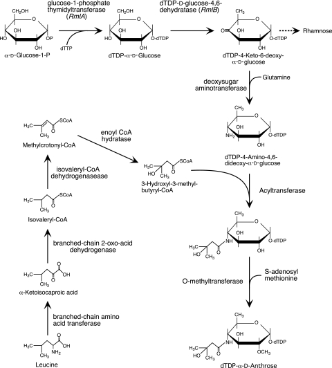
To gain insight into the biosynthesis of anthrose and eventually identify the enzymes and genes involved in anthrose synthesis, we devised a plausible anthrose biosynthetic pathway, which is illustrated in Fig. 1. We predicted that a key step in anthrose biosynthesis would be the addition of an amino group to the C-4 position of the sugar ring. Because deoxyamino sugars are usually formed by the transamination of a keto group in the sugar ring with an amino group derived from glutamine or glutamate (33), we predicted that the keto sugar is dTDP-4-keto-6-deoxy-α-d-glucose, an intermediate in the biosynthesis of rhamnose. Such a reaction would be catalyzed by a deoxysugar 4-aminotransferase. The amino group of anthrose is substituted with a branched-chain 5-carbon acyl group. Because acylation of amino nitrogens often involves an acyl coenzyme A (acyl-CoA), we predicted that 3-hydroxyl-3-methyl-butyrl-CoA is utilized in anthrose biosynthesis. This reaction would be catalyzed by a specific acyltransferase. We further proposed that 3-hydroxyl-3-methyl-butyrl-CoA is formed by the addition of water to methylcrotonyl-CoA, catalyzed by an enoyl-CoA hydratase. The methylcrotonyl-CoA, in turn, could be derived from leucine catabolism, as shown in Fig. 1 (28). Although the O methylation of the hydroxyl group at C-2 is shown as the last step in anthrose biosynthesis (Fig. 1), it could also occur before acylation of the amino nitrogen.
FIG. 1.
Plausible anthrose biosynthetic pathway. The pathway requires four dedicated enzymes and the initial substrates methylcrotonyl-CoA and dTDP-4-keto-6-deoxy-α-d-glucose, which are provided by leucine catabolism and l-rhamnose biosynthesis, respectively.
Identification of genes that could encode anthrose biosynthetic enzymes.
Since the genes encoding enzymes involved in the biosynthesis of a particular sugar are often contiguous, we attempted to identify a cluster of genes that could encode the predicted anthrose biosynthetic enzymes. We initially searched the sequenced genome of the Sterne strain of B. anthracis for genes encoding putative aminotransferases. Several genes were found and one, designated BAS3320 (25), encodes a protein that exhibits 26% amino acid identity and 46% amino acid similarity to a known 4-aminotransferase (i.e., perosamine synthase) of Vibrio cholerae (49). Flanking BAS3320, we found three genes that might encode other enzymes in the predicted biosynthetic pathway (Fig. 2). BAS3319 encodes a putative acyltransferase (actually annotated as an O-acetyltransferase), which could catalyze the transfer of a 3-hydroxy-3-methylbutamido group to the amino sugar. BAS3318 encodes a putative methyltransferase, which could catalyze the O methylation of the hydroxyl group at C-2. BAS3322 encodes an enoyl-CoA hydratase, an enzyme predicted to be involved in the biosynthesis of the side chain of anthrose.
FIG. 2.
Possible anthrose biosynthetic operon(s). B. anthracis Sterne genes BAS3322 through BAS3317 (designated genes 1 through 6, respectively) encode enzymes that could be involved in the biosynthesis of anthrose and its incorporation into BclA. The direction of transcription of the genes and the names of the encoded enzymes from genomic annotation are indicated. The annotated O-acetyltransferase is the acyltransferase in the predicted anthrose biosynthetic pathway.
In addition, this genomic region contains two genes, BAS3321 and BAS3317, which apparently encode glycosyltransferases (Fig. 2). These enzymes could be involved in the polymerization of the anthrose-containing pentasaccharide or its attachment to BclA. All six genes of the BAS3317 to -3322 gene cluster are oriented in the same direction, suggesting cotranscription of some or all of the genes. This cluster is flanked upstream by a collagenase-encoding gene, BAS3323, and a downstream gene of unknown function, BAS3316 (Fig. 2). Although the latter two genes are transcribed in the same direction as the BAS3317 to -3322 cluster, they are separated from this cluster by likely transcription terminators. Bacillus transcription terminators, which are almost always intrinsic terminators (2), can be readily identified as DNA sequences that specify an RNA hairpin, typically G+C-rich with a stem of 9 ± 2 bp, followed immediately by a ≥8-residue uridine-rich tract (13). For simplicity, genes BAS3322 through BAS3317 will hereafter be referred to as genes 1 through 6, as shown in Fig. 2.
Identification of possible promoters and transcription terminators for the putative anthrose biosynthetic genes.
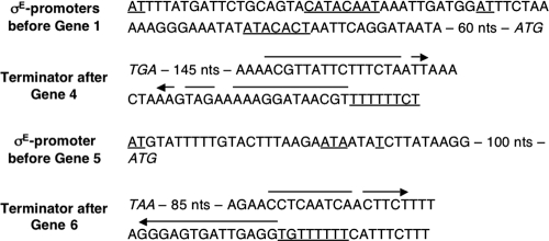
We inspected the genomic region containing genes 1 to 6 for sequences of possible promoters and intrinsic transcription terminators (25). Between the predicted transcription terminator of the collagenase-encoding gene (i.e., BAS3323) and gene 1, we identified two possible promoters with sequences closely resembling those recognized by the mother cell-specific sigma factor σE (Fig. 3) (20). We expected that the genes encoding the anthrose biosynthetic enzymes would be transcribed from a mother cell-specific promoter because the mother cell produces the components of the exosporium (21). We also identified one possible σE-specific promoter within the relatively long intergenic region between genes 4 and 5. We did not find sequences closely resembling other types of Bacillus promoters in our search. Additionally, we located two sequences of likely intrinsic transcription terminators (Fig. 3). The first is situated immediately downstream of gene 4 (i.e., upstream of the promoter between genes 4 and 5), and the second is situated immediately downstream of gene 6. These results suggest that genes 1 to 4 and genes 5 and 6 constitute two operons transcribed from σE-specific promoters.
FIG. 3.
Promoter and transcription terminator sequences. The sequences of two possible σE-specific promoters before gene 1 and one possible σE-specific promoter before gene 5 are underlined (σE-specific promoter consensus sequence = ATa-16 bases-cATAca-T). The sequences of putative intrinsic transcription terminators after genes 4 and 6 are shown. The inverted arrows and underlined T-rich regions indicate the sequences that specify terminator hairpins and U-rich tracts, the elements required for intrinsic termination of transcription. The start codon (ATG) of the gene downstream of each promoter and the stop codon (TAA or TGA) of the gene upstream of each intrinsic terminator are italicized. nts, nucleotides.
It is also worth noting that the intergenic regions within the predicted gene 1-to-4 operon are very short or, as in the case of genes 3 and 4, nonexistent. In the latter case, the genes overlap by 5 bp. Such overlapping reading frames are common for operons in B. anthracis (25). Each gene of the predicted 1-to-4 operon is preceded by a sequence resembling a strong ribosome binding site (54). These features are consistent with a gene 1-to-4 operon. In the case of the predicted gene 5-6 operon, the intergenic spacing is relatively long and there do not appear to be strong ribosome binding sites preceding the annotated open reading frames (25). For gene 5, however, the sixth codon of the annotated open reading frame is an ATG codon, which is preceded by a possible ribosome binding site. Therefore, the open reading frame of gene 5 is probably slightly shorter than annotated. We were unable to find an alternative ribosome binding site for gene 6.
Construction of mutations inactivating putative anthrose biosynthetic genes.
To determine the roles of genes 1 through 6 in anthrose biosynthesis, we constructed by allelic replacement six mutant B. anthracis strains containing chromosomal deletions that remove selected genes and promoters. Four strains contain an in-frame deletion that removes nearly all of either gene 1, 2, 3, or 4. Another strain contains a deletion that removes the DNA between the first codon of gene 5 and the last codon of gene 6. The final strain contains a deletion that removes 180 bp upstream and 8 bp downstream of the start codon of gene 1. This deletion removes both putative σE-specific promoters upstream of gene 1. The six mutations and corresponding strains are designated Δ1 through Δ4, Δ5-6, and ΔPro, respectively. The selectable marker in each construction was a spectinomycin-resistant cassette. For the Δ1 through Δ4 strains, this cassette was inserted into an intergenic region outside of the two putative anthrose operons. For the Δ5-6 and ΔPro strains, the cassette replaced the deleted DNA. A detailed description of mutant constructions is given in Materials and Methods. The mutations had no effect on cell growth or sporulation (data not shown).
Effects of the mutations on anthrose biosynthesis.
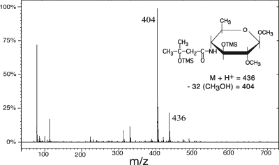
We prepared purified spores produced by the six mutant strains and, as a control, the wild-type strain. Anthrose levels of the spores were measured using GC/MS. Rhamnose levels, which do not appear to be significantly affected by the mutations described above, were also measured as an internal standard. The glycoconjugates in the spores were depolymerized by methanolysis, and after N reacetylation of the amino sugars, the resulting methyl glycosides were converted to volatile trimethylsilyl (TMS) derivatives. GC/MS of the mixtures gave chromatograms containing a large number of peaks with characteristic retention times and mass spectra corresponding to the sugar derivatives (and several other volatile components). Mass spectra of the peaks obtained using electron ionization of the compounds showed extensive fragmentation, with small, relatively common fragment ions. However, to minimize fragmentation of derivatized compounds and obtain more specific relatively high-mass-fragment ions, we used chemical ionization (CI) with acetonitrile as the reagent gas. This resulted in base peaks corresponding to the molecular ion of the derivative with a loss of methanol. Thus the positive-ion CI spectrum of anthrose shows a molecular ion at m/z 436 and a base peak at m/z 404 (M − methanol) (Fig. 4). Single-ion monitoring (SIM) of m/z 404 exhibits a high sensitivity for anthrose peaks, since other sugar derivatives yield few or no m/z 404 fragment ions. Similarly, SIM of m/z 363 can be used to identify rhamnose peaks.
FIG. 4.
Positive-ion CI mass spectrum of the TMS derivative of anthrose methyl glycoside. A molecular ion at m/z 436 and a base ion at m/z 404, which results from the loss of methanol, are specific for the anthrose derivative.
The GC/MS analyses of the wild-type and mutant spores are shown in Fig. 5. SIM of the m/z 363 (rhamnose) and 404 (anthrose) ions for all spores is displayed. The results show that anthrose is undetectable in Δ2, Δ3, and Δ4 spores, indicating that the corresponding genes are essential for anthrose synthesis. In contrast, the level of anthrose in Δ1 spores was approximately half of that detected in wild-type spores. Apparently, the activity encoded by gene 1 is involved in—but not essential for—anthrose synthesis. The level of anthrose in Δ5-6 spores was essentially the same as that in wild-type spores. Thus, genes 5 and 6 do not appear to be directly involved in anthrose synthesis. Finally, the level of anthrose in ΔPro spores was reduced to approximately 4% of the wild-type level. This residual anthrose synthesis is presumably due to low-level readthrough transcription of genes 1 through 4 from an uncharacterized upstream promoter (see below).
FIG. 5.
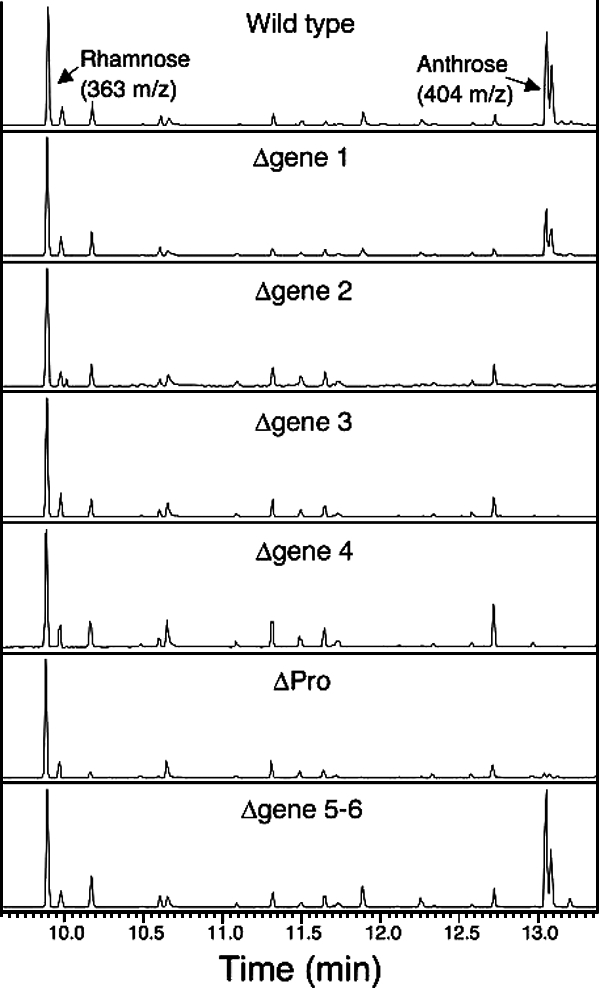
Chemical ionization GC/MS determinations of anthrose and rhamnose levels in wild-type and mutant spores. Chromatograms were produced using 108 spores of the indicated strains. TMS derivatives of methyl glycosides were analyzed using selected ion monitoring of m/z 363 for rhamnose and m/z 404 for anthrose. Note that two peaks are obtained for each sugar (in sugar-specific ratios) due to the alpha and beta forms of their methyl glycosides.
Complementation of mutations.
By design, the in-frame deletions used to inactivate putative anthrose biosynthetic genes do not create untranslated regions in the polycistronic mRNAs specified by the mutated operon. Therefore, these mutations will not cause Rho-mediated transcription polarity (36). Consistent with this expectation, spores produced by the Δ1 strain contained substantial levels of anthrose, indicating high-level expression of genes 2, 3, and 4. However, this experimental design does not preclude the possibility that secondary mutations (separate from the mutated operon) arise during mutant construction and that these secondary mutations are actually responsible for the absence of anthrose in Δ2, Δ3, and Δ4 spores. To rule out this possibility, we introduced plasmid pCLT1479, which carries the gene 1-to-4 operon, into strains Δ2, Δ3, and Δ4. We also introduced this plasmid into the Sterne strain to create an isogenic wild-type control strain. The four transformants were then used to prepare purified spores, and the level of anthrose in each spore type was measured as described above. The results showed that anthrose levels were essentially identical in the Δ2, Δ3, Δ4, and wild-type spores and that these levels were similar to those measured in untransformed wild-type spores (Fig. 5) (data not shown). Thus, the absence of anthrose in Δ2, Δ3, and Δ4 spores is due to a mutation within the gene 1-to-4 operon.
Establishing the number of genes in the putative operons.
The inspection of genomic sequences and our mutational analyses suggest that genes 1 to 6 constitute two operons, one containing genes 1 to 4 and the other genes 5 and 6. Apparent σE-specific promoters precede each putative operon, and potential intrinsic transcription terminators are downstream of genes 4 and 6. However, the terminator downstream of gene 4 is atypical, in that it has a terminator hairpin with an unusually long stem. This feature would likely reduce the efficiency of the terminator (13, 58). To determine whether genes 1 to 6 are indeed organized into two operons, we examined the transcripts specified by genes 1 to 6 in sporulating cells.
Initially, we established the time of appearance of transcripts specified by the two putative operons. Specifically, we used RT-PCR and primers specific for gene 1, 4, 5, or 6 to detect transcripts in cells at selected times during growth and sporulation. The results show an essentially identical pattern of appearance for all four genes (Fig. 6, with Tn indicating the time in hours relative to the nominal start of sporulation). Transcripts were first detected at approximately T0, transcript levels peaked at T2, and transcripts were no longer detectable after T5. For reference, we also determined the time of appearance of transcripts specified by the bclA operon, which is apparently transcribed from a σK-specific promoter (46). The bclA transcripts were first detected approximately 2 h later than those of the putative operons. In the mother cell, σE is expressed early and σK is expressed late in sporulation. Thus, our results are consistent with transcription of the putative anthrose operon(s) from one or more σE-specific promoters.
FIG. 6.
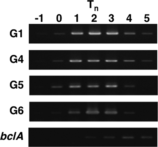
Time course of transcription of the anthrose (gene 1-to-4) and downstream operons. RT-PCR was used to detect transcripts specified by genes (G) 1, 4, 5, and 6 and, as a control, bclA. Tn indicates the sampling time, with n indicating hours relative to the start of sporulation.
To demonstrate independent transcription of the gene 1-to-4 and gene 5-6 operons, we used quantitative RT-PCR, with primer sets specific for each gene, to measure transcript levels in wild-type and ΔPro cells harvested at time T2 during sporulation (Fig. 7). The results show that the ΔPro mutation, which deletes the putative σE-specific promoters preceding gene 1, greatly reduces transcription of all genes in the gene 1-to-4 operon. In contrast, the ΔPro mutation has no measurable effect on the level of transcripts specified by genes 5 and 6. These results are consistent with two independent operons. However, in wild-type cells we also detected low levels of transcripts specified by the junction region between genes 4 and 5. This junction region includes sequences upstream and downstream of the putative intrinsic transcription terminator downstream of gene 4. Furthermore, the level of junction transcripts was greatly reduced in the ΔPro mutant. Thus, these results indicate that the intrinsic transcription terminator downstream of gene 4 is relatively weak and permits readthrough of transcripts initiated at the promoter(s) of the gene 1-to-4 operon. Finally, we were unable to detect transcripts downstream of the intrinsic terminator following gene 6, which indicates highly efficient transcription termination at the end of the gene 5-6 operon (data not shown).
FIG. 7.
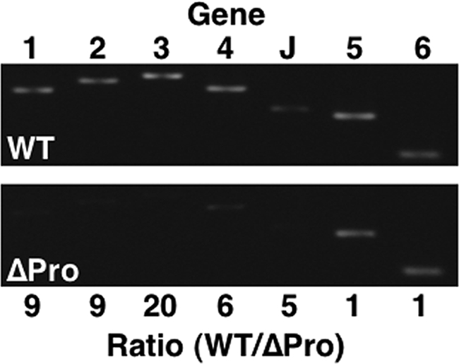
Levels of transcripts specified by the anthrose (gene 1-to-4) and downstream operons in sporulating cells of the wild-type (WT) and ΔPro strains. Quantitative RT-PCR was use to measure the levels of transcripts specified by genes (G) 1 to 6 and the junction between genes 4 and 5 (J). The WT/ΔPro transcript-level ratios are indicated for each gene and the operon junction. Transcripts were isolated from cells at stage T2.
Further confirmation that genes 1 to 4 and genes 5 and 6 form separate operons was provided by Northern blot analyses. In these experiments, we used 32P-labeled RNA probes specific for gene 3 or gene 6 and transcripts from wild-type and ΔPro cells at stage T2 of sporulation (Fig. 8). The Northern blot with the gene 3 probe and wild-type transcripts revealed a major band of 5.5 kb and a minor band of 8.3 kb. These sizes correspond to those predicted for full-length transcripts initiated at the promoter(s) upstream of gene 1 and terminated at the intrinsic terminators downstream of gene 4 and gene 6, respectively. Consistent with these assignments, both bands were absent when transcripts from ΔPro cells were analyzed. The Northern blot with the gene 6 probe again showed a minor 8.3-kb band that was present in transcripts from wild-type cells but absent from transcripts from ΔPro cells. This result confirmed that the 8.3-kb band contained gene 1-to-6 transcripts. A 5.5-kb band was not detected with the gene 6 probe, indicating that this band indeed contained gene 1-to-4 transcripts. The analysis with the gene 6 probe also revealed a major 2.5-kb band, which was present in transcripts from both wild-type and ΔPro cells. This result indicates that this band corresponds to gene 5-6 transcripts. Taken together, our results provide strong evidence for separate gene 1-to-4 and gene 5-6 operons, with minor readthrough transcription at the intrinsic terminator downstream of gene 4. (Note that the source of the ∼2.5-kb bands in the wild-type blot with the G3 probe [Fig. 8] is not known, but they may be degradation products.)
FIG. 8.
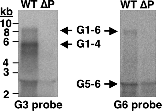
Detection of full-length transcripts specified by the anthrose (gene 1-to-4) and downstream operons. Transcripts were isolated from sporulating (T2) cells of the wild-type (WT) and ΔPro (ΔP) strains. An equal amount of RNA from each strain was used for Northern blot analysis, and the probes were specific for genes (G) 3 and 6. Transcripts that appeared to read through both operons (G1-6), the anthrose operon (G1-4), or the downstream operon (G5-6) are indicated.
Distribution of the anthrose operon and biosynthesis of anthrose in other Bacillus species.
Our results indicate that the gene 1-to-4 operon, or at least the last three genes of this operon, are essential for anthrose biosynthesis. A search of other available genomic sequences for this operon revealed that all four of the B. anthracis strains with sequenced genomes contain an identical anthrose biosynthetic operon, from the promoter region through the transcription terminator (25). Unexpectedly, we found some strains of B. cereus (i.e., E33L) and B. thuringiensis (i.e., Al Hakam and 97-27) with apparent anthrose operons that were nearly identical to the operon of B. anthracis. This near identity extended from the promoter region through the transcription terminator. For example, the encoded sequences of the anthrose biosynthetic enzymes of these strains are 97 to 100% identical to the corresponding B. anthracis enzymes (Table 1). We also found three other strains (i.e., B. cereus 14579, 10987, and G9241) with operons similar to the anthrose operon of B. anthracis in terms of gene order and in that genes 2 to 4 encode enzymes that are 80 to 84% identical to the corresponding B. anthracis enzymes (Table 1). However, these operons have promoter and terminator regions that are unrelated to those of B. anthracis, and they contain a gene 1 that encodes a 382-amino-acid protein with no recognizable similarity to the 263-amino-acid enzyme 1 of B. anthracis. The absence of the 263-amino-acid enzyme 1 does not preclude anthrose production, though, because we found that it is not essential for anthrose biosynthesis in B. anthracis.
TABLE 1.
Comparison of the anthrose biosynthetic enzymes and anthrose levels in selected Bacillus strains
| Strain | % Identity to corresponding enzyme in B. anthracis (no. of amino acids)a
|
Relative anthrose level in sporesb | |||
|---|---|---|---|---|---|
| Enzyme 1 | Enzyme 2 | Enzyme 3 | Enzyme 4 | ||
| B. anthracisc | 100 (263) | 100 (851) | 100 (376) | 100 (188) | 100 |
| B. thuringiensis Al Hakam | 98 (263) | 99 (851) | 99 (376) | 97 (188) | 42 |
| B. cereus D17 (Fri-13) | 98 (263) | NDd | 99 (376) | ND | 0 |
| B. thuringinesis 97-27 subsp. konkukian | 100 (263) | 100 (851) | 99 (376) | 100 (188) | ND |
| B. cereus E33L (ZK) | 98 (263) | 99 (851) | 98 (376) | 98 (188) | ND |
| B. thuringiensis subsp. kurstaki | 0 (382) | ND | 81 (376) | ND | 3 |
| B. cereus T | 0 (382) | ND | 81 (376) | ND | 8 |
| B. cereus ATCC 14579 | 0 (382)e | 84 (853) | 81 (376) | 82 (188) | ND |
| B. cereus ATCC 10987 | 0 (382) | 84 (853) | 81 (376) | 84 (188) | ND |
| B. cereus G9241 | 0 (382) | 84 (853) | 80 (376) | 83 (188) | ND |
Enzymes 1 to 4 are encoded by genes 1 to 4, respectively. DNA sequences are from KEGG (25) or were determined in this study (data not shown).
Relative anthrose levels per spore, with the level for B. anthracis spores set at 100.
Includes Sterne 34F2, Ames, Ames 0581, and A2012 strains. Anthrose levels were determined only for the Sterne strain.
ND, not determined.
Annotated as 400 amino acids, which appears to be incorrect.
To directly determine the ability of B. cereus and B. thuringiensis strains to synthesize anthrose, we measured the levels of anthrose in spores produced by B. thuringiensis Al Hakam, B. cereus D17 (Fri-13), B. thuringiensis subsp. kurstaki, and B. cereus T (Fig. 9). The first two strains have apparent anthrose operons nearly identical to the B. anthracis operon, while the last two strains contain operons encoding the 382-amino-acid enzyme 1 (Table 1). Additionally, it had previously been reported that spores of the last two strains did not contain anthrose (12). The GC/MS analyses of the strains showed that spores of B. thuringiensis Al Hakam contained approximately half the amount of anthrose found in B. anthracis spores. On the other hand, spores of B. cereus D17 (Fri-13) were devoid of anthrose. However, the B. cereus D17 spores also lacked rhamnose (Fig. 9), which may be directly related to the absence of anthrose as discussed below. Surprisingly, small anthrose peaks were also present in the selected ion chromatograms of B. thuringiensis subsp. kurstaki and B. cereus T spores (Fig. 9 and Table 1). The mass spectra of these small peaks were identical to the spectrum of anthrose (Fig. 4), which confirmed their identification.
FIG. 9.
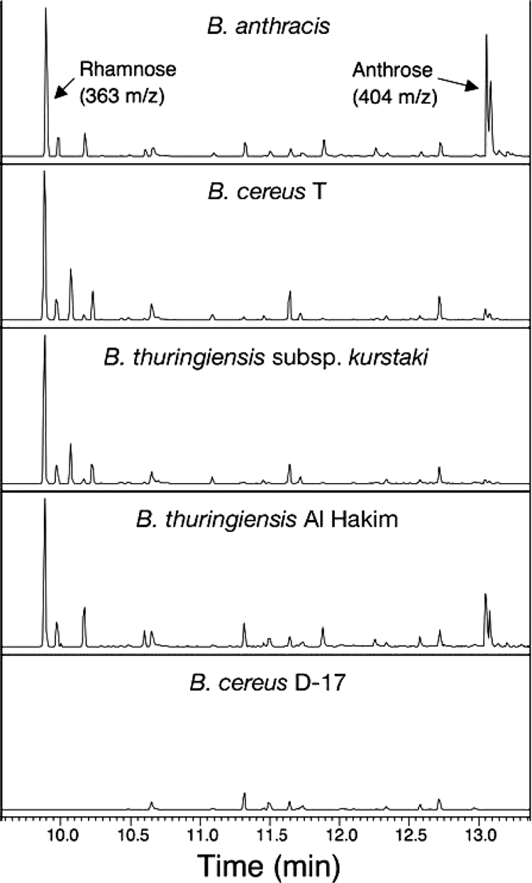
CI GC/MS determinations of anthrose and rhamnose levels in spores of selected B. cereus and B. thuringiensis strains. As described in the legend to Fig. 5, selected ion chromatograms were obtained for spores of the indicated strains.
DISCUSSION
For many years, it was thought that bacteria were unable to glycosylate proteins. However, in the last decade numerous studies have provided clear evidence for covalent attachment of carbohydrates to bacterial proteins (4, 30, 52). Several different functions for glycosylation of bacterial proteins have been proposed, including the maintenance of protein conformation, heat stability, surface recognition, resistance to proteolysis, enzymatic activity, cell adhesion, agglutination, inhibition of ice nucleation, and immune evasion (32, 44). In the case of BclA of B. anthracis, glycosylation is apparently required for maintaining protein conformation, resistance to proteolysis, cell adhesion, and surface recognition (5-7, 47). The anthrose-containing pentasaccharide attached at multiple sites within the collagen-like region of BclA is likely to play a critical role in these functions. This raises the obvious and interesting question: what is the role of the unusual sugar anthrose? The fact that B. anthracis maintains the ability to synthesize this complicated sugar, even in starving cells, suggests that anthrose performs a key function in spore survival or propagation of the bacterium.
To help elucidate the importance of anthrose, we investigated its biosynthesis. We predicted a plausible pathway for anthrose biosynthesis that requires four enzymatic activities commonly found in bacteria, (i.e., enoyl-CoA hydratase, deoxysugar aminotransferase, acyltransferase, and O-methyltransferase). Based on our hypothetical pathway, we located in the B. anthracis genomic sequence a cluster of six similarly oriented genes that could encode the four predicted enzymes and two putative glycosyltransferases. We showed experimentally that this gene cluster includes a four-gene (genes 1 to 4) operon and a downstream two-gene (genes 5 and 6) operon, each encoding one of the glycosyltransferases. Additional experiments indicated that these operons were transcribed from promoters recognized by the mother cell-specific sigma factor σE, consistent with their predicted roles within the mother cell compartment.
To confirm the predicted roles of the putative biosynthetic genes, we constructed B. anthracis strains carrying in-frame deletions that inactivate a particular gene (or genes) and then measured anthrose levels in spores produced by these mutant strains. As predicted, inactivation of either gene 3 or gene 4 results in spores devoid of anthrose, indicating that their encoded enzymes (i.e., deoxysugar aminotransferase and acyltransferase) are indeed required for anthrose biosynthesis. Similarly, inactivation of gene 2 results in spores devoid of anthrose. This result indicates that the putative glycosyltransferase encoded by gene 2 is required for the attachment of anthrose to BclA, most likely by the transfer of anthrose to the oligosaccharide side chain of BclA. In contrast, inactivation of gene 1, which encodes the enoyl-CoA hydratase, results in spores with about half as much anthrose as that found in wild-type spores. This result indicates that the enzyme encoded by gene 1 is involved in anthrose biosynthesis but that other activities in the cell can partially compensate for its absence. These activities may include other enoyl-CoA hydratases—the annotated B. anthracis genome contains multiple genes that apparently encode this enzyme (25). Another possibility is that the predicted hydration reaction occurs, at least to some extent, nonenzymatically. Based on the clear involvement in anthrose biosynthesis or transfer of each gene in the gene 1-to-4 operon, we propose that this operon be designated the antABCD operon.
Unexpectedly, deletion of the entire two-gene operon did not have a significant effect on the anthrose content of spores, indicating that genes 5 and 6 do not play an essential role in the biosynthesis of anthrose. If gene 5 does not encode the enzyme required for O methylation at position C-2 of anthrose, this methyltransferase must be encoded elsewhere in the genome. An alternative interpretation is that genes 5 and 6 are involved in anthrose biosynthesis, but their inactivation is complemented fully by other genes in the cell. Additional experiments will be required to resolve this issue.
Using the antABCD operon as a probe, we searched all available genomic sequences (25). Matches were found in the genomes of all nine B. anthracis, B. cereus, and B. thuringiensis strains in the database (not counting B. anthracis Sterne), but not in the genomes of any other bacteria. The three B. anthracis genomes contain antABCD operons and also gene 5-6 operons that are identical to the corresponding operons in the Sterne strain. Strains B. cereus E33L, B. thuringiensis Al Hakam, and B. thuringiensis 97-27 were found to contain apparent antABCD and gene 5-6 operons that are nearly identical (≥97% at the protein level) to the corresponding operons of B. anthracis. The remaining matches were somewhat more limited (i.e., 80 to 84% identity to the proteins encoded by genes 2, 3, and 4 and no sequence similarity elsewhere) and were found in the genomes of B. cereus strains 14579, 10987, and G9241. These observations suggested that anthrose biosynthesis is not restricted to B. anthracis.
To test this hypothesis, we screened four strains for their ability to produce spores containing anthrose. Two of these strains were B. thuringiensis Al Hakam and B. cereus D-17, both exhibiting ≥97% identity (at the protein level) to the antABCD operon and among the nearest relatives to B. anthracis (9, 19). We found that spores of B. thuringiensis Al Hakam contain a high level of anthrose, nearly half that of B. anthracis wild-type spores. This discovery provides clear evidence that a strain other than B. anthracis is able to synthesize anthrose. In contrast, spores of strain B. cereus D-17 were devoid of anthrose, suggesting that this strain could not synthesize this sugar. However, an alternative explanation was suggested by the observation that B. cereus D-17 spores also lack rhamnose. Rhamnose is the sugar to which anthrose is attached during oligosaccharide polymerization in B. anthracis, and rhamnose-deficient strains are unable to incorporate anthrose into spore-bound oligosaccharides (12). Perhaps a similar situation exists with B. cereus D-17; i.e., anthrose is made but not incorporated into the spore. The other two strains in our screen were B. thuringiensis subsp. kurstaki and B. cereus T, both of which possess a partial match to antABCD similar to that of B. cereus strains 14579, 10987, and G9241. We had previously reported that these strains produce spores lacking anthrose (12), and they were initially included in the screen as negative controls. However, we detected anthrose in spores of both strains, albeit at very lower levels (i.e., ≤8% of the level in B. anthracis spores). These levels were too low to be detected with the assay used in our earlier study. Thus, even strains carrying a highly divergent antABCD locus are capable of anthrose synthesis. More importantly, these results suggest that anthrose synthesis can occur in a wide variety of B. cereus and B. thuringiensis strains.
A wider distribution of anthrose may not eliminate it as a useful marker for detection of B. anthracis spores or as a target for therapeutic intervention of anthrax. It appears that high levels of anthrose are found only in spores of B. anthracis and of the nominal B. cereus and B. thuringiensis strains that are members of the B. anthracis lineage (19). These strains share virulence phenotypes, including the ability to cause severe disease in humans (19). Therefore, it would be important to detect spores of all of these pathogens. The obvious requirement is the ability to discriminate between spores with high and low levels of anthrose. The potential use of anthrose as a target for therapeutic intervention has not yet been studied.
Acknowledgments
We thank Robert T. Cartee at the UAB Gas Chromatography-Mass Spectrometry Shared Facility for Carbohydrate Research and the UAB DNA Sequencing Core for sample analyses. We also thank Evvie Allison for editorial assistance.
This work was supported by Public Health Service grant AI057699 from the National Institute of Allergy and Infectious Diseases.
Footnotes
Published ahead of print on 1 February 2008.
REFERENCES
- 1.Arnaud, M., A. Chastanet, and M. Débarbouillé. 2004. New vector for efficient allelic replacement in naturally nontransformable, low-GC-content, gram-positive bacteria. Appl. Environ. Microbiol. 706887-6891. [DOI] [PMC free article] [PubMed] [Google Scholar]
- 2.Artsimovitch, I., V. Svetlov, L. Anthony, R. R. Burgess, and R. Landick. 2000. RNA polymerases from Bacillus subtilis and Escherichia coli differ in recognition of regulatory signals in vitro. J. Bacteriol. 1826027-6035. [DOI] [PMC free article] [PubMed] [Google Scholar]
- 3.Ausubel, F. M., R. Brent, R. E. Kingston, D. D. Moore, J. G. Seidman, J. A. Smith, and K. Struhl (ed.). 1989. Current protocols in molecular biology. John Wiley & Sons, Inc., New York, NY.
- 4.Benz, I., and M. A. Schmidt. 2002. Never say never again: protein glycosylation in pathogenic bacteria. Mol. Microbiol. 45267-276. [DOI] [PubMed] [Google Scholar]
- 5.Boydston, J. A., P. Chen, C. T. Steichen, and C. L. Turnbough, Jr. 2005. Orientation within the exosporium and structural stability of the collagen-like glycoprotein BclA of Bacillus anthracis. J. Bacteriol. 1875310-5317. [DOI] [PMC free article] [PubMed] [Google Scholar]
- 6.Bozue, J., K. L. Moody, C. K. Cote, B. G. Stiles, A. M. Friedlander, S. L. Welkos, and M. L. Hale. 2007. Bacillus anthracis spores of the bclA mutant exhibit increased adherence to epithelial cells, fibroblasts, and endothelial cells but not to macrophages. Infect. Immun. 754498-4505. [DOI] [PMC free article] [PubMed] [Google Scholar]
- 7.Bozue, J. A., N. Parthasarathy, L. R. Phillips, C. K. Cote, P. F. Fellows, I. Mendelson, A. Shafferman, and A. M. Friedlander. 2005. Construction of a rhamnose mutation in Bacillus anthracis affects adherence to macrophages but not virulence in guinea pigs. Microb. Pathog. 381-12. [DOI] [PubMed] [Google Scholar]
- 8.Brittingham, K. C., G. Ruthel, R. G. Panchal, C. L. Fuller, W. J. Ribot, T. A. Hoover, H. A. Young, A. O. Anderson, and S. Bavari. 2005. Dendritic cells endocytose Bacillus anthracis spores: implications for anthrax pathogenesis. J. Immunol. 1745545-5552. [DOI] [PubMed] [Google Scholar]
- 9.Challacombe, J. F., M. R. Altherr, G. Xie, S. S. Bhotika, N. Brown, D. Bruce, C. S. Campbell, M. L. Campbell, J. Chen, O. Chertkov, C. Cleland, M. Dimitrijevic, N. A. Doggett, J. J. Fawcett, T. Glavina, L. A. Goodwin, L. D. Green, C. S. Han, K. K. Hill, P. Hitchcock, P. J. Jackson, P. Keim, A. R. Kewalramani, J. Longmire, S. Lucas, S. Malfatti, D. Martinez, K. McMurry, L. J. Meincke, M. Misra, B. L. Moseman, M. Mundt, A. C. Munk, R. T. Okinaka, B. Parson-Quintana, L. P. Reilly, P. Richardson, D. L. Robinson, E. Saunders, R. Tapia, J. G. Tesmer, N. Thayer, L. S. Thompson, H. Tice, L. O. Ticknor, P. L. Wills, P. Gilna, and T. S. Brettin. 2007. The complete genome sequence of Bacillus thuringiensis Al Hakam. J. Bacteriol. 1893680-3681. [DOI] [PMC free article] [PubMed] [Google Scholar]
- 10.Chaudry, G. J., M. Moayeri, S. Liu, and S. H. Leppla. 2002. Quickening the pace of anthrax research: three advances point towards possible therapies. Trends Microbiol. 1058-62. [DOI] [PubMed] [Google Scholar]
- 11.Crich, D., and O. Vinogradova. 2007. Synthesis of the antigenic tetrasaccharide side chain from the major glycoprotein of Bacillus anthracis exosporium. J. Org. Chem. 726513-6520. [DOI] [PMC free article] [PubMed] [Google Scholar]
- 12.Daubenspeck, J. M., H. Zeng, P. Chen, S. Dong, C. T. Steichen, N. R. Krishna, D. G. Pritchard, and C. L. Turnbough, Jr. 2004. Novel oligosaccharide side-chains of the collagen-like region of BclA, the major glycoprotein of the Bacillus anthracis exosporium. J. Biol. Chem. 27930945-30953. [DOI] [PubMed] [Google Scholar]
- 13.de Hoon, M. J., Y. Makita, K. Nakai, and S. Miyano. 2005. Prediction of transcriptional terminators in Bacillus subtilis and related species. PLoS Comput. Biol. 1e25. [DOI] [PMC free article] [PubMed] [Google Scholar]
- 14.Foster, S. J., and D. L. Popham. 2002. Structure and synthesis of cell wall, spore cortex, teichoic acids, S-layers, and capsules, p. 21-41. In A. L. Sonenshein, J. A. Hoch, and R. Losick (ed.), Bacillus subtilis and its closest relatives. From genes to cells. ASM Press, Washington, DC.
- 15.Gerhardt, P. 1967. Cytology of Bacillus anthracis. Fed. Proc. 261504-1517. [PubMed] [Google Scholar]
- 16.Gerhardt, P., and S. H. Black. 1961. Permeability of bacterial spores. II. Molecular variables affecting solute permeation. J. Bacteriol. 82750-760. [DOI] [PMC free article] [PubMed] [Google Scholar]
- 17.Green, B. D., L. Battisti, T. M. Koehler, C. B. Thorne, and B. E. Ivins. 1985. Demonstration of a capsule plasmid in Bacillus anthracis. Infect. Immun. 49291-297. [DOI] [PMC free article] [PubMed] [Google Scholar]
- 18.Guidi-Rontani, C., M. Levy, H. Ohayon, and M. Mock. 2001. Fate of germinated Bacillus anthracis spores in primary murine macrophages. Mol. Microbiol. 42931-938. [DOI] [PubMed] [Google Scholar]
- 19.Han, C. S., G. Xie, J. F. Challacombe, M. R. Altherr, S. S. Bhotika, N. Brown, D. Bruce, C. S. Campbell, M. L. Campbell, J. Chen, O. Chertkov, C. Cleland, M. Dimitrijevic, N. A. Doggett, J. J. Fawcett, T. Glavina, L. A. Goodwin, D. L. Green, K. K. Hill, P. Hitchcock, P. J. Jackson, P. Keim, A. R. Kewalramani, J. Longmire, S. Lucas, S. Malfatti, K. McMurry, L. J. Meincke, M. Misra, B. L. Moseman, M. Mundt, A. C. Munk, R. T. Okinaka, B. Parson-Quintana, L. P. Reilly, P. Richardson, D. L. Robinson, E. Rubin, E. Saunders, R. Tapia, J. G. Tesmer, N. Thayer, L. S. Thompson, H. Tice, L. O. Ticknor, P. L. Wills, T. S. Brettin, and P. Gilna. 2006. Pathogenomic sequence analysis of Bacillus cereus and Bacillus thuringiensis isolates closely related to Bacillus anthracis. J. Bacteriol. 1883382-3390. [DOI] [PMC free article] [PubMed] [Google Scholar]
- 20.Helmann, J. D., and C. P. Moran, Jr. 2002. RNA polymerase and sigma factors, p. 289-312. In A. L. Sonenshein, J. A. Hoch, and R. Losick (ed.), Bacillus subtilis and its closest relatives. From genes to cells. ASM Press, Washington, DC.
- 21.Henriques, A. O., and C. P. Moran, Jr. 2000. Structure and assembly of the bacterial endospore coat. Methods 2095-110. [DOI] [PubMed] [Google Scholar]
- 22.Hilbert, D. W., and P. J. Piggot. 2004. Compartmentalization of gene expression during Bacillus subtilis spore formation. Microbiol. Mol. Biol. Rev. 68234-262. [DOI] [PMC free article] [PubMed] [Google Scholar]
- 23.Horton, R. M., H. D. Hunt, S. N. Ho, J. K. Pullen, and L. R. Pease. 1989. Engineering hybrid genes without the use of restriction enzymes: gene splicing by overlap extension. Gene 7761-68. [DOI] [PubMed] [Google Scholar]
- 24.Inglesby, T. V., T. O'Toole, D. A. Henderson, J. G. Bartlett, M. S. Ascher, E. Eitzen, A. M. Friedlander, J. Gerberding, J. Hauer, J. Hughes, J. McDade, M. T. Osterholm, G. Parker, T. M. Perl, P. K. Russell, and K. Tonat. 2002. Anthrax as a biological weapon, 2002: updated recommendations for management. JAMA 2872236-2252. [DOI] [PubMed] [Google Scholar]
- 25.Kanehisa, M., S. Goto, M. Hattori, K. F. Aoki-Kinoshita, M. Itoh, S. Kawashima, T. Katayama, M. Araki, and M. Hirakawa. 2006. From genomics to chemical genomics: new developments in KEGG. Nucleic Acids Res. 34D354-D357. [DOI] [PMC free article] [PubMed] [Google Scholar]
- 26.Koehler, T. M., Z. Dai, and M. Kaufman-Yarbray. 1994. Regulation of the Bacillus anthracis protective antigen gene: CO2 and a trans-acting element activate transcription from one of two promoters. J. Bacteriol. 176586-595. [DOI] [PMC free article] [PubMed] [Google Scholar]
- 27.Lai, E. M., N. D. Phadke, M. T. Kachman, R. Giorno, S. Vazquez, J. A. Vazquez, J. R. Maddock, and A. Driks. 2003. Proteomic analysis of the spore coats of Bacillus subtilis and Bacillus anthracis. J. Bacteriol. 1851443-1454. [DOI] [PMC free article] [PubMed] [Google Scholar]
- 28.Massey, L. K., J. R. Sokatch, and R. S. Conrad. 1976. Branched-chain amino acid catabolism in bacteria. Bacteriol. Rev. 4042-54. [DOI] [PMC free article] [PubMed] [Google Scholar]
- 29.Mehta, A. S., E. Saile, W. Zhong, T. Buskas, R. Carlson, E. Kannenberg, Y. Reed, C. P. Quinn, and G. J. Boons. 2006. Synthesis and antigenic analysis of the BclA glycoprotein oligosaccharide from the Bacillus anthracis exosporium. Chemistry 129136-9149. [DOI] [PubMed] [Google Scholar]
- 30.Messner, P. 2004. Prokaryotic glycoproteins: unexplored but important. J. Bacteriol. 1862517-2519. [DOI] [PMC free article] [PubMed] [Google Scholar]
- 31.Mock, M., and A. Fouet. 2001. Anthrax. Annu. Rev. Microbiol. 55647-671. [DOI] [PubMed] [Google Scholar]
- 32.Moens, S., and J. Vanderleyden. 1997. Glycoproteins in prokaryotes. Arch. Microbiol. 168169-175. [DOI] [PubMed] [Google Scholar]
- 33.Nedal, A., and S. B. Zotchev. 2004. Biosynthesis of deoxyaminosugars in antibiotic-producing bacteria. Appl. Microbiol. Biotechnol. 647-15. [DOI] [PubMed] [Google Scholar]
- 34.Nicholson, W. L., N. Munakata, G. Horneck, H. J. Melosh, and P. Setlow. 2000. Resistance of Bacillus endospores to extreme terrestrial and extraterrestrial environments. Microbiol. Mol. Biol. Rev. 64548-572. [DOI] [PMC free article] [PubMed] [Google Scholar]
- 35.Nicholson, W. L., and P. Setlow. 1990. Sporulation, germination and outgrowth, p. 391-450. In C. R. Harwood and S. M. Cutting (ed.), Molecular biological methods for Bacillus. John Wiley & Sons, Ltd., West Sussex, United Kingdom.
- 36.Nudler, E., and M. E. Gottesman. 2002. Transcription termination and anti-termination in E. coli. Genes Cells 7755-768. [DOI] [PubMed] [Google Scholar]
- 37.Redmond, C., L. W. Baillie, S. Hibbs, A. J. Moir, and A. Moir. 2004. Identification of proteins in the exosporium of Bacillus anthracis. Microbiology 150355-363. [DOI] [PubMed] [Google Scholar]
- 38.Rider, T. H., M. S. Petrovick, F. E. Nargi, J. D. Harper, E. D. Schwoebel, R. H. Mathews, D. J. Blanchard, L. T. Bortolin, A. M. Young, J. Chen, and M. A. Hollis. 2003. A B cell-based sensor for rapid identification of pathogens. Science 301213-215. [DOI] [PubMed] [Google Scholar]
- 39.Saile, E., and T. M. Koehler. 2006. Bacillus anthracis multiplication, persistence, and genetic exchange in the rhizosphere of grass plants. Appl. Environ. Microbiol. 723168-3174. [DOI] [PMC free article] [PubMed] [Google Scholar]
- 40.Saile, E., and T. M. Koehler. 2002. Control of anthrax toxin gene expression by the transition state regulator abrB. J. Bacteriol. 184370-380. [DOI] [PMC free article] [PubMed] [Google Scholar]
- 41.Saksena, R., R. Adamo, and P. Kovác. 2006. Synthesis of the tetrasaccharide side chain of the major glycoprotein of the Bacillus anthracis exosporium. Bioorg. Med. Chem. Lett. 16615-617. [DOI] [PubMed] [Google Scholar]
- 42.Saksena, R., R. Adamo, and P. Kovác. 2007. Immunogens related to the synthetic tetrasaccharide side chain of the Bacillus anthracis exosporium. Bioorg. Med. Chem. 154283-4310. [DOI] [PMC free article] [PubMed] [Google Scholar]
- 43.Sambrook, J., E. F. Fritsch, and T. Maniatis. 1989. Molecular cloning: a laboratory manual, 2nd ed. Cold Spring Harbor Laboratory, Cold Spring Harbor, NY.
- 44.Schmidt, M. A., L. W. Riley, and I. Benz. 2003. Sweet new world: glycoproteins in bacterial pathogens. Trends Microbiol. 11554-561. [DOI] [PubMed] [Google Scholar]
- 45.Setlow, P. 2003. Spore germination. Curr. Opin. Microbiol. 6550-556. [DOI] [PubMed] [Google Scholar]
- 46.Steichen, C., P. Chen, J. F. Kearney, and C. L. Turnbough, Jr. 2003. Identification of the immunodominant protein and other proteins of the Bacillus anthracis exosporium. J. Bacteriol. 1851903-1910. [DOI] [PMC free article] [PubMed] [Google Scholar]
- 47.Steichen, C. T., J. F. Kearney, and C. L. Turnbough, Jr. 2005. Characterization of the exosporium basal layer protein BxpB of Bacillus anthracis. J. Bacteriol. 1875868-5876. [DOI] [PMC free article] [PubMed] [Google Scholar]
- 48.Stopa, P. J. 2000. The flow cytometry of Bacillus anthracis spores revisited. Cytometry 41237-244. [DOI] [PubMed] [Google Scholar]
- 49.Stroeher, U. H., L. E. Karageorgos, M. H. Brown, R. Morona, and P. A. Manning. 1995. A putative pathway for perosamine biosynthesis is the first function encoded within the rfb region of Vibrio cholerae O1. Gene 16633-42. [DOI] [PubMed] [Google Scholar]
- 50.Swiecki, M. K., M. W. Lisanby, C. L. Turnbough, Jr., and J. F. Kearney. 2006. Monoclonal antibodies for Bacillus anthracis spore detection and functional analyses of spore germination and outgrowth. J. Immunol. 1766076-6084. [DOI] [PubMed] [Google Scholar]
- 51.Sylvestre, P., E. Couture-Tosi, and M. Mock. 2002. A collagen-like surface glycoprotein is a structural component of the Bacillus anthracis exosporium. Mol. Microbiol. 45169-178. [DOI] [PubMed] [Google Scholar]
- 52.Szymanski, C. M., and B. W. Wren. 2005. Protein glycosylation in bacterial mucosal pathogens. Nat. Rev. Microbiol. 3225-237. [DOI] [PubMed] [Google Scholar]
- 53.Tamborrini, M., D. B. Werz, J. Frey, G. Pluschke, and P. H. Seeberger. 2006. Anti-carbohydrate antibodies for the detection of anthrax spores. Angew. Chem. Int. Ed. Engl. 456581-6582. [DOI] [PubMed] [Google Scholar]
- 54.Vellanoweth, R. L., and J. C. Rabinowitz. 1992. The influence of ribosome-binding-site elements on translational efficiency in Bacillus subtilis and Escherichia coli in vivo. Mol. Microbiol. 61105-1114. [DOI] [PubMed] [Google Scholar]
- 55.Werz, D. B., and P. H. Seeberger. 2005. Total synthesis of antigen Bacillus anthracis tetrasaccharide—creation of an anthrax vaccine candidate. Angew. Chem. Int. Ed. Engl. 446315-6318. [DOI] [PubMed] [Google Scholar]
- 56.Williams, D. D., O. Benedek, and C. L. Turnbough, Jr. 2003. Species-specific peptide ligands for the detection of Bacillus anthracis spores. Appl. Environ. Microbiol. 696288-6293. [DOI] [PMC free article] [PubMed] [Google Scholar]
- 57.Wilson, H. R., C. D. Archer, J. K. Liu, and C. L. Turnbough, Jr. 1992. Translational control of pyrC expression mediated by nucleotide-sensitive selection of transcriptional start sites in Escherichia coli. J. Bacteriol. 174514-524. [DOI] [PMC free article] [PubMed] [Google Scholar]
- 58.Wilson, K. S., and P. H. von Hippel. 1995. Transcription termination at intrinsic terminators: the role of the RNA hairpin. Proc. Natl. Acad. Sci. USA 928793-8797. [DOI] [PMC free article] [PubMed] [Google Scholar]


