Abstract
During the period of September 1997 through July 1998, two coelacanth fishes were captured off Manado Tua Island, Sulawesi, Indonesia. These specimens were caught almost 10,000 km from the only other known population of living coelacanths, Latimeria chalumnae, near the Comores. The Indonesian fish was described recently as a new species, Latimeria menadoensis, based on morphological differentiation and DNA sequence divergence in fragments of the cytochrome b and 12S rRNA genes. We have obtained the sequence of 4,823 bp of mitochondrial DNA from the same specimen, including the entire genes for cytochrome b, 12S rRNA, 16S rRNA, four tRNAs, and the control region. The sequence is 4.1% different from the published sequence of an animal captured from the Comores, indicating substantial divergence between the Indonesian and Comorean populations. Nine morphological and meristic differences are purported to distinguish L. menadoensis and L. chalumnae, based on comparison of a single specimen of L. menadoensis to a description of five individuals of L. chalumnae from the Comores. A survey of the literature provided data on 4 of the characters used to distinguish L. menadoensis from L. chalumnae from an additional 16 African coelacanths; for all 4 characters, the Indonesian sample was within the range of variation reported for the African specimens. Nonetheless, L. chalumnae and L. menadoensis appear to be separate species based on divergence of mitochondrial DNA.
The discovery of a living coelacanth off the coast of South Africa in 1938 (1) surprised the scientific community, because the fish is a member of a lineage that was thought to have gone extinct almost 80 million years ago (MYA) (2). A subsequent 14-year search for a second specimen eventually revealed the “true” home of Latimeria chalumnae in the Comores Islands in the western Indian Ocean (3). Since that discovery in 1952, more than 200 coelacanths have been captured in or near the Comores, and by 1994 the population size was thought to have dwindled to a few hundred animals (4). The discovery of coelacanths off the coast of Manado Tua Island, Sulawesi, Indonesia, was reported in 1998 (5). These coelacanths are the first individuals recorded from a location outside of the western Indian Ocean, and extensive interviews with Indonesian fishermen have revealed a history of catches in the north Sulawesi area (6). These facts and the general pattern of current flow from north Sulawesi toward the Comores (7) imply that the Indonesian specimens cannot be regarded as waifs from the Comorean population (8). Rather, the Indonesian and African Latimeria may represent members of a widespread population, members of separate but conspecific populations (perhaps with one population being recently established via long-distance dispersal), or two separate species.
Ideally, a wide range of systematic and ecological information would be used to distinguish among the three possibilities listed above. Unfortunately, the preferred habitat of the coelacanth at depths exceeding 150 m (9) precludes the regular collection of relevant populational or ecological data. Detailed knowledge of the ecology of these fish in the Comores is limited largely to the findings from six submersible expeditions (4), and the natural history of the fish in Indonesia is virtually unknown. In lieu of in-depth knowledge of population structure of two populations, systematists typically use measures of divergence in morphological and molecular characters to gauge whether populations merit specific status. However, there are several caveats to consider. In considering differences in morphology, such characters may be influenced heavily by environmental difference between two localities. Although molecular characters are rarely affected by environment, molecular divergences reflect the phylogenies of alleles, and this does not always correspond to the history of the populations in which the genes are found. Furthermore, the number of molecular differences that are expected between two sibling species varies widely depending on the gene studied and the taxa under consideration. Finally, although molecular divergence data may be useful for dating the timing of separation between two populations, rates of molecular evolution vary across lineages, so estimates of the time of divergence of two alleles must consider a range of substitution rates.
Based on a preliminary comparison of external morphological measurements from the Sulawesi specimen with those of L. chalumnae from literature reports, Erdmann et al. (6) concluded that the Indonesian coelacanth is morphologically extremely similar to L. chalumnae. Those authors suggested further that the specific status of the Indonesian fish would be resolved only after careful consideration of the results of both an ongoing genetic comparative analysis and a detailed morphological examination planned by the Indonesian Institute of Sciences.
Pouyaud et al. (10) described the Indonesian coelacanth as a new species, Latimeria menadoensis, based on both molecular and morphological grounds. Briefly, those authors sequenced 1,829 bp from parts of two mitochondrial genes of the Indonesian coelacanth and reported those sequences to be 4.85% divergent from cytochrome b and 2.85% divergent from 12S rRNA sequences published from a Comorean specimen (11). They estimated the time since the last common ancestor to be 1.22 million years based on cytochrome b and 1.42 million years based on 12S rRNA sequences. Additionally, Pouyaud et al. (10) reported nine morphological traits that purportedly differentiate L. menadoensis from L. chalumnae (2).
We have collected 4,823 bp of mitochondrial DNA sequence from the same specimen sampled by Pouyaud et al. (10) and have examined the purported morphological differences between the Indonesian and Comorean fishes.
Methods
Gill tissue from the second known coelacanth captured off Manado Tua [MZB10003; Coelacanth Conservation Council (CCC) no. 175] was preserved in ethanol immediately after death. DNA was obtained by digestion of the sample with proteinase K in sodium chloride/Tris/EDTA and 1% SDS. The lysate was purified by two extractions with phenol and chloroform followed by two extractions with chloroform. Extracted DNA was precipitated by using NaCl and ethanol. DNA was resuspended in distilled water. DNA fragments were PCR-amplified from genomic DNA by using the primers shown in Table 1.
Table 1.
The pairs of primers used to amplify and sequence the coelacanth mitochondrial DNA
| Name | Location | Sequence |
|---|---|---|
| MVZ05 | 14,293–14,320 | CGAAGCTTGATATGAAAAACCATCGTTG |
| LcProR | 15,534–15,508 | AATAGTTTAATTAGAATTTTAGCTTTGGGAGC |
| LCCYTB2.5 | 14,768–14,787 | GGCAACCGTCATCACAAAC |
| LCCYTB2.3 | 15,332–15,313 | TGCTACAAGGGCTCAGAATA |
| mt1148 | 15,411–15,436 | ACCTACTTCAGCCTATTCCTCATCCT |
| mt1704 | 15,966–15,943 | TAAAAAGCCCTTCCCCTCACTAAA |
| mt1662 | 15,925–15,949 | CTGGCATCTGGTTTTGGGTTTAGTG |
| mt2387 | 240–221 | CCTTGGGGGTGTGGCTGGAC |
| 12SH | 70–90 | AAAGGTTTGGTCCTAGCCTT |
| 12eR | 821–798 | AGAAAATGTAGCCCATTTCTTCCC |
| 12Sa | 507–531 | AAACTGGGATTAGTATCCCCACTAT |
| 16SH | 1,342–1,321 | GCTAGACCATKATGCAAAAGGTA |
| mt3245 | 1,098–1,115 | CCACGAAAGCGGGTCATT |
| mt4110 | 1,963–1,941 | ATCTTTCGTGGTTGCATTCCTGT |
| 16SC | 1,668–1,686 | TAUGGCCTAAAAGCTGCCAC |
| 16SDR | 2,631–2,606 | CTCCGGTCTGAACTCAGATCACGTAG |
| mt4461 | 2,315–2,338 | ACTGGCCCTATTGTCTTTGGTTGG |
| mt4919 | 2,771–2,746 | TACGGGAGCGGTTGTGTTCTTCTTTA |
The location of the primer indicates the number of the first and last base of the primer when aligned to the sequence of Zardoya and Meyer (11). All sequences are given 5′ to 3′.
PCR products were cleaned by using WizardPreps (Promega), and cycle-sequencing reactions were performed by using Applied Biosystems rhodamine dye-terminated nucleotides. Unused dyes and primers were removed by passing the sample through Sephadex G50 columns. Sequencing reactions were analyzed on an Applied Biosystems Prism 377 Automated Sequencer. Sequences from the Indonesian coelacanth were compared with the published sequence (11) of the entire mitochondrial genome of an African coelacanth (GenBank accession no. U82228, CCC no. 138). Sequences were aligned by eye.
Results
The region sequenced contained the genes for cytochrome b, 12S rRNA, 16S rRNA, the control region, four tRNAs (threonine, proline, phenylalanine, and valine), and a portion of the glutamate tRNA. A total of 4,828 bases was sequenced. One hundred and eighty-five base substitutions were detected between the Indonesian sample and the African sequence (3.8% different); 162 of the substitutions were transitions, and 23 were transversions. There were 11 sites that required gaps to maintain alignment between the two sequences. Six of the 11 indel sites were a single extra base in the African coelacanth’s mitochondrial DNA. Three sites had 1 extra base in the Indonesian sample, one indel appeared to contain 2 extra bases in the Indonesian sample, and one indel represented a 22-base duplication in the Indonesian sequence. Two of the indels occurred in the control region (including the 22-base indel), with the rest occurring in the tRNA and rRNA genes. The data were separated into three units: cytochrome b, the control region, and structural RNA genes. In the 1,143 bases of cytochrome b, the Indonesian sample differed from the African sequence at 52 sites (4.5% different); 51 of the substitutions were transitions; 6 of the changes were in the first position of the codon; 6 changes were in the second position; and 40 changes were in the third position. In the 806 bases of the control region, there were 49 changes (6.1% divergent); 41 changes were transitions. In the 2,879 bases of structural RNA genes, 84 sites had base substitutions between the Indonesian and African samples (2.9% divergent). In these genes there were 70 transitions and 14 transversions.
The region that we sequenced included the 1,829 bases that were reported by Pouyaud et al. (10). Because the same individual was the source of DNA for both studies, any differences between the sequences are the result of errors in one of the sequences. There were six sites in which our sequence from the Indonesian coelacanth differed from the sequence presented in Pouyaud et al. In five of six cases the sequence presented in this paper differed from Pouyaud et al., but was identical to the sequence of the Comorean fish reported by Zardoya and Meyer (11). In the other case, a difference between the African fish and the Indonesian fish was not detected by Pouyaud et al., but was confirmed in the present study by three separate sequencing reactions from two different PCR products. We conclude that there are six errors in the sequence presented by Pouyaud et al. (10). Interestingly, it appears that the Comorean fish sequence published in Pouyaud et al. differs at two sites from the sequence in GenBank (positions 486 and 913 of Pouyaud et al.’s figure 1). As a result of this error, two differences between the Indonesian and African fish were missed. Overall, the sequence divergence presented by Pouyaud et al. appears to be overestimated by two mutations (five spurious differences, but one difference undetected because of a sequencing error and two differences undetected because of an incorrect version of the Zardoya and Meyer sequence).
We have used a molecular clock to estimate the date of the split between the gene sequences, although the clock is difficult to calibrate because Latimeria has no close living relatives. We used rates of divergence published for tetrapods (the closest relatives of coelacanths that are sufficiently species-rich to allow for the determination of accurate rates of molecular evolution) to estimate a date of the most recent common ancestor of the mitochondrial genomes of the African and the Indonesian fish. To calculate upper and lower limits for the date of separation, substitutions were assumed to occur by a Poisson process with a constant rate. Maximum likelihood was used to infer the best value of the mean of the Poisson distribution. The likelihood-ratio statistic was used to calculate the highest and lowest values of the mean that were consistent with the data. Different regions of the mitochondrial genome are known to accumulate substitutions at different rates. The mutation rate for the Poisson process was assumed to be the mean of the published rates for the three sequence classes (control region, cytochrome b, and structural RNA) weighted by the number of bases in the data set that belonged to each of the classes. Using a substitution rate of 0.38 for rRNA genes, 0.77 for cytochrome b (12), and 2.0 for the control region (13, 14) (all rates in percent sequence divergence per million years of separation), we calculate the date of the most recent common ancestor to be 5.5 MYA. Because of sampling variation, even if these rates are correct, the most recent common ancestor could have been as late as 4.7 MYA or as early as 6.3 MYA. If all of the loci evolve quickly in coelacanths [2% per million years (MY)], the common ancestor could have lived as recently as 1.8 MYA. Conversely, if the rate of molecular evolution in coelacanths is similar to slow rates reported for vertebrates (0.38%/MY for rRNAs, 0.12%/MY for cytochrome b, and 1%/MY for control region; ref. 13), the two sequences may have been diverging for 11.0 million years (Table 2).
Table 2.
Estimated times of divergence of the mitochondrial genomes of Indonesian and Comorean Latimeria
| Estimate of branch length, % divergence | Substitution rate, % divergence/MY | Estimated time of divergence, MYA |
|---|---|---|
| 3.52 | Slow | 8.3 |
| 3.52 | Best | 4.7 |
| 3.52 | Fast | 1.8 |
| 4.06 | Slow | 9.6 |
| 4.06 | Best | 5.5 |
| 4.06 | Fast | 2.0 |
| 4.66 | Slow | 11.0 |
| 4.66 | Best | 6.3 |
| 4.66 | Fast | 2.3 |
The three estimates of branch length correspond to the lowest, best, and highest branch lengths consistent with the data as calculated by assuming that mutations follow a Poisson process (15). For each branch length, three substitution rates were used (as described in the text), resulting in nine estimates of the time of divergence for the mitochondrial genomes of the Indonesian and African coelacanths.
Pouyaud et al. (10) reported a level of sequence divergence similar to the level that we found for cytochrome b and 12S rRNA, but the estimated time of divergence was 1.4 million years based on 12S rRNA and 1.2 million years based on cytochrome b. Both of these dates are more recent than the most recent date consistent with our data. There are several reasons for the discrepancy between the two estimates. Pouyaud et al. used higher published rates of substitution than we used for the best estimate; that study assumed that the rRNA genes were evolving at 1%/MY and the cytochrome b was evolving at 2%/MY. Pouyaud et al. apparently misinterpreted these published rates as the rate of evolution along each branch, so the rates employed in the paper were actually 2%/MY for 12S rRNA and 4%/MY for cytochrome b. These rates are much higher than are typically estimated for poikilothermic vertebrates (16).
Schliewen et al. (17) found that there was very little genetic variation among African coelacanths. Only two haplotypes were detected in the mitochondrial control region among 16 coelacanths from the Comores and one from Mozambique (17). The two sequences of 261 bp differed by only one transition (0–0.4% divergence among individuals). In contrast, our results indicate 49 differences between the single Indonesian and Comorean specimens in the 806 bp sequenced in the control region (6.1% divergence). Given the small census size of the African populations and their low genetic diversity, it is unlikely that the divergence between the Indonesian fish and the African samples is the result of mitochondrial polymorphism in a large population.
Although there is uncertainty in the estimated time of divergence, our best estimate is that the mitochondrial genomes of the Latimeria samples from the Comores and Indonesia have been separated for 4.7–6.3 million years. However, given that the genetic markers that we examined are maternally inherited, our result does not completely rule out the possibility of a close connection between the African and Indonesian populations, if the connection were provided by male-mediated gene flow. Nonetheless, the observed level of differentiation between the samples implies either a long history of genetic separation or a large population size. The results of Schliewen et al. (17), which show almost no mitochondrial variation among the African fish, are inconsistent with the latter possibility. Sequence divergence indicates that neither the Indonesian nor African population is likely to be a recently established colony.
In view of the substantial sequence divergence between the populations of coelacanths, it is likely that the Indonesian population represents a distinct species of living coelacanth, L. menadoensis. However, despite this substantial molecular divergence, a preliminary morphological examination and limited morphometric comparison have shown that the Indonesian specimen has a very similar allometry and overall external morphology to L. chalumnae (6). This apparent contradiction perhaps is to be expected; the coelacanth lineage has shown surprisingly few morphological changes throughout its 360-million year history, and Latimeria is remarkably similar to its nearest fossil relative Macropoma, despite up to 80 million years of separation (18).
In contrast to the conclusions of Erdmann et al. (6), Pouyaud et al. (10) list nine morphological measurements from L. menadoensis that they claim differentiate it from L. chalumnae. Their conclusions are based on a comparison with Forey’s recent diagnosis of L. chalumnae (2), which, in turn, was based on measurements from five Comorean specimens, two of which were fetuses. However, a comparison with two other literature reports (19, 20) of an additional 16 adult specimens (>100 cm total length) clearly shows that four of these measurements (head length and body height in proportion to standard length, and the number of fin rays in the second dorsal fin and in the ventral caudal lobe) are within the range reported for L. chalumnae (Table 3). A fifth measurement, the number of fin rays in the supplementary caudal lobe, is apparently mistakenly compared by Pouyaud et al. (10) with Forey’s (2) diagnosis for the number of neural arches in the caudal fin, because Forey’s description of L. chalumnae does not include the number of fin rays in the supplementary caudal lobe. The remaining four measurements were not reported in the two additional studies and cannot be verified or rejected as real morphological differences with L. chalumnae without direct comparison with further specimens.
Table 3.
Comparison of selected morphological measurements from the Indonesian specimen (MZB10003) with those of L. chalumnae reported by various workers
| Morphological measurement | Pouyaud et al. (10) (Sulawesi; n = 1) | Forey (2) (Comores; n = 5) | McAllister and Smith (19) (Comores; n = 15) | Heemstra et al. (20) (Madagascar; n = 1) |
|---|---|---|---|---|
| Body length* | 23% | 24–26% | 21–26% | — |
| Predorsal length (PD1/PD2)* | 38%/60% | 40%/63–65% | — | |
| Body depth* | 20% | 27% | 20–30% | — |
| Length caudal peduncle* | 31% | 26–28% | — | — |
| Length caudal fin* | 16% | 15% | — | — |
| Fin rays, 2nd dorsal fin | 27 | 29–31 | 27–31 | 27 |
| Fin rays, ventral caudal lobe | 24 | 21–22 | — | 24 |
| Fin rays, supplementary caudal lobe | 30 | —† | — | 24 |
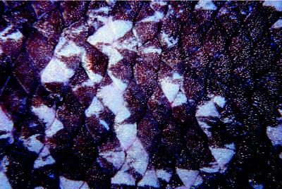
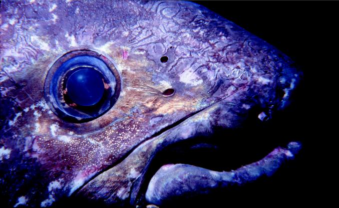
The case for morphological differentiation of L. menadoensis is much more tenuous than originally reported (10) and will be resolved only by a detailed morphological comparison of the Sulawesi specimen with similar-sized specimens of L. chalumnae. One important morphological character that certainly should be considered in future examinations is that of scale ornamentation; Forey suggests that this is one of the best characters available in coelacanth systematic investigations (2). Erdmann et al.’s report (5) of a unique, gold-flecked appearance to the Indonesian fish’s scales (Figs. 1 and 2), apparently a prismatic effect of light reflecting off the scale denticles, may indicate an important morphological difference in the scale ornamentation of the Sulawesi coelacanth.
Figure 1.
Shown is a photograph of the scales of the second known coelacanth captured off Manado Tua, Sulawesi, Indonesia (MZB 10003). The area depicted is on the left side, near the lateral line and under the second dorsal fin. The photograph was taken in water while the fish was alive. The gold flecks are prismatic reflections off the scale denticles.
Figure 2.
Gold prismatic reflections off the denticles of its scales are visible under the eye of this coelacanth, captured on July 30, 1998, off of Manado Tua, Sulawesi, Indonesia (MZB 10003). The photograph was taken while the fish was alive.
Although the extent of morphological differentiation between the Indonesian and Comorean coelacanths is still unclear, significant molecular sequence divergence between these two populations is evident and raises important questions about coelacanth dispersal and biogeography. With no fossil record of the age or original geographic range of Latimeria, it is difficult to postulate historical biogeographic explanations of the molecular divergence of these populations. Recently, Springer (21) suggested that the most recent ancestor of Latimeria was likely distributed continuously along the deep coasts of Africa through Eurasia, but that the collision of India with Eurasia (and the subsequent siltation caused by the formation of major rivers there) created a coelacanth habitat disjunction that allowed populations on either side of India to diverge. Although this is certainly a plausible scenario, these geologic events occurred significantly earlier (Eocene through mid-Miocene, 15–50 MYA) than our molecular clock estimates indicate these populations began diverging.
Springer’s hypothesis may be reconciled with our data if slower rates of molecular evolution are considered. The rate of substitutions in cytochrome b has been studied in extensively in sharks and found to be seven to eight times slower than in mammals (22) (although within the range of rates for all tetrapods used in Table 2). Sharks and coelacanths may have similar rates of molecular evolution because they are both large, poikilothermic vertebrates with long generation times (15, 23). Estimated rates of evolution are unavailable for shark rRNA genes and control region. However, using only our data from cytochrome b and a rate of evolution of 0.27%/MY (calculated from shark sequences; ref. 24), the best estimate of the divergence time between L. chalumnae and L. menadoensis is 16.6 MYA, which is consistent with Springer’s biogeographic hypothesis.
If we restrict our estimated range of times of divergence to those based on rates of evolution from tetrapods, explanations of the split between L. menadoensis and L. chalumnae must center on events in the late Miocene through the Pleistocene. It is possible that the massive tectonic changes that led to the formation of the Indo-Australian Arc and resultant separation of the Indian and Pacific Oceans caused a barrier to gene flow between these populations during the Miocene (25, 26). The proposed Mindanao-Sulawesi land bridge, in conjunction with various proposed closures/constrictions of the Makassar Strait between Borneo and Sulawesi, may have resulted in isolation of a north Sulawesi coelacanth population in a Sulawesi/Sulu Sea basin during the Miocene and Pliocene (27, 28). More recently, a combination of eustatic changes and possible tectonic movements may have resulted in constriction of the Makassar Strait during the Pleistocene (29, 30).
Today, no obvious physical barrier to dispersal between Sulawesi and the western Indian Ocean exists; Indonesian throughflow water moves from the Sulawesi Sea into the Makassar Strait, eventually reaching the Indian Ocean in the westward-moving South Equatorial Current (7). However, biological factors also must be considered. Living coelacanths are ovoviviparous, giving birth to large (>36 cm), precocial pups (31). Typical marine models of planktonic larval dispersal clearly do not apply. Furthermore, vast expanses of deep (>3,000 m) water are likely a barrier to dispersal of the adults, which appear neither active pelagic swimmers nor well protected against large pelagic or deep-water predators (32). Benthic migrations through this cold, deep water are also highly unlikely because of physiological limitations (17).
Given these apparent biological and geological restrictions to dispersal, it is perhaps not surprising that the Comorean and Indonesian populations represent distinct evolutionary lineages of Latimeria. This finding clearly does not preclude the existence of further living species of coelacanths in the area between Sulawesi and the Comores or elsewhere, and genetic studies of any such population will help unravel the mysteries of coelacanth distribution and evolution.
Acknowledgments
We gratefully acknowledge the support of Dr. M. K. Moosa and Dr. Ono of P3O LIPI, and we thank the Indonesian Institute of Sciences for allowing us the opportunity to conduct these investigations. Thanks also to Pak Subiyanto, S. Jewett, and the Smithsonian Institution for assistance in both preservation of the tissues and in arranging the CITES paperwork. This work was supported by the Alfred W. Roark Centennial Professorship to D.H., a National Science Foundation Predoctoral Fellowship to M.H., and the National Science Foundation (INT-9704616) and the National Geographic Society (Grant 6349-98) to M.E. and R.C. Thanks also to Om Lameh for bringing to us the first known specimen of L. menadoensis. We thank two anonymous reviewers for comments that helped us improve this manuscript.
Abbreviations
- MY
million years
- MYA
million years ago
Footnotes
This paper was submitted directly (Track II) to the PNAS office.
Data deposition: The sequence reported in this paper has been deposited in the GenBank database (accession no. AF176901).
References
- 1.Smith J L B. Nature (London) 1939;143:455–456. [Google Scholar]
- 2.Forey P L. History of the Coelacanth Fishes. London: Chapman & Hall; 1998. [Google Scholar]
- 3.Smith J L B. Nature (London) 1953;171:99–101. doi: 10.1038/171099a0. [DOI] [PubMed] [Google Scholar]
- 4.Hissmann K, Fricke H, Schauer J. Cons Biol. 1998;12:759–765. [Google Scholar]
- 5.Erdmann M V, Caldwell R L, Moosa M K. Nature (London) 1998;395:335. [Google Scholar]
- 6.Erdmann M V, Caldwell R L, Jewett S L, Tjakrawidjaja A. Environ Biol Fish. 1999;54:445–451. [Google Scholar]
- 7.Gordon A L. Nature (London) 1998;395:634. [Google Scholar]
- 8.Forey P L. Nature (London) 1998;395:319–320. [Google Scholar]
- 9.Fricke H, Hissmann K, Schauer J, Reinicke O, Lutz K, Plante R. Environ Biol Fish. 1991;32:287–300. [Google Scholar]
- 10.Pouyaud L, Wirjoatmodjo S, Rachmatika I, Tjakrawidjaja A, Hadiaty R, Hadie W. C R Acad Sci. 1999;322:261–267. doi: 10.1016/s0764-4469(99)80061-4. [DOI] [PubMed] [Google Scholar]
- 11.Zardoya R, Meyer A. Genetics. 1997;146:995–1010. doi: 10.1093/genetics/146.3.995. [DOI] [PMC free article] [PubMed] [Google Scholar]
- 12.Caccone A, Milinkovitch M C, Sbordoni V, Powell J R. Syst Biol. 1997;46:126–144. doi: 10.1093/sysbio/46.1.126. [DOI] [PubMed] [Google Scholar]
- 13.Faber J E, Stepien C A. In: Molecular Systematics of Fishes. Kocher T D, Stepien C A, editors. New York: Academic; 1997. pp. pp.129–144. [Google Scholar]
- 14.Brown W M, George M, Wilson A C. Proc Natl Acad Sci USA. 1979;76:1967–1971. doi: 10.1073/pnas.76.4.1967. [DOI] [PMC free article] [PubMed] [Google Scholar]
- 15.Hillis D M, Mable B K, Moritz C. In: Molecular Systematics. 2nd ed. Hillis D M, Moritz C, Mable B K, editors. Sunderland, MA: Sinauer; 1996. pp. 515–543. [Google Scholar]
- 16.Martin A P, Palumbi S R. Proc Natl Acad Sci USA. 1993;90:4087–4091. doi: 10.1073/pnas.90.9.4087. [DOI] [PMC free article] [PubMed] [Google Scholar]
- 17.Schliewen U, Fricke H, Schartl M, Epplen J T, Pääbo S. Nature (London) 1993;363:405. [Google Scholar]
- 18.Forey P L. Sci Progress Oxford. 1990;74:53–67. [Google Scholar]
- 19.McAllister D E, Smith C L. Naturaliste Can. 1978;105:63–76. [Google Scholar]
- 20.Heemstra P C, Freeman A L, Wong H Y, Hensley D A, Rabesandratana H D. S Afr J Sci. 1996;92:150–151. [Google Scholar]
- 21.Springer V G. Environ Biol Fish. 1999;54:453–456. [Google Scholar]
- 22.Martin A P, Naylor G J P, Palumbi S R. Nature (London) 1992;357:153–155. doi: 10.1038/357153a0. [DOI] [PubMed] [Google Scholar]
- 23.Rand D M. Trends Ecol Evol. 1994;9:125–131. doi: 10.1016/0169-5347(94)90176-7. [DOI] [PubMed] [Google Scholar]
- 24.Cantatore P, Roberti M, Pesole G, Ludovico A, Milella F, Gadaleta M N, Saccone C. J Mol Evol. 1994;39:589–597. doi: 10.1007/BF00160404. [DOI] [PubMed] [Google Scholar]
- 25.Audley-Charles M G. In: Wallace’s Line and Plate Tectonics. Whitmore T C, editor. Oxford: Clarendon; 1991. pp. 24–35. [Google Scholar]
- 26.Hall R. In: Tectonic Evolution of Southeast Asia. Hall R, Blundell D, editors. London: Geological Society; 1996. , Spec. Pub. No. 106, pp. 153–184. [Google Scholar]
- 27.McManus J W. Proc Fifth Int Coral Reef Symp. 1985;4:133–138. [Google Scholar]
- 28.Hamilton W. U S Geological Survey Prof Paper. 1979;1078:1–345. [Google Scholar]
- 29.Audley-Charles M G. In: Biogeographical Evolution of the Malay Archipelago. Whitmore C, editor. Oxford: Clarendon; 1981. pp. 5–25. [Google Scholar]
- 30.Katili J A. Tectonophysics. 1978;45:289–322. [Google Scholar]
- 31.Heemstra P C, Greenwood P H. Proc R Soc London Ser B. 1992;249:49–55. [Google Scholar]
- 32.Fricke H, Hissmann K, Schauer J, Reinicke O, Kasang L, Plante R. Environ Biol Fish. 1991;32:287–300. [Google Scholar]


