Abstract
Many previous reports suggest total hip arthroplasty performs suboptimally in young patients with osteonecrosis. We retrospectively compared the performance of metal-on-metal articulation in a select group of 107 patients with 112 hips (98 uncemented and 14 cemented stems) 60 years of age or younger with either osteonecrosis (27 patients, 30 hips) or primary osteoarthritis (80 patients, 82 hips). We evaluated all patients with patient-generated Harris hip score forms and serial radiographs. Five mechanical complications were caused by impingement, two with pain, two dislocations, and one liner dissociation. At a minimum followup of 2.2 years (mean, 5.5 years; range, 2.2–11.7 years), we observed no osteolysis or aseptic loosening in the osteonecrosis group, whereas one osteoarthritic hip had cup revision for loosening (none showed evidence of osteolysis). None of the stems were loose. Patients with osteonecrosis or primary osteoarthritis were similar in clinical and radiographic performance. The patients with metal-on-metal hip arthroplasty for osteonecrosis had no revisions for aseptic loosening, but did have one liner change in a cup for painful impingement.
Level of Evidence: Level III, therapeutic study. See the Guidelines for Authors for a complete description of levels of evidence.
Introduction
Osteonecrosis (ON) is predominantly a disease of the young, causing secondary arthritis [22]. Few reports suggest longevity of THA for ON is inferior when compared with THA for osteoarthritis (OA) [24, 28, 32]. Young, more active patients have higher revision rates with THA performed for any disease, but failure rates with ON are reportedly even higher for reasons not clearly understood [23, 24].
Metal-on-metal articulations in younger active patients have a low rate of wear and osteolysis [30, 31]. Low wear has been the most important factor in long-term performance of metal-on-metal articulations [35].
We asked whether metal-on-metal articulation, combined with noncemented fixation of the stem and cup, would provide equivalent survival and clinical scores in patients aged 60 years or younger with ON compared with primary OA.
Materials and Methods
We retrospectively compared clinical outcomes from clinical examination, medical records, and radiographs in 129 selected patients (135 hips) who were 60 years or younger with ON and OA and who had Metasul® (Zimmer, Inc., Warsaw, IN) metal-on-metal articulation. The group is selected, in part, because of inconsistent availability of the implant until 1999. In addition, from 1999 to 2000, there was contamination of the Sulzer InterOp metal shell (Sulzer Orthopedics, Austin, TX) used by us, which required revision in 14 patients; these patients were eliminated from this study group. Nine of the 129 patients (nine hips) had surgery between 1991 and 1993; 33 patients (35 hips) had surgery between 1993 and 1998 in an investigational device exemption for Metasul®; and 87 patients (91 hips) had surgery 1999 to 2003 after the Metasul® articulation was approved by the US Food and Drug Administration. The total number of hips operated on by us from 1999 to 2003 was 1016, so the 91 metal-on-metal hips in that same period represent 9% of our volume. Of the 129 patients potentially available, three (three hips) died, five (five hips) were lost to followup, and 14 patients (15 hips) were eliminated from the study because they had revision surgery from a failed recalled InterOp cup (Sulzer Orthopedics, Austin, TX) [20]. The final study group therefore had 107 patients (112 hips), of which 80 patients (82 hips, two bilateral) had primary OA and 27 (30 hips, three bilateral) had ON. The patients with OA were 7 years older on average (p < 0.0005) than those with ON with no other differences between the two groups (Table 1). All patients were contacted for recall for clinical and radiographic examination with five patients lost to followup. We obtained prior Institutional Review Board consent for review of the records.
Table 1.
Patient demographic and preoperative clinical data according to group
| Variable | Osteonecrosis | Primary osteoarthritis | p Value |
|---|---|---|---|
| Number of patients (hips) | 27 (30) | 80 (82) | |
| Mean age, years (SD) | 44.7 (±6.9) | 51.67 (±6.7) | < 0.0005 |
| Male patients (hips) | 22 (25) | 56 (57) | |
| Mean followup, years (range) | 5.5 (2.25–11.7) | 5.35 (1.1–13.2) | 0.2 |
| Female patients (hips) | 5 (5) | 24 (25) | |
| Weight, kg (SD) | 80.6 (± 12.43) | 87.95 (± 21.8) | 0.181 |
| Height, m (SD) | 1.71 (± 0.10) | 1.76 (± 0.10) | 0.108 |
| Body mass index (SD) | 27.53 (± 4.96) | 28.21 (± 6.18) | 0.675 |
| Dorr bone type | A = 14, B = 16, C = 0 | A = 31, B = 50, C = 1 | 0.56 |
| Preoperative Harris hip score | 45.92 (± 10.26) | 46.15 ( ± 15.25) | 0.96 |
SD = standard deviation.
The causes of ON were idiopathic (48%), posttraumatic (26%), alcohol abuse (15%), corticosteroids (7%), and sickle cell disease (4%). There were six hips in Stage 3 of ON with an obvious sequestrum and segmental collapse of the femoral head and 24 hips in Stage 4 with severe femoral head deformity and secondary OA using a staging system designed by Ficat [14]. Only one patient with bilateral ON had received surgical treatment in the past in the form of core decompression.
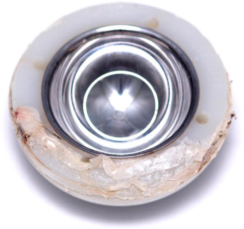
Patients were operated on with a traditional posterior approach as previously described [6]. All procedures were performed by the senior surgeons (LDD, WTL). Epidural anesthesia was augmented with general anesthesia to keep the average arterial pressure between 60 and 80 mmHg. Between 1991 and 1998, we used 12 cemented Weber cups (Sulzer Medica, Winterthur, Switzerland) and 32 cementless APR cups (Anatomic Porous Replacement; Zimmer) (Table 2). The APR cup was a 3.5-mm wall thickness titanium shell with a cancellous structured titanium porous coating. These 44 cups were coupled with 44 APR stems (11 cemented and 33 cementless). Between 1999 and 2003, we used 28 cementless InterOp cups (Sulzer) and 40 cementless Converge cups (Zimmer) with 67 cementless stems and one cemented APR stem. The InterOp was changed to the Converge in 2001 because of the recall of the InterOp in 2000 [20]. The InterOp metal shell and the Converge metal shell did not differ from the APR metal shell in thickness or porous surface. The only difference in the InterOp and Converge cups was the locking mechanism for the acetabular insert. The Metasul® acetabular insert for all cups had an articulation surface of cobalt-chromium metal, which was inlaid into a polyethylene hemisphere having the locking mechanism for the metal shell. This metal insert was manufactured separately and then embedded into the polyethylene [35]. This insert was the same for all metal shells used with the exception of the locking mechanism of the polyethylene into the metal shell. Metasul® (Zimmer) was designed to have better clearance of surfaces to avoid excess friction, promote lubrication, and allow clearance of debris [29, 35]. The cobalt-chromium metal used in the acetabular surface of the Metasul® liner and the femoral head was Protasul-21 WF (Zimmer) cobalt-chromium alloy, a high carbon wrought forged alloy. The diameter of the femoral head used in this study was 28 mm. The clearance between the femoral and acetabular articulation surfaces was a mean 120 μm (range, 70–170 μm). The Metasul® insert had an elevated metal edge where it was inlaid. This protruding edge could cause impingement of the metal neck and cup (Fig. 1).
Table 2.
Various implant combinations used during the study period
| Implant | Osteonecrosis (n = 30) | Primary osteoarthritis (n = 82) |
|---|---|---|
| Cup design | ||
| Weber | 4* | 8* |
| APR | 8 | 24 |
| Interop | 10 | 18 |
| Converge | 8 | 32 |
| Stem type | ||
| APR | 30 (2*) | 82 (10*) |
* Cemented fixation; APR = anatomic porous replacement.
Fig. 1.
Retrieved Metasul® liner with a protruded edge of the inlay has damage to the polyethylene caused by impingement.
We obtained data on pain and functional outcome preoperatively and at the final followup with a patient self-assessment form (patient-generated Harris hip score [16]; Orthographics, Salt Lake City, UT). We determined activity separately by asking the patients their activity level; activity was graded as unlimited community ambulation (more than eight blocks), active community ambulation (can walk up to eight blocks), limited community ambulation (can walk two blocks), and household ambulation (limited to household activities) [12]. Patients completed their forms either by mail or during followup in the office. Ninety-six of the 107 patients (101 hips) were seen at final followup, and 11 (11 hips) were graded by forms returned by mail. We reviewed the medical records for revisions, complications, and clinical scores.
We obtained anteroposterior pelvic radiographs and lateral radiographs (iliac oblique views) preoperatively and at each followup visit. The preoperative radiograph was used to determine the diagnosis. We used the 6-week postoperative radiographs as the baseline for comparison with final followup radiographs for fixation and osteolysis. Wear could not be measured from the radiographs because it was not possible to distinguish between the edge of the femoral head and the metal articulation surface of the acetabular liner. One of us (ZW) measured inclination of the cup using the technique of Callaghan et al. [4] and anteversion using the modified technique of Ackland et al. [1, 11]. Femoral radiolucent lines and osteolysis were recorded in each of seven Gruen zones [15] on the anteroposterior and lateral radiographs [17]. Calcar resorption was a focal radiolucent area that was seen immediately under the collar of the stem and was located between the cortex of the calcar and the medial edge of the stem. Fixation and osteolysis of the cup were measured by the zones of DeLee and Charnley [8]. Cup loosening was diagnosed when there was: (1) a circumferential radiolucent line of 1 mm or wider; (2) appearance of a new radiolucent line in any zone; (3) progression of a radiolucent line; or (4) migration of the cup by more than 2 mm of vertical or horizontal shift or a change in inclination of more than 5° [33].
We used the Student’s t-test to compare demographic parameters such as age, height, weight, and body mass index and for pre- and postoperative Harris hip scores, pain and function scores, and number of years of followup in both groups. Chi square test was used to compare the categorical data (gender, bone type [9], patient self-assessment score, and postoperative level of activity) in the two groups. A probability value of p ≤ 0.05 was considered significant. All data were analyzed using SPSS software (SPSS Inc, Chicago, IL).
Results
At a mean followup of 5.5 years, the postoperative score was similar for patients with ON and those with OA (93.73 ± 8.2 versus 93.12 ± 8.5, respectively). Mean pain scores at the final followup were also similar (40.48 ± 5.78 for ON versus 40.74 ± 5.76 for OA) and only three patients with ON (11.11%) and six patients with OA (7.5%) had self-described mild to moderate pain. There was no difference in the mean functional scores for either group (44.64 ± 2.78 for ON versus 43.93 ± 4.71 for OA) with only two patients in each group requiring an assistive device for walking. Seventeen patients (20 hips) out of 27 (30 hips) with ON reported their outcome as excellent in the “patient self-assessment form” compared with 60 patients (62 hips) out of 80 (82 hips) patients with OA. Overall clinical outcome and functional activity were similar for all measured parameters (Tables 3, 4).
Table 3.
Clinical outcome in patients with osteonecrosis and primary osteoarthritis at final followup
| Outcome (clinical grade) | Patients in the osteonecrosis group (hips) | Patients in the primary osteoarthritis group (hips) | p Value |
|---|---|---|---|
| Excellent | 17 (20) | 60 (62) | 0.58 |
| Good | 8 (8) | 15 (15) | |
| Fair | 2 (2) | 5 (5) |
Table 4.
Functional activity in patients with osteonecrosis and primary osteoarthritis at final followup
| Outcome (functional activity) | Patients in the osteonecrosis group (hips) | Patients in the primary osteoarthritis group (hips) | p Value |
|---|---|---|---|
| Unlimited ambulator | 25 (28) | 68 (70) | 0.32 |
| Active community ambulator | 2 (2) | 10 (10) | |
| Limited community ambulator | 0 | 2 (2) |
Radiographic results were also similar between the OA and ON hips. The mean cup inclination for the ON group was 39° ± 5.9° compared with 38° ± 5.7° for OA; the mean cup anteversion for ON hips was 18.2° ± 5.4° and 17.9° ± 5.1° for OA hips. No pelvic osteolysis was observed in either of the groups. Femoral osteolysis was seen in Zones 3 and 4 in one of 82 hips (1.22% in the OA group and none of the ON hips) and calcar lysis of 3 mm x 3 mm or less was observed in two hips with ON and in three with OA.
There were no loose stems in either group and one loose cup in the OA group. Femoral radiolucent lines, confined to Zones 3, 4, and 5, were seen in two hips with ON and eight with OA. Radiolucent lines were more prevalent around the cup with seven cups in the ON group and 20 in the OA group affected. These incomplete radiolucent lines were in three zones in one hip with ON and in three hips with OA; in two zones in three hips with ON and eight hips with OA; and in one zone in three hips with ON and nine hips with OA. Other than the revised cup, no cup had a complete radiolucent line in all three zones or progressive lines, and no cup showed migration.
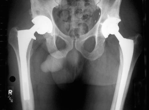
Overall, there were six acetabular revisions with four metal shell replacements and two liner changes. There were no stem revisions. We revised one cup in the OA group for aseptic loosening. This cup had bone graft for protrusio acetabuli and had three zone circumferential radiolucent lines on the postoperative radiograph. We revised two cups in the OA group for dislocation. There were two liner changes, one in each group, for painful impingement at 2 years and 8.5 years postoperatively. One patient with OA had a liner dissociation, which occurred at 7.7 years followup (Fig. 2). In this hip, the acetabular component was malpositioned with 30° of inclination and 2° of anteversion, which resulted in impingement and failure of the locking mechanism. This hip had a revision of the cup to a position that prevented impingement. The only other reoperation was in one patient with ON in whom a periprosthetic fracture occurred distal to the femoral component as a result of a traffic accident; it was successfully fixed with open reduction and internal fixation.
Fig. 2.
Anteroposterior pelvic radiograph shows a disassociated Metasul® liner in the right THA at 7.7 years postoperatively.
Discussion
Traditionally, ON of the femoral head as the reason for THA has had outcomes inferior to the hips with OA as the cause of the operation [24, 28]. Specifically, the ON hips have had more loosening of components [24, 26, 28]. We asked whether the outcome of THA was different in patients 60 years old or younger with ON compared with OA when using noncemented implant fixation and metal-on-metal articulation (Metasul®).
Our study was limited in that it was not randomized and has nonconsecutive patient selection with a smaller sample size. These patients were operated on at various time periods, depending on availability of Metasul® implants. The study did, however, include all of the patients 60 years or younger operated on by us who had ON and OA and a Metasul® insert. The study included both cementless and cemented cups, which have different propensities for osteolysis. However, we had no case of osteolysis around the cup in either group so this did not influence results. Finally, wear cannot be measured in metal-on-metal articulation so we could not compare it with series using polyethylene articulations.
We found no difference in the clinical or radiographic outcome for the length of our followup. Our mean followup was 5.35 years for OA and 5.5 years for ON with the longest followup being 13.2 years and 11.7 years, respectively. There was no osteolysis of the pelvis in either group and only one OA hip showed osteolysis in the femur. Fixation was secure in all cemented or noncemented hips and failures in both groups were caused by impingement except for one OA hip with a technical error, which was revised.
The importance of our data is reflected in the similarity of outcomes in ON and OA hips. The results of cemented THAs with metal-on-polyethylene articulation in ON have not generally been satisfactory in studies reported with a similar age group and followup as our mean 5.5 years [5, 7, 10, 24, 26, 28]. One study [7] reported an overall failure rate of 37% at a mean followup of 7.6 years in 28 cemented arthroplasties; a second study [28] reported unsatisfactory results in 14 of 29 ON hips (48%) compared with 16 of 63 (25%) in patients with OA, and a third study [26] had a poor outcome in 11 of 12 cemented ON hips. At a long-term followup of 15 years, Ortiguera et al. [24] had nine of 18 failed hips in 35 patients as compared with four of 17 in patients with OA.
In contrast, cementless arthroplasties with metal-on-polyethylene articulation have had better survival than cemented arthroplasties for ON. One study [23] reported no difference in fixation at 3 years among 52 matched hips; a second [25] reported loosening of 3% of acetabular components in 35 ON hips at 7.5 years; and a third [19] had 98% survival of 100% cementless hips at 10 years.
Failure with cemented components may be related to changes in cancellous bone structure and remodeling with ON. Defective cancellous bone might not support the interdigitation of cement and the increased load placed on it. The framework of cancellous bone in ON is apparently weak. Arlot et al. [2] studied the histomorphometry of iliac bone in 77 patients with ON and normal kidney function and found osteomalacia in nine patients with the remaining 68 patients having reduced trabecular bone volume, a reduced calcification rate, and a thin osteoid seam indicating defective osteoblastic apposition and healing. Calder et al. [3] described extensive osteocyte death and an abnormal remodeling capacity in the proximal femur in ON, and proposed premature loosening of implants in patients with ON may be related to this presence of abnormal cancellous bone at the implant-bone and cement-bone interfaces.
Metasul® metal articulation also protects against bone failure by a low volume of particles, which prevents changes in osteolysis. In retrieval analysis of 118 second-generation Metasul® articulations for up to 8 years, there was volumetric wear of 0.3 mm3 per year, which is 60 times less than metal-on-polyethylene articulation [31]. Osteolysis with Metasul® metal-on-metal articulation has been rarely observed in studies with no osteolysis in 39 cementless arthroplasties (20 ON, 19 OA hips) in one study [21]; in a second study [13], none at 5 to 12 years followup of 56 cemented and cementless arthroplasties; in a third [27], none in 106 cementless arthroplasties at a mean 6.4 years; and in the fourth [18], one of 68 hips with pelvic osteolysis needing revision and another hip with two small areas of focal femoral lesions around a stable stem.
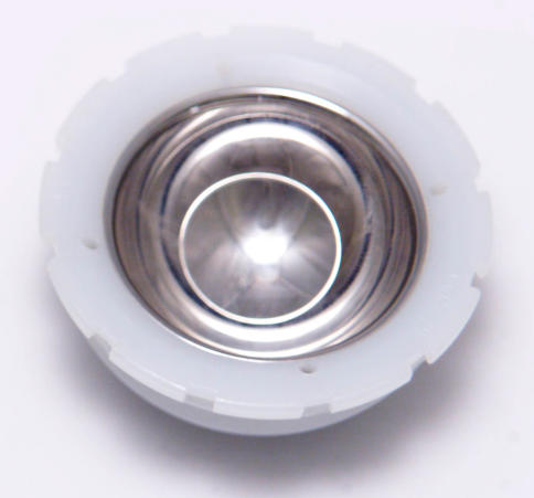
We observed several mechanical complications from impingement. Two dislocations, two liner changes, and one liner disassociation had evidence of impingement at revision with indentations on the femoral neck and acetabular liner. The Metasul® liners used in our patients had an inlay with a prominent metal rim. In addition, we used a 28-mm head on a 12 to 14 taper femoral neck giving a head-neck ratio of 2.0, which is a risk for impingement [34]. Metasul® liners have been redesigned to have a buried metal inlay in a chamfered polyethylene rim (Fig. 3), and we use a 32-mm or larger femoral head (36-mm+ Durom; Zimmer) when we implant Metasul® articulations.
Fig. 3.
The current Metasul® liner has a buried metal inlay and a chamfered polyethylene rim to reduce impingement.
THA for arthritis secondary to ON of the femoral head did not result in higher failure rates than OA with modern cementless implants. We continue to use cementless fixation and this metal-on-metal articulation for patients 60 years of age or younger with either ON or OA as a cause of their arthritic hip.
Footnotes
One or more of the authors (LDD) have received benefits from Zimmer Inc for conducting this study.
Each author certifies that his or her institution has approved the human protocol for this investigation, that all investigations were conducted in conformity with ethical principles of research, and that informed consent for participation in the study was obtained.
References
- 1.Ackland MK, Bourne WB, Uhthoff HK. Anteversion of the acetabular cup. Measurement of angle after total hip replacement. J Bone Joint Surg Br. 1986;68:409–413. [DOI] [PubMed]
- 2.Arlot ME, Bonjean M, Chavassieux PM, Meunier PJ. Bone histology in adults with aseptic necrosis. Histomorphometric evaluation of iliac biopsies in seventy-seven patients. J Bone Joint Surg Am. 1983;65:1319–1327. [PubMed]
- 3.Calder JD, Pearse MF, Revell PA. The extent of osteocyte death in the proximal femur of patients with osteonecrosis of the femoral head. J Bone Joint Surg Br. 2001;83:419–422. [DOI] [PubMed]
- 4.Callaghan JJ, Salvati EA, Pellicci PM, Wilson PD Jr, Ranawat CS. Results of revision for mechanical failure after cemented total hip replacement, 1979 to 1982. A two to five-year follow-up. J Bone Joint Surg Am. 1985;67:1074–1085. [PubMed]
- 5.Chandler HP, Reineck FT, Wixson RL, McCarthy JC. Total hip replacement in patients younger than thirty years old. A five-year follow-up study. J Bone Joint Surg Am. 1981;63:1426–1434. [PubMed]
- 6.Cohen J, Bindelglass DF, Dorr LD. Total hip replacement using the APR II system. Techniques Orthop. 1991;6:40–58. [DOI]
- 7.Cornell CN, Salvati EA, Pellicci PM. Long-term follow-up of total hip replacement in patients with osteonecrosis. Orthop Clin North Am. 1985;16:757–769. [PubMed]
- 8.DeLee JG, Charnley J. Radiological demarcation of cemented sockets in total hip replacement. Clin Orthop Relat Res. 1976;121:20–32. [PubMed]
- 9.Dorr LD, Faugere MC, Mackel AM, Gruen TA, Bognar B, Malluche HH. Structural and cellular assessment of bone quality of proximal femur. Bone. 1993;14:231–242. [DOI] [PubMed]
- 10.Dorr LD, Takei GK, Conaty JP. Total hip arthroplasties in patients less than forty-five years old. J Bone Joint Surg Am. 1983;65:474–479. [PubMed]
- 11.Dorr LD, Wan Z. Ten years of experience with porous acetabular components for revision surgery. Clin Orthop Relat Res. 1995;319:191–200. [PubMed]
- 12.Dorr LD, Wan Z, Gruen T. Functional results in total hip replacement in patients 65 years and older. Clin Orthop Relat Res. 1997;336:143–151. [DOI] [PubMed]
- 13.Dorr LD, Wan Z, Longjohn DB, Dubois B, Murken R. Total hip arthroplasty with use of the Metasul metal-on-metal articulation. Four to seven-year results. J Bone Joint Surg Am. 2000;82:789–798. [DOI] [PubMed]
- 14.Ficat RP. Idiopathic bone necrosis of the femoral head. Early diagnosis and treatment. J Bone Joint Surg Br. 1985;67:3–9. [DOI] [PubMed]
- 15.Gruen TA, McNeice GM, Amstutz HC. ‘Modes of failure’ of cemented stem-type femoral components: a radiographic analysis of loosening. Clin Orthop Relat Res. 1979;141:17–27. [PubMed]
- 16.Harris WH. Traumatic arthritis of the hip after dislocation and acetabular fractures: treatment by mold arthroplasty. An end-result study using a new method of result evaluation. J Bone Joint Surg Am. 1969;51:737–755. [PubMed]
- 17.Johnston RC, Fitzgerald RH Jr, Harris WH, Poss R, Muller ME, Sledge CB. Clinical and radiographic evaluation of total hip replacement. A standard system of terminology for reporting results. J Bone Joint Surg Am. 1990;72:161–168. [PubMed]
- 18.Kim SY, Kyung HS, Ihn JC, Cho MR, Koo KH, Kim CY. Cementless Metasul metal-on-metal total hip arthroplasty in patients less than fifty years old. J Bone Joint Surg Am. 2004;86:2475–2481. [DOI] [PubMed]
- 19.Kim YH, Oh SH, Kim JS, Koo KH. Contemporary total hip arthroplasty with and without cement in patients with osteonecrosis of the femoral head. J Bone Joint Surg Am. 2003;85:675–681. [DOI] [PubMed]
- 20.Manning DW, Ponce BA, Chiang PP, Harris WH, Burke DW. Isolated acetabular revision through the posterior approach: short-term results after revision of a recalled acetabular component. J Arthroplasty. 2005;20:723–729. [DOI] [PubMed]
- 21.Migaud H, Jobin A, Chantelot C, Giraud F, Laffargue P, Duquennoy A. Cementless metal-on-metal hip arthroplasty in patients less than 50 years of age: comparison with a matched control group using ceramic-on-polyethylene after a minimum 5-year follow-up. J Arthroplasty. 2004;19(Suppl 3):23–28. [DOI] [PubMed]
- 22.Mont MA, Hungerford DS. Non-traumatic avascular necrosis of the femoral head. J Bone Joint Surg Am. 1995;77:459–474. [DOI] [PubMed]
- 23.Mont MA, Seyler TM, Plate JF, Delanois RE, Parvizi J. Uncemented total hip arthroplasty in young adults with osteonecrosis of the femoral head: a comparative study. J Bone Joint Surg Am. 2006;88S:104–109. [DOI] [PubMed]
- 24.Ortiguera CJ, Pulliam IT, Cabanela ME. Total hip arthroplasty for osteonecrosis: matched-pair analysis of 188 hips with long-term follow-up. J Arthroplasty. 1999;14:21–28. [DOI] [PubMed]
- 25.Piston RW, Engh CA, De Carvalho PI, Suthers K. Osteonecrosis of the femoral head treated with total hip arthroplasty without cement. J Bone Joint Surg Am. 1994;76:202–214. [DOI] [PubMed]
- 26.Ranawat CS, Atkinson RE, Salvati EA, Wilson PD Jr. Conventional total hip arthroplasty for degenerative joint disease in patients between the ages of forty and sixty years. J Bone Joint Surg Am. 1984;66:745–752. [PubMed]
- 27.Saito S, Ryu J, Watanabe M, Ishii T, Saigo K. Midterm results of Metasul metal-on-metal total hip arthroplasty. J Arthroplasty. 2006;21:1105–1110. [DOI] [PubMed]
- 28.Saito S, Saito M, Nishina T, Ohzono K, Ono K. Long-term results of total hip arthroplasty for osteonecrosis of the femoral head. A comparison with osteoarthritis. Clin Orthop Relat Res. 1989;244:198–207. [PubMed]
- 29.Santavirta S, Bohler M, Harris WH, Konttinen YT, Lappalainen R, Muratoglu O, Rieker C, Salzer M. Alternative materials to improve total hip replacement tribology. Acta Orthop Scand. 2003;74:380–388. [DOI] [PubMed]
- 30.Schmalzried TP, Peters PC, Maurer BT, Bragdon CR, Harris WH. Long-duration metal-on-metal total hip arthroplasties with low wear of the articulating surfaces. J Arthroplasty. 1996;11:322–331. [DOI] [PubMed]
- 31.Sieber HP, Rieker CB, Kottig P. Analysis of 118 second-generation metal-on-metal retrieved hip implants. J Bone Joint Surg Br. 1999;81:46–50. [DOI] [PubMed]
- 32.Stulberg BN, Singer R, Goldner J, Stulberg J. Uncemented total hip arthroplasty in osteonecrosis: a 2- to 10-year evaluation. Clin Orthop Relat Res. 1997;334:116–123. [DOI] [PubMed]
- 33.Udomkiat P, Wan Z, Dorr LD. Comparison of preoperative radiographs and intraoperative findings of fixation of hemispheric porous-coated sockets. J Bone Joint Surg Am. 2001;83:1865–1870. [DOI] [PubMed]
- 34.Usrey MM, Noble PC, Rudner LJ, Conditt MA, Birman MV, Santore RF, Mathis KB. Does neck/liner impingement increase wear of ultrahigh-molecular-weight polyethylene liners? J Arthroplasty. 2006;21:65–71. [DOI] [PubMed]
- 35.Weber BG. Experience with the Metasul total hip bearing system. Clin Orthop Relat Res. 1996;329S:69–77. [DOI] [PubMed]


