Abstract
Two regioisomers with C3 or D3 symmetry of water-soluble carboxylic acid C60 derivatives, containing three malonic acid groups per molecule, were synthesized and found to be equipotent free radical scavengers in solution as assessed by EPR analysis. Both compounds also inhibited the excitotoxic death of cultured cortical neurons induced by exposure to N-methyl-d-aspartate (NMDA), α-amino-3-hydroxy-5-methyl-4-isoxazolepropionic acid (AMPA), or oxygen-glucose deprivation, but the C3 regioisomer was more effective than the D3 regioisomer, possibly reflecting its polar nature and attendant greater ability to enter lipid membranes. At 100 μM, the C3 derivative fully blocked even rapidly triggered, NMDA receptor-mediated toxicity, a form of toxicity with limited sensitivity to all other classes of free radical scavengers we have tested. The C3 derivative also reduced apoptotic neuronal death induced by either serum deprivation or exposure to Aβ1–42 protein. Furthermore, continuous infusion of the C3 derivative in a transgenic mouse carrying the human mutant (G93A) superoxide dismutase gene responsible for a form of familial amyotrophic lateral sclerosis, delayed both death and functional deterioration. These data suggest that polar carboxylic acid C60 derivatives may have attractive therapeutic properties in several acute or chronic neurodegenerative diseases.
Since their discovery in 1985, the pure carbon spheres of C60 (buckminsterfullerene) have generated increasing interest from many different branches of science and engineering, culminating in presentation of the 1996 Nobel Prize in Chemistry to Kroto, Smalley, and Curl for their identification of these unique molecules. Subsequently, investigation into the chemical and physical properties of C60 (and larger fullerenes) has yielded an extensive amount of information about C60, including its avid reactivity with free radicals (1). Buckminsterfullerenes, for example, are capable of adding multiple radicals per molecule; the addition of as many as 34 methyl radicals to a single C60 sphere has been reported, leading Krusic et al. (1) to characterize C60 as a “radical sponge.” However, native C60 is soluble in only a limited number of organic solvents, such as toluene or benzene. We have been interested in the possibility that the potent innate antioxidant properties of C60 could be harnessed for use in biological systems by adding functional groups aimed at enhancing its water solubility.
Glutamate receptor-mediated excitotoxicity has been implicated in the pathogenesis of neuronal loss in the central nervous system in several disease states, including hypoxia-ischemia, epilepsy, and trauma (2–5). Oxygen or nitric oxide radicals are produced as a consequence of glutamate receptor overstimulation (6–10), and free radical scavengers have been shown to attenuate, but not to block, excitotoxic neuronal death (11–15). We recently reported promising neuroprotective effects of antioxidant polyhydroxylated derivatives of C60 on cultured cortical neurons (16). However, further testing revealed considerable synthesis lot-to-lot variability in both water solubility and biological effects, presumably reflecting uncontrolled differences in the number and location of hydroxyl and hemiketal moieties ending up on the C60 shell. To refine this strategy, we have turned to malonic acid derivatives of C60, (C63((COOH)2)3), synthesized and purified as two specific regioisomers with C3 and D3 symmetry (Fig. 1) and demonstrated that they are effective neuroprotective antioxidants in vitro and in vivo. Although a recent commentary in Science by C. Holden (17) suggested that, “Buckyballs have not lived up to their early promise… . (in applications),” our findings suggest that water-soluble derivatives of C60 may have a novel application as neuroprotective agents.
Figure 1.
Structures of carboxyfullerenes showing the paired carboxyl groups on the C60 sphere. The three-dimensional models demonstrate the polar distribution of the carboxyl groups on C3 and the equatorial distribution on D3.
METHODS
Synthesis and Characterization of Malonic Acid (Carboxy) Derivatives of C60.
Malonic acid derivatives of C60 were synthesized as described by Lamparth and Hirsch (18). Briefly, diethyl bromomalonate was added to a solution of C60 in toluene, followed by the addition of 1,8-diazobicyclo[5,4,0]undec-7-ene, which resulted in a color change from violet to dark red. After stirring for 4 days, the solvent was removed in vacuo, and the blackish residue was chromatographed on silica gel (270–230 mesh) using toluene-hexane (1:1 by volume) as eluent. The unreacted C60 was obtained first, followed by a brown band that corresponds to the diester. The eluent was changed to toluene-hexane (4:1). A brown band (tetraester) was obtained after a narrow yellow band (tetraester, para addition) followed by a deep red band (tetraester). The eluent was changed again to toluene-hexane (9:1), and a red band was collected that corresponds to the D3 isomer as the major component. Toluene (100%) then was used to elute three bands. The third band corresponded to semipure C3 as the major component. The fractions containing semipure C3 or D3 were rechromatographed on silica gel (230–400 mesh) using toluene as the mobile phase to give the purified C3 or D3 isomers. To a solution of either C3 or D3 (100 mg in 100 ml toluene, ≈ 0.1 mmol) was added NaH (80%, 60 mg, 2 mmol), and the mixture was refluxed for 1 hr. After the heating source was removed, MeOH (5 ml) was added immediately to quench the reaction. Red powder soon precipitated and was collected by centrifugation. The powder was washed with toluene twice and hexane four times. The red solid was dissolved in water to which HCl (4 M) was added. A red amorphous precipitate was formed immediately, which was collected by centrifugation. The solid again was washed with HCl (4 M), and then by water twice. The solid was dissolved in MeOH, and the solvent was removed in vacuo to give the powdery pure isomer acid (red for C3 and brownish red for D3).
EPR Spectroscopy: Radical Reactivity and Membrane Interactions of Carboxyfullerenes.
The reactivity of carboxyfullerenes with oxygen radicals was assessed by EPR spectroscopy. Aqueous samples were loaded into a quartz flat cell (60 × 10 × 0.25 mm) and analyzed on a Bruker 200 X-band spectrometer, with experimental conditions as described in the legend for Fig. 2. Signal averaging was performed on some samples.
Figure 2.
Water-soluble carboxylic C60 compounds are effective free radical scavengers. EPR spectra of hydroxyl (⋅OH) (from 100 μM H2O2 with 10 μM Fe2+ via the Fenton reaction) and superoxide O2⨪ (from 25 μM xanthine + 10 mU/ml xanthine oxidase) radicals with 100 mM 5,5-dimethyl-1-pyrolline-N-oxide (DMPO) as the spin-trapping agent: ⋅OH in the presence of DMPO alone, in the presence of 4 μM C3, or in the presence of 4 μM D3, O2⨪ in DMPO alone, in the presence of 40 μM C3, or in the presence of 40 μM D3. The arrow indicates a spurious signal due to an unknown radical in the cavity. The sample was analyzed in a quartz flat cell (60 × 10 × 0.25 mm) in a Bruker 200, X-band EPR spectrometer. Settings were power, 1.6 mW; modulation, 1 G; field modulation, 100 Hz; receiver gain, 3.2 × 10⁵.
A second set of experiments was designed to evaluate the degree of membrane interaction of the two carboxyfullerene isomers. Mouse brain lipids were extracted (19) and aliquoted into test tubes. Spin-labeled lipids, 5-doxyl or 16-doxyl ketostearic acids, were added at a ratio of 1:50, and dried under N2. Tris saline (25 mM, pH 7.4), with or without C3 or D3, was added to each tube and vortexed. C3 and D3 were tested at concentrations of 10–300 nmol (1:30–1:1 compared with lipids). Settings for the above EPR experiments were power = 1.6 mW, modulation = 1 G, field modulation = 100 Hz, receiver gain = 3.2 × 10⁵. Low-temperature (77°K) EPR also was performed on H2O2-oxidized carboxyfullerenes to determine whether oxidation produces a paramagnetic (radical) species.
Media and Reagents.
Cell culture reagents and supplies were obtained from standard sources. Laboratory reagents were purchased from Sigma. Media stock consisted of minimal essential media without glutamine, containing 20 mM glucose.
Generation of Cell Cultures.
Mouse neocortical cultures were prepared as neuronal-astrocyte cocultures or as pure neuronal cultures (<1% astrocytes) from Swiss–Webster mice as described previously (20–21).
Determination of in Vitro Neuroprotection by Carboxyfullerenes.
Stock solutions of purified C3 or D3 carboxyfullerenes (25 mM or 50 mM) were freshly prepared in sterile water. Brief exposure to N-methyl-d-aspartate (NMDA) was carried out as described (16). The culture media was exchanged twice with Hepes, bicarbonate-buffered balanced salt solution (9), and then NMDA (200 μM) was added alone or with each of the carboxyfullerenes (30 μM-1 mM) for 10 min. Exposure was terminated by exchanging the medium four times with media stock. The cells were returned to the 37°C (5% CO2) incubator for 24 hr, when injury was assessed. To determine whether carboxyfullerenes could block NMDA receptor-mediated calcium entry, cells were exposed to NMDA for 10 min in the presence of tracer 45Ca2+ (0.5 μCi/well; New England Nuclear), and the amount of intracellular 45Ca2+ determined as previously described (22).
Cultures were exposed to α-amino-3-hydroxy-5-methyl-4-isoxazole propionic acid (AMPA) (8 μM), with or without carboxyfullerenes, in media stock containing MK-801 (10 μM MK-801; Merck, Sharp and Dohm) to block activation of NMDA receptors by released endogenous glutamate. Combined oxygen, glucose deprivation was performed as described (23). Cultures were placed in an anoxic chamber (O2 < 2 mm Hg), and the media was exchanged three times with a balanced salt solution (BSS0) lacking glucose and oxygen. After 60 min, the cells were washed back into oxygen, glucose-containing medium and returned to the aerobic culture incubator for 24 hr.
To induce apoptosis, pure neuronal cultures containing less than 1% astrocytes were deprived of serum on day in vitro 7 by exchanging the medium with serum-free media stock (20). Washed controls were returned to medium with serum. Exposure to Aβ1–42 was performed on day in vitro 10 mixed cultures by application of 20 μM Aβ1–42 (K-Biologicals, Rancho Cucamonga, CA) for 48 hr.
Cell death was assessed by phase contrast microscopy, assay of lactate dehydrogenase in the bathing medium, and manual counting of dead cells stained with propidium iodide (10 μM for 7 min).
Confocal Microscopy and Assessment of Reactive Oxygen Species.
Cultures were loaded with 15 μM dihydrorhodamine 123 (Molecular Probes) at the onset of serum deprivation. Cells were imaged 0.5, 4, and 8 hr later on a laser scanning confocal microscope (Noran), using Exλ = 488 nm, Emλ > 515 nm as previously described (9, 24).
In Vivo Neuroprotection in a Mouse Model of Amyotrophic Lateral Sclerosis.
Mice from the G93A SOD1 G1 strain developed by Gurney et al. (25) as a model for familial amyotrophic lateral sclerosis (FALS) were treated with carboxyfullerene beginning at 73 ± 2 days of age. Carboxyfullerene containing a mixture of isomers (>85% C3, 10% D3, with trace amounts of other Tris malonic acid C60 adducts) was dissolved in sterile 0.9% saline, and loaded into mini-osmotic pumps (no. 2004, Alza, 28-day delivery) as per manufacturer’s instructions. Pumps were soaked in sterile normal saline for 40 hr and implanted into the peritoneum through a small midline incision. Before implantation of the second pump 4 weeks later, the depleted first pump was removed, and the volume remaining transferred to a tube and measured to verify that drug delivery had occurred at the specified rate of 6.6 μl/day, to deliver 15 mg/kg per day. Mice were maintained in the colony until moribund, determined by inability of the mouse to right itself in 30 sec after being placed on its side. This correlated well with inability to drink spontaneously or to respond to offered food or water. Determination of whether an animal was moribund was determined by a blinded observer, and the date of death recorded.
Motor performance was assessed using a scale developed by Basso et al. (26) to evaluate function after spinal cord injury. Mice were videotaped weekly starting at 89 days of age, and their gait scored by a blinded observer using this 21-point scale, which rates spontaneous ambulation, including leg position and coordination. Mice were evaluated more informally daily.
RESULTS
Characterization of Carboxyfullerenes.
Mass spectroscopy of the two purified trisadduct regioisomer esters demonstrated a peak in each sample at 1,194/1,195 mass units, as previously described (18). Identity and purity of both the intermediate ester and the final malonic acid product of each regioisomer were confirmed by 13C NMR spectroscopy and mass spectrometry (18). The final malonic acids, C3 and D3, were soluble in water to at least 75 mM. Control experiments verified that the compounds did not interfere with the colorimetric lactate dehydrogenase assay, and solutions to at least 25 mM failed to alter the pH of the experimental solutions.
Carboxyfullerenes Are Potent Free Radical Scavengers.
Both C3 and D3 isomers were unusually potent scavengers of hydroxyl radical (⋅OH) and superoxide anion (O2⨪) in solution. They were able to completely eliminate ⋅OH at concentrations as low as 4–5 μM (Fig. 2), 10- to 100-fold less than required for most other free radical scavengers. Both isomers also effectively scavenged superoxide radical (O2⨪), although 10-fold higher concentrations were required to do this (Fig. 2).
Neuroprotection Against Excitotoxic Injury in Vitro: The C3 and D3 Regioisomers Show Differential Efficacy.
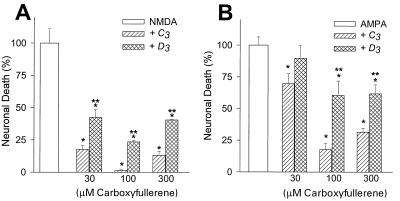
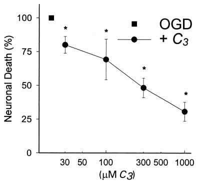
Both the C3 and D3 isomers produced a dose-dependent decrease in death of cortical neurons exposed to NMDA or AMPA (Fig. 3 A and B), although the C3 compound was both more potent and more effective than D3 (Fig. 3 A and B). The C3 compound provided essentially complete neuroprotection against NMDA receptor-mediated neuronal death. Although the degree of protection afforded by C3 against AMPA-induced neurotoxicity was generally somewhat less, coapplication of C3 still reduced neuronal death by over 80%. The concentration of carboxyfullerene required to produce maximal protection against these excitotoxic insults varied somewhat with the degree of injury; i.e., insults that resulted in 100% neuronal death required slightly higher concentrations of carboxyfullerene for maximal neuroprotection. The C3 isomer also reduced neuronal death after combined oxygen-glucose deprivation for 45–60 min (Fig. 4), an injury mediated in large part by NMDA receptors (23); the D3 compound was not tested. To exclude the possibility that C3 could antagonize NMDA receptors, the cellular uptake of 45Ca2+ from the bathing medium induced by NMDA was assessed; coapplication of 30–300 μM C3 did not affect Ca2+ uptake (data not shown).
Figure 3.
The C3 isomer was a more effective neuroprotective agent than the D3 isomer. C3 provided better protection than D3 against excitotoxic neuronal death induced by exposure to NMDA (200 μM for 10 min, A) or AMPA (8 μM for 24 hr, B). In all figures, the data were normalized to the injury condition (NMDA, AMPA, etc.) without C3, and were expressed as percent of the injury without C3. ∗, P < 0.05 vs. untreated injury condition, using ANOVA followed by Student–Newman–Keuls test for multiple comparisons. Values are mean ± SEM, n = 8–16 cultures per condition. ∗, different from excitotoxin alone at P < 0.05, using ANOVA followed by Student–Newman–Keuls test for multiple comparisons. ∗∗, different from C3.
Figure 4.
C3-reduced neuronal cell death produced by 60 min of combined oxygen-glucose deprivation; n = 8–12 per condition. The data were normalized to oxygen-glucose deprivation without C3 and were expressed as percent of this injury. ∗, P < 0.05 vs. oxygen-glucose deprivation without C3, using ANOVA followed by Student–Newman–Keuls test for multiple comparisons.
C3 and D3 Regioisomers Show Different Membrane Interactions.
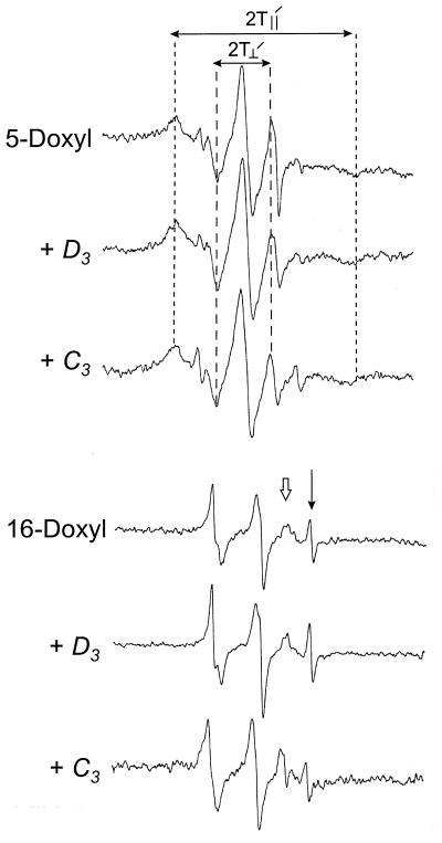
We hypothesized that the difference in biological activity between the isomers might reflect a difference in dipole moment and resultant ability to intercalate into lipid bilayers. To test the hypothesis that C3 can enter cell membranes better than D3, we used EPR spectroscopy of nitroxide spin-labels incorporated into a mixture of mouse brain lipids (Fig. 5). Three parameters of carboxyfullerene-lipid interaction were determined from the EPR measurements: the order parameter (S), correlation time (tc), and lipid/aqueous partition factor. C3 decreased the order parameter (measured with the spin-labeled 16-doxyl lipid) more than D3, suggesting that C3 interacts with the interior of the bilayer to a greater degree than D3. This is supported by the observed changes in tc and the partition factor for C3, which indicate substantial entry of C3 into the bilayer. On the other hand, D3 caused opposite changes in tc and the partition factor, which imply interaction by D3 with the headgroup region of the bilayer, but little entry into the membrane. The results, taken together, are consistent with greater intercalation of C3 compared with D3 into brain lipid membranes (Table 1).
Figure 5.
EPR spectra of spin-labeled lipids (5- or 16-doxyl ketostearic acid) incorporated into mouse brain lipid micelles, with either C3 or D3. Both isomers produce a shift in the order parameter (S) of the 5-doxyl group (see Table 1), but C3 produced a greater change in S. C3 also altered the lipid (⇓)/aqueous (↓) partition factor, detected by 16-doxyl ketostearic acid, to a greater extent than D3. Both results suggest that C3 enters the lipid bilayer to a greater extent than D3.
Table 1.
Results of EPR spectroscopy of nitroxide spin-labels incorporated into a mixture of mouse brain lipids
| Compound | Order parameter* | Correlation time, ns† | Lipid/aqueous partition factor‡ |
|---|---|---|---|
| Control | 0.88 ± 0.01 | 5.1 ± 0.5 | 0.8 ± 0.2 |
| +C3 | 0.84 ± 0.01 | 5.7 ± 0.5 | 1.0 ± 0.2 |
| +D3 | 0.86 ± 0.01 | 4.5 ± 0.5 | 0.6 ± 0.2 |
Order parameter, S = a(T∥′ − T⊥′)/(T∥ − T⊥), where T∥′ is the measured hyperfine splitting for the parallel orientation in 5-doxyl ketostearic acid spin label/phospholipids, and T⊥′ the perpendicular orientation. We assume a = 1, and T∥ − T⊥ = 25 G.
Correlation time, t = 6.5 × 10⁻¹⁰ wo[(ho/h−1)1/2 − 1], where wo is the width of the mid-field line in gauss, ho and h−1 are peak height of mid- and high-field lines on the first derivative absorption measured for 5-doxyl ketostearic acid.
Lipid/aqueous partition factor, f = hL/hA, where hL and hA are the peak height of lipid (⇓) and aqueous phases (↓) measured for 16-doxyl ketostearic acid. We used 1/2 of the full height of hA for a standard comparison.
Carboxyfullerenes Limit Apoptotic Neurodegeneration from Serum Deprivation or Aβ1–42.
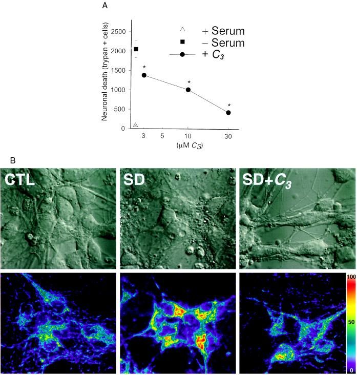
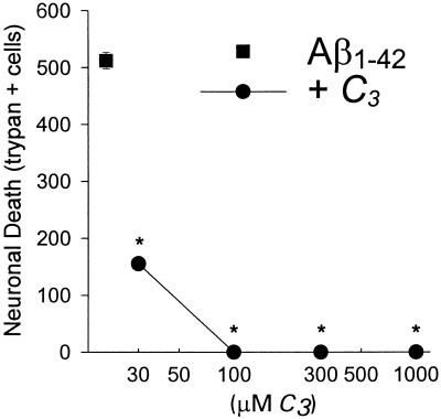
To examine whether the carboxyfullerenes also could inhibit neuronal apoptosis, we turned to two paradigms: (i) serum deprivation, and (ii) exposure to the Alzheimer disease amyloid peptide, Aβ1–42. Cortical neurons cultured without glia underwent apoptosis 24–48 hr after removal of serum (Fig. 6 A and B Top). As with sympathetic neurons deprived of nerve growth factor, this serum deprivation-induced neuronal apoptosis was associated with an enhancement of intracellular free radical production (Fig. 6B, Bottom), detectable by oxidation of dihydrorhodamine to fluorescent rhodamine 123. Application of C3 at the onset of serum deprivation reduced subsequent free radical production and apoptosis (Fig. 6 A and B). In addition, application of C3 also reduced apoptotic death of cortical neurons induced by 24–48 hr exposure to Aβ1–42 (Fig. 7).
Figure 6.
C3 attenuated neuronal death (A) and dihydrorhodamine oxidation (B) induced by serum deprivation. Cell death was determined by manual cell counts of trypan-stained neurons 48 hr after onset of serum deprivation. ∗, P < 0.05 vs. serum deprivation, using ANOVA followed by Student–Newman–Keuls test for multiple comparisons. Values are mean ± SEM, n = 4–8 per condition. Figure is representative of two additional replicates. Confocal images (B) of cortical neurons showing Nomarski images (Upper) of neurons before (CTL) and 8 hr after the onset of serum deprivation (SD). Neurons in the SD condition demonstrate typical apoptotic features, including membrane blebbing and condensation of nuclear contents. (Lower) Concurrent photomicrographs show increased fluorescence due to oxidation of preloaded, nonfluorescent dihydrorhodamine to fluorescent rhodamine 123. Rhodamine fluorescence is quantified with a linear pseudocolor scale corresponding to arbitrary fluorescence intensity units.
Figure 7.
C3-blocked neurodegeneration produced by application Aβ1–42, (20 μM). Manual cell counts of trypan 48 hr after application of Aβ1–42 were graphed as mean ± SEM, n = 10–16 per condition.
Carboxyfullerenes Are Neuroprotectve in a Mouse Model of FALS.
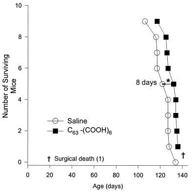
Mice that received C3 carboxyfullerene via intraperitoneal mini-osmotic pumps for 2 months, beginning just after 2 months of age, showed delayed onset of symptoms, improved functional performance, and delayed death as compared with saline-treated controls. C3-treated mice exhibited a delay in deterioration of about 10 days, and scored 4.6 ± 2.2 points (mean ± SEM, P = 0.048, by t test) better than controls on weekly testing. Death in the treated mice also was delayed by 9.0 ± 3.3 days (mean ± SEM, P = 0.023, by t test) (Fig. 8).
Figure 8.
Survival curves for FALS mice treated with continuous intraperitoneal infusion of saline or 15 mg/kg per day C3 carboxyfullerene via implanted mini-osmotic pumps for 2 months, starting at age 2 months. Animals treated with carboxyfullerene had increased survival (P = 0.023 by t test). Three surgical deaths occurred during pump implantation, two in the C3-treated group (after 1 month of treatment, before symptom onset), and one in the control group (with initial pump placement). All procedures conformed to Washington University institutional guidelines for animal welfare.
DISCUSSION
Carboxyfullerenes effectively reduced neuronal death resulting from exposure to glutamate receptor agonists, NMDA or AMPA. C60 derivatives are the only class of antioxidant compounds that we have worked with to date that can fully block intense, rapidly triggered, NMDA receptor-mediated excitotoxicity in our cortical neuronal cultures. In this system, many benchmark scavengers show little ability to attenuate rapidly triggered NMDA receptor-mediated excitotoxicity, and the previous best scavenger we have tested, 21-aminosteroids, reduced neuronal death by less than half (16). Indeed, the C3 compound provided a level of neuroprotective efficacy comparable to that of NMDA receptor antagonists. The C3 isomer also protected neurons from combined oxygen glucose deprivation injury, an insult mediated in part through overactivation of NMDA receptors. Ca2+ flux studies confirmed that carboxyfullerenes are not NMDA receptor antagonists.
Previous studies in our cultures have indicated that the above neuronal deaths, induced by the addition of exogenous excitotoxins, or by oxygen-glucose deprivation, are necrosis deaths (21, 27–28). Apoptosis, which occurs normally during nervous system development, also has been implicated in several forms of pathological neuronal loss (29–31), and oxidative stress has been implicated as both a trigger and a mediator of apoptosis (32–34). To examine whether the carboxyfullerenes also could inhibit neuronal apoptosis, we tested them in two apoptotic insults: (i) serum deprivation and (ii) exposure to Aβ1–42. Degeneration of cultured cortical neurons after removal of trophic factors (serum deprivation) is a delayed event with morphological features consistent with an apoptotic process, including cell body shrinkage, fragmentation of neuronal processes, and chromatin condensation (20–21). Exposure of neurons to Aβ1–42 also results in a delayed injury with features of apoptosis (35). Carboxyfullerenes were able to block neuronal death in both of these apoptotic injuries. Thus, our data support the emerging concept that free radicals contribute to neuronal death in excitotoxic insults as well as injuries that result in apoptosis.
Data from our EPR studies confirmed that the carboxyfullerenes retained the potent free radical scavenging ability of the parent C60 molecule. The higher potency of both carboxyfullerenes as ⋅OH scavengers versus O2⨪ scavengers may reflect the greater ability of C60 to accept multiple ⋅OH compared with O2⨪ moieties. We speculate that after the first ⋅OH radical is added onto C60, the newly generated hydroxyl carboxyfullerene radical may take up another ⋅OH to form a diol, a nonradical diamagnetic adduct. If an odd number of ⋅OH radicals are added to a pentagon of the fullerene framework, a three-membered ring epoxide might be formed (2), due to loss of H by reaction with another nearby ⋅OH, thus preserving the diamagnetic character of the compound. Addition of two O2⨪ radicals on the same pentagon, on the other hand, would be less favorable, due to generation of an adduct with two negative charges in close proximity.
The excellent neuroprotective efficacy of the carboxyfullerenes may reflect their ability to react with superoxide radical in addition to hydroxyl radical. We speculate that because the half-life of ⋅OH is extremely short (10−9 sec) it may be difficult for compounds that are only capable of scavenging ⋅OH to achieve sufficient concentration near the site of ⋅OH generation to out-compete endogenous targets of ⋅OH attack (lipids, proteins, DNA, and other macromolecules). On the other hand, O2⨪, which can be a major source of ⋅OH via the Haber–Weiss reaction, is a relatively stable radical, with a τ1/2 generally measured in seconds. It may be that the ability of carboxyfullerenes to eliminate O2⨪ before its conversion to ⋅OH is an important aspect of their biologically relevant antioxidant properties.
While the two carboxyfullerene isomers show similar free radical scavenging potency when compared under similar “test tube” experimental conditions, they demonstrated different neuroprotective potency and efficacy in cortical cultures. We hypothesized that the C3 isomer, because of its amphiphilic (dipole) nature, might intercalate into biological membranes better than the D3 isomer, which lacks a dipole moment (with carboxyl groups evenly spaced around the C60 equator). EPR studies using spin-labels in aqueous dispersions of mouse brain lipids demonstrated that the C3 isomer intercalates more deeply into the lipid bilayer than the D3 isomer, as indicated by the higher lipid/aqueous partition factor for the 16-doxyl compound, in which the spin-label is at carbon 16 in the bilayer. Results for the 5-doxyl-labeled lipid suggest that both C3 and D3 interact with the surface of the bilayer, although, again, the increased correlation time seen with the C3 isomer suggest it is actually embedded in the bilayer. While we have not yet been able to confirm these results in live cells, it is likely that entry of C3 into membranes would improve its ability to limit lipid peroxidation and to gain access to the interior of the cell.
Finally, to test C3 for in vivo efficacy, we administered the compound systemically to transgenic mice carrying a human disease gene for FALS, an adult onset neurodegenerative disorder characterized by progressive motor neuron death and motor weakness. Mice overexpressing the mutant human Cu, Zn superoxide dismutase (SOD1) gene (G93A) appear normal at birth, but go on to develop hind limb motor impairment at about 100–110 days of age, and progress over the next 2–3 weeks to paralysis and death accompanied by degeneration of lower motorneurons (25). Transgenic mice carrying other human disease SOD1 mutations also have developed comparable features (36–37). While the exact connection between the mutant gene and selective degeneration of the motor system has not yet been defined, participation of free radicals in the neurodegenerative process is suspected (38). Supporting this connection, administration of the free radical scavenger, vitamin E, delayed the onset of symptoms, although not death, in G93A SOD1 transgenic mice (39).
Neuronal degeneration in this mouse FALS model occurs in a sharply compressed fashion. Pathological evidence has suggested that lower motor neuron degeneration becomes detectable at 8 weeks of age and then progresses over the next several weeks to near-complete motor neuron loss. Functional impairment of motor function is apparent only 2–3 weeks before death. The 10-day delay in symptom onset induced by C3 represents a 15% increase in symptom-free life, and the 8-day improvement in survival is nearly half the 2–3 week symptomatic period. Greater beneficial effects might be produced in situations where neurodegeneration is less aggressive.
Present data thus provide substantial extension of our earlier suggestion that C60 derivatives might constitute antioxidant compounds useful in biological systems. The unique efficacy of the C3 carboxyfullerene against excitotoxic necrosis, as well as its powerful protective effects against two forms of neuronal apoptosis, provides support for the evolving idea that oxidative stress is a critical downstream mediator in disparate necrotic and apoptotic neuronal deaths. In addition, the present direct comparison between C3 and D3 regioisomers suggests that amphiphilicity is a desirable feature, increasing intercalation into brain membranes and neuroprotective efficacy. Finally, we provide here a direct demonstration that C60 derivatives can indeed function as neuroprotective drugs in vivo.
Acknowledgments
We thank Scott Schweikart for excellent technical assistance with the FALS mice. This work was supported by National Institutes of Health Grants NS 32636 (L.L.D.), NS 30337 (D.W.C.), and NS 31248 (Teepu Siddique), a grant from Hoffman La Roche (L.L.D., T.-S.L., and D.W.C.), and from the National Science Council of Taiwan (T.-Y.L.).
ABBREVIATIONS
- NMDA
N-methyl-d-aspartate
- AMPA
α-amino-3-hydroxy-5-methyl-4-isoxazolepropionic acid
- FALS
familial amyotrophic lateral sclerosis
References
- 1.Krusic P J, Wasserman E, Keizer P N, Morton J R, Preston K F. Science. 1991;254:1183–1185. doi: 10.1126/science.254.5035.1183. [DOI] [PubMed] [Google Scholar]
- 2.McGeer E G, McGeer P L. Nature (London) 1976;263:517–524. doi: 10.1038/263517a0. [DOI] [PubMed] [Google Scholar]
- 3.Rothman S M, Olney J W. Ann Neurol. 1986;19:105–111. doi: 10.1002/ana.410190202. [DOI] [PubMed] [Google Scholar]
- 4.Choi D W. Neuron. 1988;1:623–634. doi: 10.1016/0896-6273(88)90162-6. [DOI] [PubMed] [Google Scholar]
- 5.McIntosh T, Soares H, Hayes R, Simon R. In: Frontiers in Excitatory Amino Acid Research. Cavallo E A, Lehman J, Turski L, editors. New York: Liss; 1988. pp. 653–656. [Google Scholar]
- 6.Garthwaite J, Charles S L, Chess-Williams R. Nature (London) 1988;336:385–388. doi: 10.1038/336385a0. [DOI] [PubMed] [Google Scholar]
- 7.Dawson V L, Dawson T M, London E D, Bredt D S, Snyder S H. Proc Natl Acad Sci USA. 1991;88:6368–6371. doi: 10.1073/pnas.88.14.6368. [DOI] [PMC free article] [PubMed] [Google Scholar]
- 8.Lafon-Cazal M, Pietri S, Culcasi M, Bockaert J. Nature (London) 1993;364:535–537. doi: 10.1038/364535a0. [DOI] [PubMed] [Google Scholar]
- 9.Dugan L L, Sensi S L, Canzoniero L M T, Handran S D, Rothman S M, Lin T T, Goldberg M P, Choi D W. J Neurosci. 1995;15:6377–6388. doi: 10.1523/JNEUROSCI.15-10-06377.1995. [DOI] [PMC free article] [PubMed] [Google Scholar]
- 10.Reynolds I J, Hastings T G. J Neurosci. 1995;15:3318–3327. doi: 10.1523/JNEUROSCI.15-05-03318.1995. [DOI] [PMC free article] [PubMed] [Google Scholar]
- 11.Dykens J A, Stern A, Trenkner E. J Neurochem. 1987;49:1222–1228. doi: 10.1111/j.1471-4159.1987.tb10014.x. [DOI] [PubMed] [Google Scholar]
- 12.Chan P H, Chu L, Chen S F, Carlson E J, Epstein C J. Acta Neurochirurgica. 1990;51:245–247. doi: 10.1007/978-3-7091-9115-6_82. [DOI] [PubMed] [Google Scholar]
- 13.Monyer H, Hartley D M, Choi D W. Neuron. 1990;5:121–126. doi: 10.1016/0896-6273(90)90302-v. [DOI] [PubMed] [Google Scholar]
- 14.Yue T-L, Gu J-L, Lysko P G, Cheng H-Y, Barone F C, Feuerstein G. Brain Res. 1992;574:193–197. doi: 10.1016/0006-8993(92)90816-r. [DOI] [PubMed] [Google Scholar]
- 15.Chow H S, Lynch J L, Rose K, Choi D W. Brain Res. 1994;639:102–108. doi: 10.1016/0006-8993(94)91769-8. [DOI] [PubMed] [Google Scholar]
- 16.Dugan L L, Gabrielsen J K, Yu S P, Lin T S, Choi D W. Neurobiol Dis. 1996;3:129–135. doi: 10.1006/nbdi.1996.0013. [DOI] [PubMed] [Google Scholar]
- 17.Holden C. Science. 1996;273:1495. [Google Scholar]
- 18.Lamparth I, Hirsch A. J Chem Soc Chem Commun. 1994;1994:1727–1728. [Google Scholar]
- 19.Hara A, Radin N S. Anal Biochem. 1978;90:420–427. doi: 10.1016/0003-2697(78)90046-5. [DOI] [PubMed] [Google Scholar]
- 20.Dugan L L, Bruno V M G, Amagasu S M, Giffard R G. J Neurosci. 1995;15:4545–4555. doi: 10.1523/JNEUROSCI.15-06-04545.1995. [DOI] [PMC free article] [PubMed] [Google Scholar]
- 21.Koh J Y, Gwag B J, Lobner D, Choi D W. Science. 1995;268:573–575. doi: 10.1126/science.7725105. [DOI] [PubMed] [Google Scholar]
- 22.Hartley D M, Kurth M C, Bjerknes L, Choi D W. J Neurosci. 1993;13:1993–2000. doi: 10.1523/JNEUROSCI.13-05-01993.1993. [DOI] [PMC free article] [PubMed] [Google Scholar]
- 23.Goldberg M P, Choi D W. J Neurosci. 1993;13:3510–3524. doi: 10.1523/JNEUROSCI.13-08-03510.1993. [DOI] [PMC free article] [PubMed] [Google Scholar]
- 24.Dugan L L, Creedon D J, Johnson E M, Holtzman D M. Proc Natl Acad Sci USA. 1997;94:4086–4091. doi: 10.1073/pnas.94.8.4086. [DOI] [PMC free article] [PubMed] [Google Scholar]
- 25.Gurney M E, Haifeng P, Chiu A Y, Dal Canto M C, Polchow C Y, Alexander D D, Caliendo J, Hentati A, Kwon Y W, Deng H-X, Chen W, Zhai P, Sufit R L, Siddique T. Science. 1994;264:1772–1775. doi: 10.1126/science.8209258. [DOI] [PubMed] [Google Scholar]
- 26.Basso D M, Beattie M S, Bresnahan J C. J Neurotrauma. 1995;12:1–10. doi: 10.1089/neu.1995.12.1. [DOI] [PubMed] [Google Scholar]
- 27.Gwag B J, Koh J K, Chen M M, Dugan L L, Behrens M, Lobner D, Choi D W. NeuroReport. 1995;7:93–96. [PubMed] [Google Scholar]
- 28.Choi D W. Curr Opin Neurobiol. 1996;6:667–672. doi: 10.1016/s0959-4388(96)80101-2. [DOI] [PubMed] [Google Scholar]
- 29.Heron A, Pollard H, Dessi F, Moreau J, Lasbennes F, Ben-Ari Y, Charriaut-Marlangue C. J Neurochem. 1993;61:1973–1976. doi: 10.1111/j.1471-4159.1993.tb09843.x. [DOI] [PubMed] [Google Scholar]
- 30.MacManus J P, Buchan A M, Hill I E, Rasquinha I, Preston E. Neurosci Lett. 1993;164:89–92. doi: 10.1016/0304-3940(93)90864-h. [DOI] [PubMed] [Google Scholar]
- 31.Du C, Hu R, Csernansky C A, Hsu C Y, Choi D W. J Cereb Blood Flow Metab. 1996;16:195–201. doi: 10.1097/00004647-199603000-00003. [DOI] [PubMed] [Google Scholar]
- 32.Hockenberry D M, Oltvai Z N, Yin X M, Milliman C L, Korsmeyer S J. Cell. 1993;75:241–251. doi: 10.1016/0092-8674(93)80066-n. [DOI] [PubMed] [Google Scholar]
- 33.Ratan R R, Murphy T H, Baraban J M. J Neurochem. 1994;62:376–379. doi: 10.1046/j.1471-4159.1994.62010376.x. [DOI] [PubMed] [Google Scholar]
- 34.Greenlund L J, Deckwerth T L, Johnson E M., Jr Neuron. 1995;14:303–315. doi: 10.1016/0896-6273(95)90287-2. [DOI] [PubMed] [Google Scholar]
- 35.Koh J Y, Yang L L, Cotman C W. Brain Res. 1990;533:315–320. doi: 10.1016/0006-8993(90)91355-k. [DOI] [PubMed] [Google Scholar]
- 36.Wong P C, Pardo C A, Borchelt D R, Lee M K, Copeland N G, Jenkins N A, Sisodia S S, Cleveland D W, Price D L. Neuron. 1995;14:1105–1116. doi: 10.1016/0896-6273(95)90259-7. [DOI] [PubMed] [Google Scholar]
- 37.Ripps M E, Huntley G W, Hof P R, Morrison J H, Gordon J W. Proc Natl Acad Sci USA. 1995;92:689–692. doi: 10.1073/pnas.92.3.689. [DOI] [PMC free article] [PubMed] [Google Scholar]
- 38.Wiedau-Pazos M, Goto J J, Rabizadeh S, Gralla E B, Roe J A, Lee M K, Valentine J S, Bredesen D E. Science. 1996;271:515–518. doi: 10.1126/science.271.5248.515. [DOI] [PubMed] [Google Scholar]
- 39.Gurney M E, Cuttings F B, Zhai P, Doble A, Taylor C P, Andrus P K, Hall E D. Ann Neurol. 1996;39:147–157. doi: 10.1002/ana.410390203. [DOI] [PubMed] [Google Scholar]


