Abstract
This paper entails a clinical review of outcomes and complications of 19 consecutive paediatric patients having undergone elastic stable intramedullary nailing for diaphyseal forearm fractures over a one year period. The mean age of patients was 9 years. The majority were male with a ratio of 17:2. In this group there were two patients with grade 1 open fractures. Four of the fractures required open reduction due to difficulty in reduction and soft tissue interposition. All fractures went on to osseous union with minimal deformity and full recovery. There were three complications which included one EPL rupture requiring delayed repair, one EPB partial rupture repaired at time of surgery, and one superficial radial nerve injury. Two patients also presented with nails penetrating the skin prior to removal. Elastic stable intramedullary nails offer good fixation to control deformity in midshaft forearm fractures for paediatric patients. However there is a high rate of possible complications around the radial insertion point.
Résumé
L’étude clinique des complications à propos d’une série consécutive de 19 patients nécessitant l’utilisation d’un enclouage élastique stable intra médullaire pour fracture de la diaphyse des os de l’avant-bras a été réalisée. Cette étude a été réalisée sur une période d’un an. L’âge moyen des patients était de 9 ans avec une majorité de garçons (17 garçons et 2 filles). Dans ce groupe, trois patients présentaient une fracture ouverte de grade I. 4 patients ont nécessité une réduction à ciel ouvert secondaire à une interposition tissulaire. Trois complications ont été constatées, deux ruptures tendineuses et une lésion superficielle du nerf radial. 2 patients présentaient également une lésion cutanée secondaire en regard des clous. L’enclouage intra médullaire élastique stable permet une excellente fixation de la déformation de la fracture des deux os de l’avant bras chez les enfants mais cependant, le taux de complications est relativement élevé au niveau du point d’insertion radial.
Introduction
Over recent years the use of elastic stable intra-medullary nails has dramatically increased with the introduction of a variety of nails for paediatric fractures. The forearm fractures, especially with both bones involved, are increasingly being treated with intra-medullary devices to prevent displacement during the healing phase. Many devices have been used such as K-wires [8, 10] and Steinmann pins [7], as well as many designs of flexible nails [4, 6].
In the past many of these fractures would have been treated by manipulation and a carefully moulded plaster as described by Sir John Charnley. This would then be followed by a careful period of observation for any fracture displacement.
The use of an intra-medullary device is indicated for open fractures, unstable fractures or irreducible fractures. This technique is preferable in many circumstances to open reduction and plating of the forearm bones as it prevents stripping of the soft tissues; also, there is little in the way of surgical scar tissue and is therefore cosmetically acceptable.
However, intra-medullary nails do have complications and it may be preferable to treat many of these fractures with manipulation and casting followed by close observation.
We present the results and complications of 19 consecutive paediatric patients who underwent elastic stable intra-medullary nailing for diaphyseal forearm fractures.
Methods and results
All patients treated with elastic stable intra-medullary nails over a one year period were reviewed for clinical outcome and radiographs reviewed for any residual deformity.
Nineteen consecutive patients were identified during this time. All patients underwent intra-medullary stabilisation for diaphyseal fractures of both forearm bones under the care of the on-call orthopaedic team on the next available trauma list. During all procedures stabilisation was carried out to both the ulna and the radius. The insertion point for the radial nail was either between the first and second extensor compartments or through a dorsal approach.
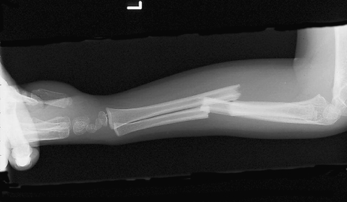
Patients ranged from the age of 4 years 4 months to 15 years with a mean distribution of 9 years. The male to female distribution was 17:2. The typical presentation is demonstrated on the radiograph in Fig. 1.
Fig. 1.
Lateral radiograph of a mid-shaft forearm paediatric fracture
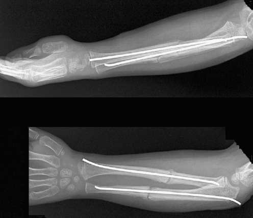
Two of the 19 patients presented with an open fracture. A further four patients required open reduction due to a failure in closed reduction and soft tissue interposition. One of the patients requiring open reduction was a secondary procedure at two weeks following failed attempt at closed manipulation and treatment with a moulded plaster. All patients after either open or closed reduction underwent stabilisation to both forearm bones with the use of elastic stable intra-medullary nails as demonstrated in Fig. 2.
Fig. 2.
Radiograph showing standard method of fixation
All patients remained in hospital overnight until comfortable for discharge. Patients were placed in an above elbow cast and reviewed initially at weekly intervals for wound inspections and to ensure satisfactory position of the nails and the fracture. The patients were removed from the cast when osseous union was achieved.
Complications as a result of the procedure included: one neuropraxia involving the superficial radial nerve, which resolved after several weeks with no long term complication; one partial rupture of extensor pollicus brevis tendon identified and repaired at the time of removal of the intramedullary flexible nails; and one complete rupture of extensor pollicus longus tendon, which was recognised early and a primary repair was performed. In this case the insertion point was through a dorsal insertion at the second and third extensor compartment interval. All of the above complications occurred due to the nails being inserted through stab incisions at the radial insertion point.
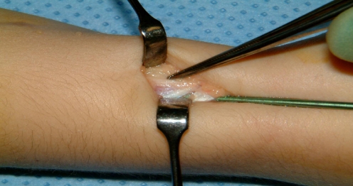
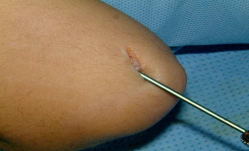
All other procedures were performed through an open procedure between the first and second extensor compartments to identify all structures prior to insertion of the nail (see Figs. 3 and 4).
Fig. 3.
Insertion of radial nail at 1st and 2nd extensor interval through open procedure with direct vision of structures
Fig. 4.
Insertion of ulna nail through open procedure
Two patients also presented with protrusion of the wires through the skin although they had been buried during the procedure. These nails required removal 2–3 weeks prior to the planned date of removal.
At follow-up clinics all patients went on to osseous union and regained a full range of movement after rehabilitation. There were no cases of delayed union, non-union or mal-union.
After removal of the nails all patients regained full function and all complications resolved. The patients were discharged from the clinic after full recovery.
Discussion
Elastic stable intra-medullary nails are now commonly used for the treatment of paediatric forearm fractures and their use has minimised the surgical scarring previously caused by open reduction and plating.
We should like to emphasise that the majority of these fractures should be treated by closed manipulation and moulded casting. Previous papers have shown excellent results in children under the age of ten years with closed treatment, with poorer results and increased incidence of re-displacement in children over the age of ten years [5].
Further studies have looked at the position of immobilsation after closed manipulation. One such paper showed excellent results with no displacements when the elbow was immobilised in an extended position and a 17.6% re-displacement with the elbow immobilised in a flexed position [2].
In this modern age there is an increasing tendency towards operative fixation as new implants and techniques become available. We should not lose our ability to perform closed reduction and apply a moulded cast in these fractures.
The procedure of inserting intra-medullary nails is not without the possibility of complication. In this series of patients we have reported a complication rate of 16%. This is similar to the complication rate reported by Lascombes in 1990 [6]. A further paper comparing intra-medullary versus plate fixation reported similar complications of thumb neuropathies, rod migration and skin infections [3]. This paper also reported major complications such as re-fracture and non-union.
All our complications occurred by insertion through either a dorsal approach or through a stab incision. The nail should be inserted on the radial aspect between the first and second compartments after careful dissection to identify the superficial radial nerve and extensor tendons. The nails should not be inserted from a dorsal entry point as suggested in some publications [1, 9] as, in our experience, complications are more likely to occur.
Conclusion
We believe that the technique of elastic stable intra-medullary nails should be reserved for those fractures that have failed the test of closed manipulation and moulded casting, as well as open fractures of the forearm.
We would also like to stress the importance of the radial insertion point in order to limit the number of possible complications.
References
- 1.Barry M, Paterson JMH (2004) Aspects of current management: Flexible intramedullary nails for fractures in children. J Bone Joint Surg Br 86-B:947–953 [DOI] [PubMed]
- 2.Bochang C, Jie Y, Zhigang W, Weigl D, Bar-On E, Katz K (2005) Immobilisation of forearm fractures in children: extended versus flexed elbow. J Bone Joint Surgery Br 87-B:994–996 [DOI] [PubMed]
- 3.Fernandez FF, Egenholf M, Carsten C, Holz F, Schneider S, Wentzensen A (2005) Unstable diaphyseal fractures of both bones of the forearm in children: plate fixation versus intramedullary nailing. Injury 36(10):1210–1216 [DOI] [PubMed]
- 4.Huber RI, Keller HW, Iluber PM, Rehm KE (1996) Flexible intramedullary nailing as fracture treatment in children. J Pediatr Orthoped 16:602–605 [DOI] [PubMed]
- 5.Kay MD, Smith C, Oppenheim WL (1986) Both-bone midshaft forearm fractures in children. J Pediatr Orthoped 6:306–310 [DOI] [PubMed]
- 6.Lascombes P, Prevot J, Ligier JN, Metazieau JP, Poncelet T (1990) Elastic stable intramedullary nailing in forearm shaft fractures in children: 85 cases. J Pediatr Orthoped 10:167–171 [PubMed]
- 7.Pugh DMW, Galpin RD, Carey TP (2000) Intramedullary Steinmann pin fixation of forearm fractures in children: long term results. Clin Orthop Relat R 376:39–48 [DOI] [PubMed]
- 8.Shoemaker SD, Comstock CP, Mubarak SJ, Wenger DR, Chambers FIG (1999) Intramedullary Kirshner wire fixation of open or unstable forearm fractures in children. J Pediatr Orthoped 19:329–337 [DOI] [PubMed]
- 9.Wiss DA (1998) Fractures: master techniques in orthopaedic surgery. Lippincott Williams & Wilkins, Philadelphia
- 10.Yung SH, Lam CY, Choi KY et al (1998) Percutaneous intramedullary Kirshner wiring for displaced diaphyseal forearm fractures in children. J Bone Joint Surg Br 80-B:91–94 [DOI] [PubMed]


