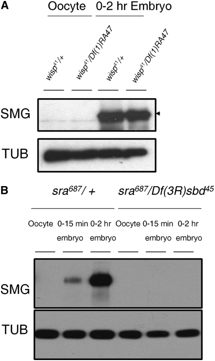
Figure 4.—
SMG protein is translated in wisp mutant embryos, but not in sra embryos. (A) Total protein extracts of ovarian oocytes (left) and laid 0- to 2-hr embryos (right) from wisp41/Df(1)RA47 or wisp41/+ females were separated by SDS–PAGE, blotted, and probed for Smaug (SMG) and α-tubulin (TUB; loading control). SMG protein is translated normally in laid embryos from wisp mutant females; there is no statistically significant difference between control and mutant embryos in the amount of SMG signal intensity, normalized to TUB signal intensity, in five independent replicates (matched pairs t-test: t-ratio = −0.205, 4 d.f., P = 0.848). (B) Total protein extracts of oocytes, 0- to 15-min embryos and 0- to 2-hr embryos from sra687/+ or sra687/Df(3R)sbd45 females were isolated, blotted, and probed for SMG and α-tubulin (TUB; loading control). SMG protein is not detected in embryos from sra mutant females.

