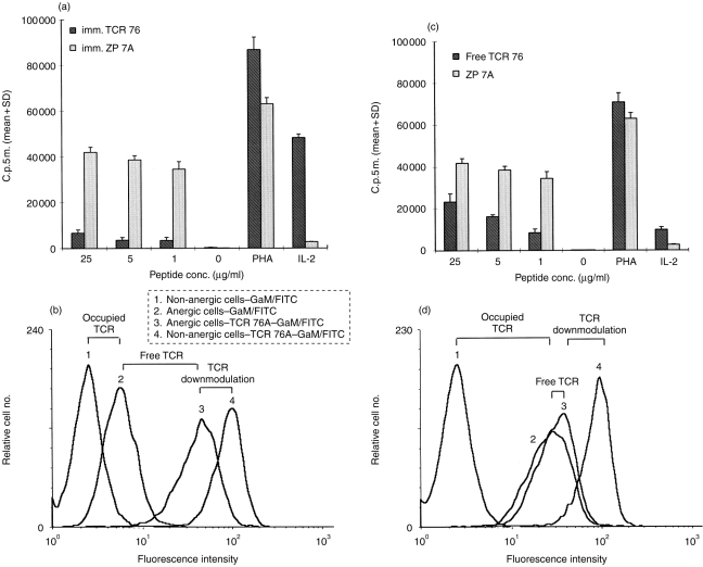
Figure 2.
Induction of anergy by anticlonotype mAb TCR 76. (a) Unresponsiveness is not due to cell death or apoptosis. Immobilized TCR 76 (anergic cells) or control mAb ZP 7A (non-anergic cells) were incubated overnight with H.243 T cells. After removal of the cells from the anergic stimulus, the cells were challenged with DRB1*0401-matched APC, and PHA or increasing concentrations of peptide. In addition, T cells were incubated with IL-2 in the absence of APC. Proliferation was assessed by [3H]thymidine incorporation. Each value represents the mean counts per 5 min of triplicate cultures+standard deviation. (b) Unresponsiveness is not caused by TCR blockade or downmodulation. Non-anergic H.243 T cells resulting from incubation with immobilized ZP 7A (curves 1 and 4) or anergic T cells resulting from incubation with immobilized TCR 76 (curves 2 and 3) were stained directly with GAM–FITC (curves 1 and 2) or indirectly with TCR 76 and GAM–FITC (curves 3 and 4). Curve 1 reflects background fluorescence level. Curve 2reflects the number of TCR molecules occupied with anticlonotype mAb after removal from the anticlonotype mAb coated plates. Curve 3 reflects the total number of TCR molecules on anergic cells and curve 4 reflects the total number of TCR molecules on non-anergic cells. (c) and (d) represent similar experiments performed with free TCR 76 instead of immobilized TCR 76.

