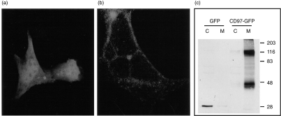
Figure 5.
Membrane localization of mouse CD97. HEK 293 cells were transfected either with the GFP vector as a control or with the mouse CD97-GFP fusion protein vector. (a) Under a confocal fluorescence microscope, simple GFP protein was shown to be present in the cytoplasm of the cells. (b) In contrast to simple GFP, CD97-GFP fusion protein was shown to be localized to the plasma membrane. (c) Western blot analysis of cytosolic (C) and membrane (M) fractions of GFP or CD97-GFP vector transfected HEK 293 cell lysates. Simple GFP was a 27 000 MW protein which was detected only in the cytosolic fraction. Mouse CD97-GFP fusion proteins were detected only in the membrane fraction. Two distinct fusion protein bands, with apparent molecular weights of 116 000 and 47 000, respectively, were recognizable. Sizes and positions of protein molecular weight markers (in kilodaltons) are shown on the right side.

