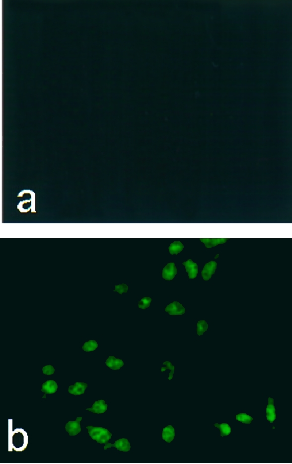
Figure 3.

Immunofluorescence staining of intracellular Bence–Jones proteins (BJPs). After incubation of LLC‐PK1 cells with 1·0 μm of non‐cytotoxic (a) or cytotoxic (b) BJP, the cells were stained with fluorescein isothiocyanate (FITC)‐labelled anti‐human κ‐chain goat immunoglobulin G (IgG), essentially as described.19
