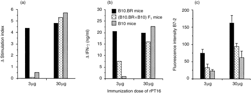
Figure 2.

Antigen dose-dependent H2 control of cellular responses to the recombinant 16 000-MW protein of Mycobacterium tuberculosis (rPT16) of immune lymph node (LN) cells in vitro. Mice (n = 5) of the B10.BR, (B10.BR × B10)F1 and B10 strains were immunized in foot pads with rPT16 prepared in Freund’s incomplete adjuvant (FIA), and 7-day primed LN cells were cultured for 3 days in the presence of 0·5 µg/ml of rPT16. Mean (n = 3) values of (a) cell proliferation and (b) culture supernatant interferon-γ (IFN-γ) levels. Δ = Stimulation index (SI) or IFN-γ values of experimental group after subtraction of values obtained from phosphate-buffered saline (PBS)/FIA-injected mice. (c) Mean fluorescence intensity for B7-2 staining of B cells after subtracting the values obtained from PBS/FIA-injected mice: double staining with fluorescein isothiocyanate (FITC)-labelled CD45R/B220 (RA3-6B2) and phycoerythrin (PE)-conjugated CD86/B7-2 (GL1) monoclonal antibodies (mAbs).
