

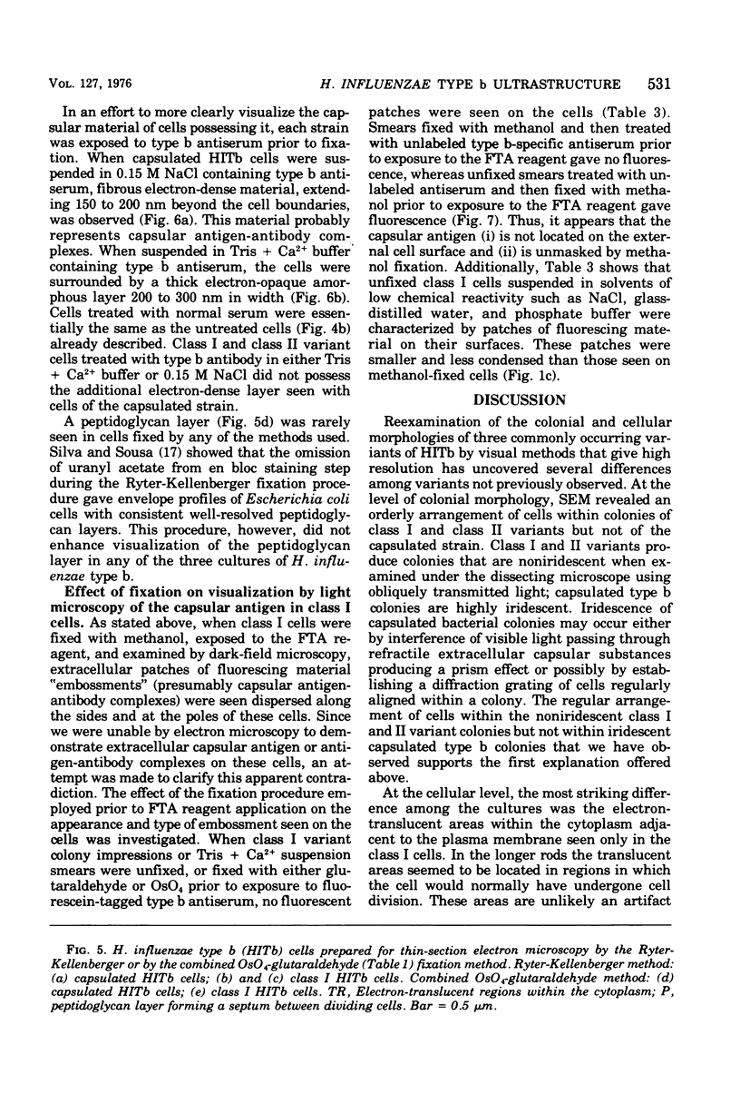
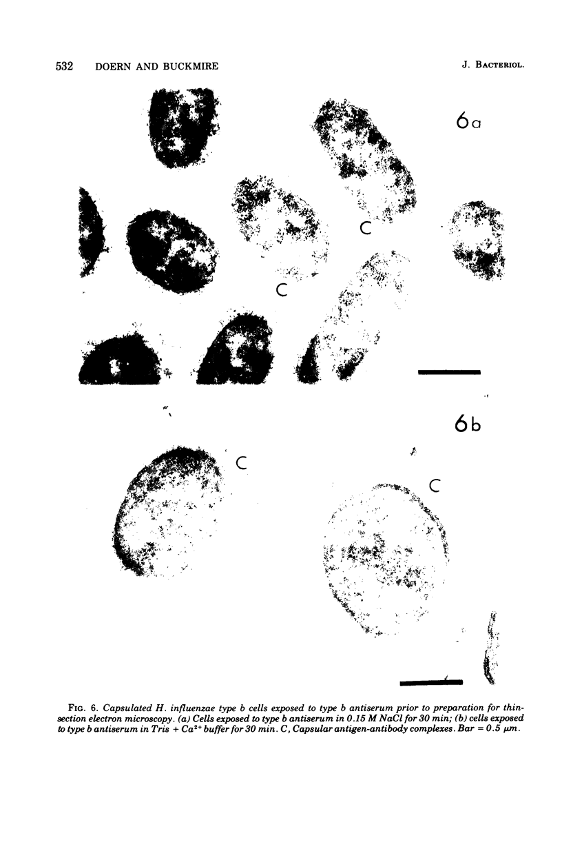
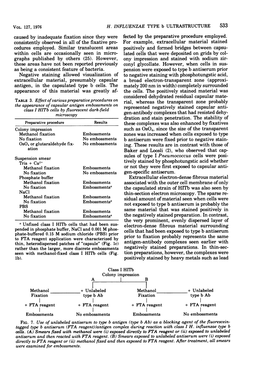
Abstract
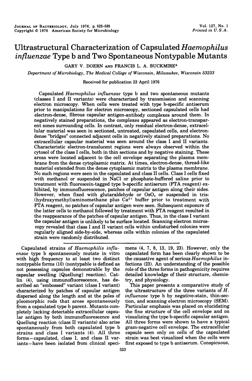
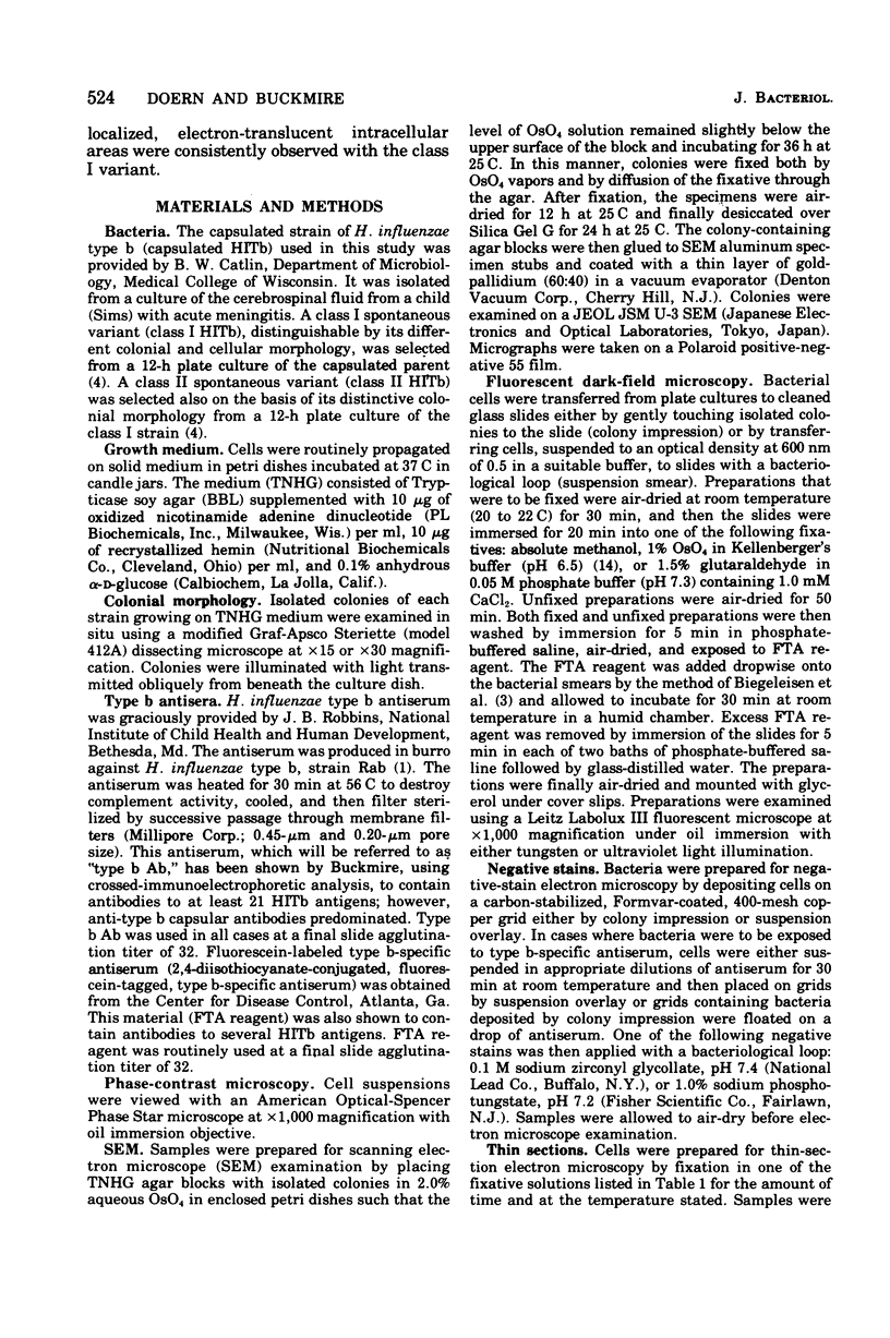
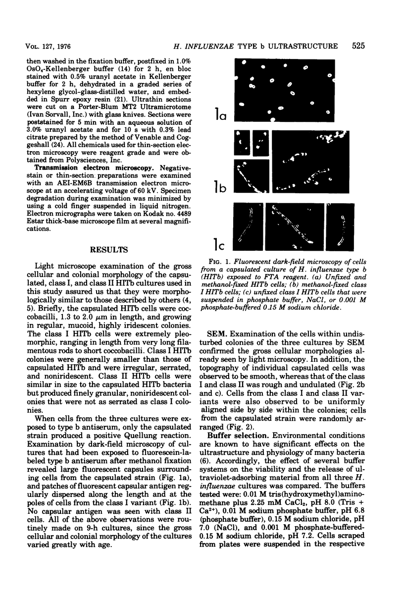
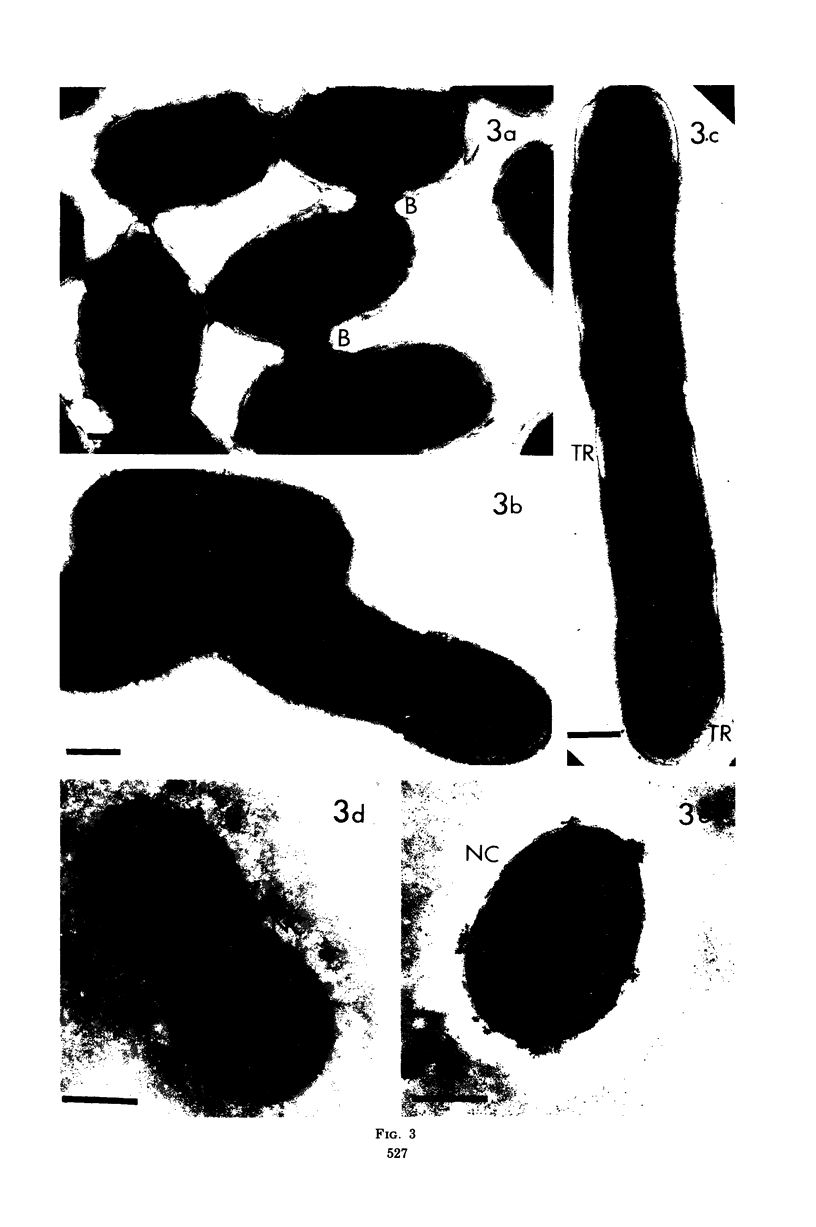
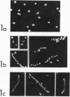
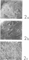
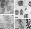
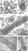
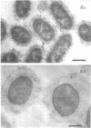
Capsulated Haemophilus influenzae type b and two spontaneous mutants (classes I and II variants) were characterized by transmission and scanning electron microscopy. When cells were treated with type b-specific antiserum prior to manipulations for electron microscopy, sectioned capsulated cells had electron-dense, fibrous capsular antigen-antibody complexes around them. In negatively stained preparations, the complexes appeared as electron-transparent zones surrounding cells. In contrast, only residual electron-dense, extracellular material was seen in sectioned, untreated, capsulated cells, and electron-dense "bridges" connected adjacent cells in negatively stained preparations. No extracellular capsular material was seen around the class I and II variants. Characteristic electron-translucent regions were always observed within the cytosol of the class I cells, both in thin sections and by negative staining. These areas were located adjacent to the cell envelope separating the plasma membrane from the dense cytoplasmic matrix. At times, electron-dense, thread-like material extended from the dense cytoplasmic matrix to the plasma membrane. No such regions were seen in the capsulated and class II cells. Class I cells fixed with methanol or suspended in NaCl or phosphate-buffered saline prior to treatment with fluorescein-tagged type b-specific antiserum (FTA reagent) exhibited, by immunofluorescence, patches of capsular antigen along their sides. However, when fixed with glutaraldehyde or OsO4 or suspended in tris-(hydroxymethyl)aminomethane plus Ca2+ buffer prior to treatment with FTA reagent, no patches of capsular antigen were seen. Subsequent exposure of the latter cells to methanol followed by treatment with FTA reagent resulted in the reappearance of the patches of capsular antigen. Thus, in the class I variant the capsular antigen is unlikely to be surface located. Scanning electron microscopy revealed that class I and II variant cells within undisturbed colonies were regularly aligned side-by-side, whereas cells within colonies of the capsulated strain were randomly distributed.
Full text
PDF
Images in this article
Selected References
These references are in PubMed. This may not be the complete list of references from this article.
- BIEGELEISEN J. Z., Jr, MITCHELL M. S., MARCUS B. B., RHODEN D. L., BLUMBERG R. W. IMMUNOFLUORESCENCE TECHNIQUES FOR DEMONSTRATING BACTERIAL PATHOGENS ASSOCIATED WITH CEREBROSPINAL MENINGITIS. I. CLINICAL EVALUATION OF CONJUGATES ON SMEARS PREPARED DIRECTLY FROM CEREBROSPINAL FLUID SEDIMENTS. J Lab Clin Med. 1965 Jun;65:976–989. [PubMed] [Google Scholar]
- Baker R. F., Loosli C. G. The ultrastructure of encapsulated Diplococcus pneumoniae type 1 before and after exposure to type specific antibody. Lab Invest. 1966 Apr;15(4):716–730. [PubMed] [Google Scholar]
- Catlin B. W. Haemophilus influenzae in cultures of cerebrospinal fluid. Noncapsulated variants typable by immunofluorescence. Am J Dis Child. 1970 Sep;120(3):203–210. doi: 10.1001/archpedi.1970.02100080087005. [DOI] [PubMed] [Google Scholar]
- Chandler C. A., Fothergill L. D., Dingle J. H. The Pattern of Dissociation in Hemophilus influenzae. J Bacteriol. 1939 Apr;37(4):415–427. doi: 10.1128/jb.37.4.415-427.1939. [DOI] [PMC free article] [PubMed] [Google Scholar]
- Corpe W. A., Salton M. R. Properties of surface materials released from cells of Chromobacterium violaceum. Biochim Biophys Acta. 1966 Jul 27;124(1):125–135. doi: 10.1016/0304-4165(66)90320-5. [DOI] [PubMed] [Google Scholar]
- Feigin R. D., Shackelford P. G., Keeney R. Hemophilus influenzae abscess associated with septicemia. Am J Dis Child. 1971 Jun;121(6):534–535. doi: 10.1001/archpedi.1971.02100170116021. [DOI] [PubMed] [Google Scholar]
- HSU K. C., RIFKIND R. A., ZABRISKIE J. B. FLUORESCENT, ELECTRON MICROSCOPIC, AND IMMUNOELECTROPHORETIC STUDIES OF LABELED ANTIBODIES. Science. 1963 Dec 13;142(3598):1471–1473. doi: 10.1126/science.142.3598.1471. [DOI] [PubMed] [Google Scholar]
- ROGERS K. B., ZINNEMANN K., FOSTER W. P. The isolation and identification of Haemophilus spp, from unusual lesions in children. J Clin Pathol. 1960 Nov;13:519–524. doi: 10.1136/jcp.13.6.519. [DOI] [PMC free article] [PubMed] [Google Scholar]
- RYTER A., KELLENBERGER E., BIRCHANDERSEN A., MAALOE O. Etude au microscope électronique de plasmas contenant de l'acide désoxyribonucliéique. I. Les nucléoides des bactéries en croissance active. Z Naturforsch B. 1958 Sep;13B(9):597–605. [PubMed] [Google Scholar]
- Reyn A., Birch-Andersen A., Lapage S. P. An electron microscope study of thin sections of Haemophilus vaginalis (Gardner and Dukes) and some possibly related species. Can J Microbiol. 1966 Dec;12(6):1125–1136. doi: 10.1139/m66-154. [DOI] [PubMed] [Google Scholar]
- SELL S. H., CHEATHAM W. J., YOUNG B., WELCH K. Hemophilus influenzae in respiratory infections. I. Typing by immunofluorescent techniques. Am J Dis Child. 1963 May;105:466–469. [PubMed] [Google Scholar]
- SMITH C. W., METZGER J. F. Demonstration of a capsular structure on Listeria monocytogenes. Pathol Microbiol (Basel) 1962;25:499–506. doi: 10.1159/000161305. [DOI] [PubMed] [Google Scholar]
- Silva M. T., Sousa J. C. Ultrastructure of the cell wall and cytoplasmic membrane of gram-negative bacteria with different fixation techniques. J Bacteriol. 1973 Feb;113(2):953–962. doi: 10.1128/jb.113.2.953-962.1973. [DOI] [PMC free article] [PubMed] [Google Scholar]
- Snyder S. N., Brunjes S. Hemophilus influenzae meningitis in adults. Review of the literature and report of 18 cases. Am J Med Sci. 1965 Dec;250(6):658–667. [PubMed] [Google Scholar]
- Springer E. L., Roth I. L. The ultrastructure of the capsules of Diplococcus pneumoniae and Klebsiella pneumoniae stained with ruthenium red. J Gen Microbiol. 1973 Jan;74(1):21–31. doi: 10.1099/00221287-74-1-21. [DOI] [PubMed] [Google Scholar]
- Spurr A. R. A low-viscosity epoxy resin embedding medium for electron microscopy. J Ultrastruct Res. 1969 Jan;26(1):31–43. doi: 10.1016/s0022-5320(69)90033-1. [DOI] [PubMed] [Google Scholar]
- Trump B. F., Bulger R. E. New ultrastructural characteristics of cells fixed in a glutaraldehyde-osmium tetroxide mixture. Lab Invest. 1966 Jan;15(1 Pt 2):368–379. [PubMed] [Google Scholar]
- VENABLE J. H., COGGESHALL R. A SIMPLIFIED LEAD CITRATE STAIN FOR USE IN ELECTRON MICROSCOPY. J Cell Biol. 1965 May;25:407–408. doi: 10.1083/jcb.25.2.407. [DOI] [PMC free article] [PubMed] [Google Scholar]
- Zollinger W. D., Kasper D. L., Veltri B. J., Artenstein M. S. Isolation and characterization of a native cell wall complex from Neisseria meningitidis. Infect Immun. 1972 Nov;6(5):835–851. doi: 10.1128/iai.6.5.835-851.1972. [DOI] [PMC free article] [PubMed] [Google Scholar]