The cloning, expression, purification and crystallization of recombinant Clostridium perfringens β2-toxin is described. The crystals diffracted to 2.9 Å resolution.
Keywords: β2-toxin, Clostridium perfringens
Abstract
Clostridium perfringens is a Gram-positive sporulating anaerobic bacterium that is responsible for a wide spectrum of diseases in animals, birds and humans. The virulence of C. perfringens is associated with the production of several enterotoxins and exotoxins. β2-toxin is a 28 kDa exotoxin produced by C. perfringens. It is implicated in necrotic enteritis in animals and humans, a disease characterized by a sudden acute onset with lethal hemorrhagic mucosal ulceration. The recombinant expression, purification and crystallization of β2-toxin using the batch-under-oil technique are reported here. Native X-ray diffraction data were obtained to 2.9 Å resolution on a synchrotron beamline at the F2 station at Cornell High Energy Synchrotron Source (CHESS) using an ADSC Quantum-210 CCD detector. The crystals belong to space group R3, with a dimer in the asymmetric unit; the unit-cell parameters are a = b = 103.71, c = 193.48 Å, α = β = 90, γ = 120° using the hexagonal axis setting. A self-rotation function shows that the two molecules are related by a noncrystallographic twofold axis with polar angles ω = 90.0, ϕ = 210.3°.
1. Introduction
Clostridium perfringens is an anaerobic Gram-positive spore-forming bacteria that has an arsenal of virulence factors associated with a wide variety of diseases that affect most domestic animal species and humans (Songer, 1996 ▶; Niilo, 1986 ▶).
C. perfringens is widespread in the environment and frequently inhabits the intestines of animals. It does not exhibit adherence and invasive properties towards healthy intestinal mucosa (Songer, 1996 ▶). Alteration of the physiological equilibrium of the intestine and resident microflora arising from antibiotic therapy, management-related stress, ingestion of soil and feces-contaminated feed or improperly fermented silage or haylage allows the colonization of toxigenic clostridia, leading to enterotoxemia and hemorrhagic enteritis (Herholz et al., 1999 ▶; Niilo, 1986 ▶).
The C. perfringens bacterium has been shown to produce at least 15 different toxins, five of which (α-, β-, ∊- and ι-toxins and enterotoxin) are responsible for tissue lesions and host death (Daube, 1992 ▶; Songer, 1996 ▶; Petit et al., 1999 ▶). The ι-, α-, β- and ∊-toxins have been used to classify C. perfringens into five isotypes or toxin-types (A, B, C, D and E), where each type carries a different combination of toxin genes (Al-khaldi et al., 2004 ▶). The α-toxin gene (cpa) had been found in all five toxin-types, while the β-toxin gene (cpb) is only found in types B and C. The ∊-toxin gene (etx) can be found in types B and D. The ι-toxin gene (iA) has been shown to be present in type E only. In addition to the above-mentioned major toxins, there are other toxins that play an important role in many human and animal diseases. C. perfringens toxin-type A strains can produce an enterotoxin (cpe) and have been mostly associated with outbreaks of food poisoning.
Recently, a variant of the β-toxin known as β2 (cpb2 gene) has been associated with enteric diseases in a wide range of animals, including swine, cattle, poultry, sheep, horses and dogs (Klaasen et al., 1999 ▶; Garmory et al., 2000 ▶; Waters et al., 2005 ▶). Since its recognition as a separate toxin-producing strain, β2-toxigenic C. perfringens has also been isolated from various avian and aquatic species (Boujon et al., 2005 ▶). Fisher et al. (2005 ▶) suggested a potential role of β2-toxin as an accessory toxin with enterotoxin in C. perfringens antibiotic-associated diarrhea and sporadic diarrhea.
During bacterial invasion, bacterial toxins exhibit certain inhibitory mechanisms on the host system. They act by inhibiting protein synthesis or by disrupting cellular membranes. By forming membrane pores, the toxins can deliver toxic components to specific cellular sites (Tilley & Saibil, 2006 ▶). These pore-forming toxins are classified according to whether they form α-helical channels or β-barrels. C. perfringens is implicated in necrotic and hemorrhagic enteritis and enterotoxemia in various species of animals. The pathogen colonizes the intestinal mucosa, multiplies and causes damage to the target cells (Manteca et al., 2001 ▶).
We have initiated a study aimed at determining the three-dimensional structure of β2-toxin in order to understand its function from a structural perspective. In this study, we describe the purification of β2-toxin from a recombinant bacterial system, crystallization of the full-length molecule and preliminary crystal characterization.
2. Methods and results
2.1. PCR amplification of the β2-toxin gene
Chromosomal DNA of C. perfringens was used to amplify the β2-toxin gene using specific primers. The primers β2-Forward (β2-F) and β2-Reverse (β2-R) span the β2 ORF but exclude the signal peptide. The β2-F and β2-R primers together with DNA from the field strain of C. perfringens were used to amplify a 704 bp fragment.
The sequences of the β2-toxin primers were as follows: forward primer, 5′-CGAATTCCAAAGAAATCGACGCTTAT-3′; reverse primer, 5′-CCGCTCGAGTGCACAATACCCTTCACC-3′. The PCR reaction contained 0.2 mM dNTP, 1.5 mM MgCl2, 0.1 µmol each of forward and reverse primers, 5 µl 10× PCR buffer, 2.5 U Platinum Pfx DNA polymerase, appropriate template DNA and autoclaved distilled water to make a total volume of 50 µl. PCR was performed under standard conditions, i.e. denaturation at 367 K for 15 s, annealing at 330 K for 30 s and elongation at 345 K for 1 min for 35 cycles in a Peltier Thermo Cycler (MJ Research, MA, USA). The amplified β2-toxin gene was isolated by agar gel electrophoresis and purified using a Quantum Prep Freeze ’N Squeeze DNA Gel Extraction Spin Column (Bio-Rad, Hercules, CA, USA).
2.1.1. Cloning and expression of the β2-toxin gene in pGEX-4T
A 704 bp PCR product was digested with XhoI and EcoRI. Following digestion, this fragment was subcloned into pGEX-4T vector which was linearized using the same restriction enzymes. Transformation was carried out using 5 µl ligation mix (pGEX-4T + digested β2-toxin gene). Competent Escherichia coli DH5α strain cells were transformed via electroporation using a BioRad Genepulser at 2400 V, 25 µF and 200 Ω for 0.70 ms. Transformants containing the complete plasmid were identified by plasmid isolation and digestion using XhoI and EcoRI restriction enzymes. The transformed cells were cultivated on Luria–Bertani medium containing 100 µg ml−1 ampicillin.
2.1.2. Overexpression of pGEX-4T
pGEX-4T/DH5α cells were inoculated in 200 ml Luria–Bertani medium supplemented with 100 µg ml−1 ampicillin. The cells were incubated overnight in shaker flasks at 310 K. Subsequently, this seed culture was used to inoculate 4 l Luria–Bertani medium supplemented with ampicillin (100 µg ml−1). Cells were grown at 310 K and β2-toxin expression was induced by the addition of 1 mM isopropyl β-d-thiogalactopyranoside at an OD600 of 0.3. Upon reaching an OD600 of 0.8, cells were harvested by centrifugation at 10 000g for 30 min and the resulting cell pellet was stored at 253 K.
2.1.3. Purification of the recombinant β2-toxin
The resulting pellet was resuspended in 200 ml lysis buffer pH 7.3 containing 1× phosphate-buffered saline (PBS). The cell suspension was lysed using ultrasonic treatment (three 20 s pulses with 1 min intervals) and then treated with 1% protease-inhibitor mixture (2 mM phenylmethylsulfonyl fluoride, 25 mM iodoacetamide, 5 µg ml−1 aprotinin, 10 µg ml−1 leupeptin, 10 µg ml−1 pepstatin A). The sonicated cells were then centrifuged for 1 h at 14 000 rev min−1 and 277 K. The β2-protein present in the supernatant was separated from the pellet following centrifugation. The supernatant containing the soluble proteins was subjected to chromatography on a glutathione Sepharose 4B column (Amersham Biosciences, NJ, USA) according to the manufacturer’s protocol.
2.1.4. Glutathione Sepharose affinity chromatography
A 2 ml glutathione Sepharose bed volume was prepared. 200 ml soluble protein sonicate was applied onto the column. The supernatant was loaded at a flow rate of 0.2 ml min−1. The column was washed with 20 ml 1× PBS for a total of three washes. The GST-β2 fusion protein was eluted from the column at room temperature using 2 ml glutathione elution buffer (10 mM reduced glutathione in 50 mM Tris–HCl pH 8.0). The elution buffer was incubated on the column for 15 min and the elute was then collected in a separate tube. The elution was repeated three times to remove all the bound GST-β2 fusion protein.
2.1.5. Thrombin cleavage of GST-β2 fusion protein
The affinity-purified β2 fusion protein has a GST tag attached to it. The purified protein was treated with a site-specific protease, human thrombin, in order to cleave the GST tag and obtain a purified recombinant protein. Thrombin was added at a concentration of 10 units per milligram of fusion protein and the reaction was incubated at room temperature for 12–16 h. The cleaved protein was passed through the GST affinity column and purified β2-toxin was collected.
2.1.6. SDS–PAGE
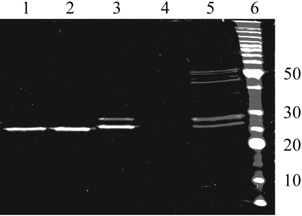
SDS–PAGE analysis of the purified recombinant β2-toxin was performed using 12% polyacrylamide gels according to the method of Laemmli (1970 ▶) using Bio-Rad equipment (Bio-Rad, Hercules, CA, USA). The proteins were visualized by staining with Coomassie brilliant blue (Fig. 1 ▶). The concentration of the recombinant β2-toxin was determined using a BCA Protein Assay kit (Pierce, IL, USA).
Figure 1.
SDS–PAGE analysis of cloned β2-toxin from C. perfringens in vector pGEX-4T. Lanes 1 and 2, purified β2-toxin. Lane 3, GST and β2-toxin after thrombin cleavage. Lane 4, GST-column flowthrough. Lane 5, BL21 cell lysate. Lane 6, molecular-weight markers (kDa).
2.2. Dynamic light scattering
Dynamic light-scattering measurements were made using a Viscotek 802 optical system equipped with a 60 mW diode laser at a wavelength of 830 nm. Ten light-scattering runs of 10 s each indicated that the sample was monodisperse and had a single mass distribution at a radius of 3.2 nm, with an approximate molecular weight of 51 kDa. The predicted molecular weight suggested the presence of a dimer in solution.
2.3. Crystallization of C. perfringens β2-toxin
The initial search for crystallization condition was carried out at the Hauptman–Woodward Institute (HWI) in Buffalo, New York. A purified C. perfringens β2-toxin (800 µl, 10 mg ml−1 in 10 mM Tris buffer) was sent to HWI for screening of preliminary crystallization conditions. Small crystals were observed in some conditions containing PEG 3350 and 0.2 M potassium sulfate, 0.2 M sodium sulfate, 0.2 M lithium sulfate, 0.2 M magnesium acetate or 0.2 M ammonium sulfate. These promising conditions and further X-ray diffraction screening were pursued at the Pennsylvania State University macromolecular crystallographic facility. The crystallization experiments were performed using the batch-under-oil method with 96-well microbatch plates from Hampton Research. Custom grids were designed with the leads from the initial screening. Controls were set up for each condition to confirm that the crystals observed were indeed those of the protein. A dye test (Hampton Izit) was used to further confirm that the crystals grown were of protein.
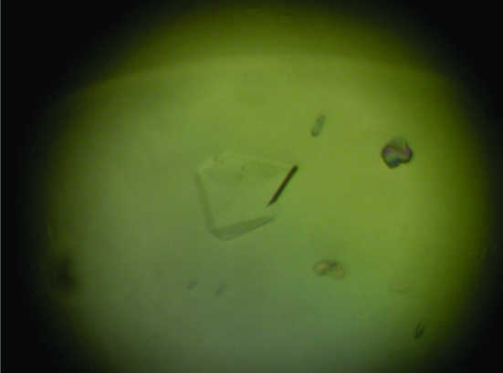
Each well in the microbatch plate contained 4 µl crystallization buffer (0.2 M lithium sulfate, 28–36% PEG 3350) and 4 µl protein solution (concentration 10 mg ml−1) layered under 20 µl mineral oil. Triangular prismatic or rhombohedron-shaped crystals appeared in conditions comprised of 32–36% PEG 3350 after one week (Fig. 2 ▶) and in conditions comprised of 28–30% PEG 3350 in about two weeks.
Figure 2.
Triangular prism-shaped crystal of β2-toxin measuring about 200 µm.
These crystals were well defined, with the long axis of the rhombus measuring about 200 µm in length. However, the crystals were extremely fragile and shattered easily when picked up with a nylon loop. An additive screen obtained from Hampton Research was used to address this problem. It was observed that when used as an additive, approximately 12% 1,1,1,3,3,3-hexafluoro-2-propanol made the crystals sufficiently robust for handling.
2.4. X-ray screening, data collection and processing
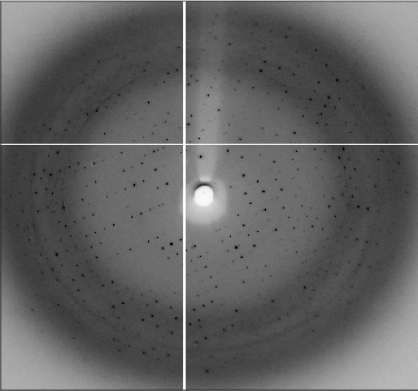
The oil above the crystallization drop was carefully removed prior to crystal mounting. Single crystals were picked up in a nylon loop (0.2 µm) and frozen in a stream of cold nitrogen (93 K). Since the crystals were grown in a buffer containing a high percentage of PEG 3350 (28–36%), no other cryoprotectant was necessary. The initial crystal screening for X-ray diffraction showed diffraction spots to 4 Å. Our attempts to improve diffraction by gradual dehydration by increasing the PEG concentration to 38% and 40% were not effective. Soaking the crystals in 5–10% glycerol prior to freezing improved the diffraction to 3.07 Å. Diffraction to 2.9 Å was observed when a cryo-annealing technique was employed in which the frozen crystal was thawed for about 10 s and frozen again. However, longer thawing and refreezing caused the diffraction to deteriorate. X-ray diffraction data were collected to 2.9 Å resolution at the synchrotron beamline at the F2 station at CHESS, Cornell on an ADSC Quantum-210 CCD detector (Fig. 3 ▶). The overall R merge was high at 12.9% and the R merge value in the highest resolution bin was 57.8% owing to the overall data quality and weak diffraction at high resolution. The preliminary X-ray crystallographic information is listed in Table 1 ▶. The data were integrated using DENZO and scaled and merged using SCALEPACK (Otwinowski, 1993 ▶).
Figure 3.
X-ray diffraction image from a β2-toxin crystal, showing diffraction spots to 2.9 Å resolution.
Table 1. Data-collection statistics.
| Unit-cell parameters (Å, °) | a = 108.13, b = 108.13, c = 195.52, α = 90, β = 90, γ = 120 |
| Space group | R3 |
| Matthews coefficient (Å3 Da−1) | 3.93 |
| No. of molecules per ASU | 2 |
| Solvent content (%) | 68.69 |
| Resolution of native data (Å) | 2.9 |
| No. of reflections observed | 108011 |
| No. of unique reflections | 19279 |
| Linear merging R factor (%) | 12.9 |
| Polar angle ω defining NCS (°) | 90.0 |
| Polar angle ϕ defining NCS (°) | 210.3 |
2.5. Noncrystallographic symmetry (NCS)
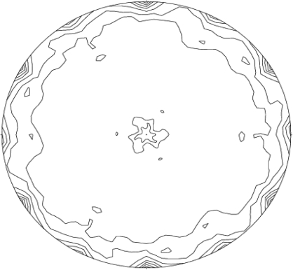
A self-rotation function calculation was carried out using the program POLARRFN (Collaborative Computational Project, Number 4, 1994 ▶). The axis of noncrystallographic twofold symmetry relating the two monomers was clearly seen in the κ = 180° section (Fig. 4 ▶). Native data from 3.5 to 8 Å and a Patterson radius of 28 Å were found to be most suitable after trials using other possible values for these parameters. The highest peak after the origin peaks were on the κ = 180° section and corresponded to the NCS twofold. There were no additional peaks seen at other κ sections, ruling out any other possibility. The polar angles defining the NCS twofold are ω = 90.0, ϕ = 210.3°. Through crystallographic symmetry, the NCS twofold is close to being parallel to the y axis. However, processing the X-ray data in R32 leads to very high merging R values and the rejection of a high number of reflections.
Figure 4.
Stereographic projection of the κ = 180° section of the self-rotation function. The peaks around the outer edge are the positions of noncrystallographic twofolds. The polar angle ω varies from 0 to 180° at the center to 90 to 270° at the edge; ϕ varies from 0 to 360° around the circle. The polar angles defining the NCS twofold are ω = 90.0, ϕ = 210.3°.
3. Conclusions
C. perfringens β2-toxin was successfully cloned, expressed and purified using glutathione sepharose affinity chromatography and crystallized using the batch-under-oil technique. Preliminary crystallographic analysis showed that the crystal obtained belongs to the primitive rhombohedral space group R3, with unit-cell parameters a = 108.13, b = 108.13, c = 195.52 Å in the hexagonal setting. As no homologous protein structures are available in the Protein Data Bank, it is not possible to use molecular replacement for structure solution. Attempts are under way to solve the phase problem using the multiple isomorphous replacement method. We are also in the process of cloning and purifying a selenomethionine derivative of the protein in order to solve the structure using the multiwavelength anomalous dispersion method. Structure determination of the β2-toxin will provide a molecular picture of the residues responsible for its antigenicity and membrane association and provide clues in our efforts to develop a better dairy vaccine.
Acknowledgments
We would like to thank the staff at Hauptman–Woodward Medical Research Institute for the initial robotic screening. Special thanks are given to the staff, especially Irina Kriksunov, at the F2 station for help with data collection at CHESS.
References
- Al-khaldi, S. F., Myers, K. M., Rasooly, A. & Chizhikov, V. (2004). Mol. Cell. Probes, 18, 359–367. [DOI] [PubMed] [Google Scholar]
- Boujon, P., Henzi, M., Penseyres, J. H. & Belloy, L. (2005). Vet. Rec.156, 746–747. [DOI] [PubMed] [Google Scholar]
- Collaborative Computational Project, Number 4 (1994). Acta Cryst. D50, 760–763. [Google Scholar]
- Daube, G. (1992). Ann. Med. Vet.136, 5–30.
- Fisher, D. J., Miyamoto, K., Harrison, B., Akimoto, S., Sarker, M. R. & McClane, B. A. (2005). Mol. Microbiol.56, 747–762. [DOI] [PubMed] [Google Scholar]
- Garmory, H. S., Chanter, N., French, N. P., Bueschel, D., Songer, J. G. & Titball, R. W. (2000). Epidemiol. Infect.124, 61–67. [DOI] [PMC free article] [PubMed] [Google Scholar]
- Herholz, C., Miserez, R., Nicolet, J., Frey, J., Popoff, M. R., Gibert, M., Gerber, H. & Straub, R. (1999). J. Clin. Microbiol.37, 358–361. [DOI] [PMC free article] [PubMed] [Google Scholar]
- Klaasen, H. L., Molkenboer, M. J., Bakker, J., Miserez, R., Hani, H., Fery, J., Popoff, M. R. & van den Bosch, J. F. (1999). FEMS Immunol. Med. Microbiol.24, 325–332. [DOI] [PubMed] [Google Scholar]
- Laemmli, U. K. (1970). Nature (London), 227, 680–685. [DOI] [PubMed] [Google Scholar]
- Manteca, C., Daube, G., Pirson, V., Limbourg, B., Kaeckenbeeck, A. & Mainil, J. G. (2001). Vet. Microbiol.81, 21–32. [DOI] [PubMed] [Google Scholar]
- Niilo, L. (1986). Can. Vet. J.29, 658–664.
- Otwinowski, Z. (1993). Proceedings of the CCP4 Study Weekend. Data Collection and Processing, edited by L. Sawyer, N. Isaacs & S. Bailey, pp. 56–62. Warrington: Daresbury Laboratory.
- Petit, L., Gilbert, M. & Popoff, M. R. (1999). Trends Microbiol.7, 104–110. [DOI] [PubMed] [Google Scholar]
- Songer, J. G. (1996). Clin. Microbiol. Rev.9, 216–234. [DOI] [PMC free article] [PubMed] [Google Scholar]
- Tilley, S. J. & Saibil, H. R. (2006). Curr. Opin. Struct. Biol.16, 1–7. [DOI] [PubMed] [Google Scholar]
- Waters, M., Raju, D., Garmory, H. S., Popoff, M. R. & Sarker, M. R. (2005). J. Clin. Microbiol.8, 4002–4009. [DOI] [PMC free article] [PubMed] [Retracted]


